Received on April 3, 2015. Revised on May 26, 2015. Accepted on June 4, 2015.
CC This is an open access article distributed under the terms of the Creative Commons Attribution Non-Commercial License (http://creativecommons.org/licenses/by-nc/4.0) which permits unrestricted non-commercial use, distribution, and reproduction in any me- dium, provided the original work is properly cited.
*Corresponding Authors. Jeehee Youn, Doo-Jin Paik, Department of Anatomy and Cell Biology, College of Medicine, Hanyang University, 222 Wangsimni-ro, Sungdong-gu, Seoul, Korea. Tel: 82-2-2220-0604; Fax: 82-2-2281-7841; E-mail: jhyoun@hanyang.ac.kr, paikdj@hanyang.ac.kr Abbreviations: Egr, Early growth response; NP-KLH, 4-hydroxy-3-nitrophenylacetyl-keyhole limpet hemocyanin; TD, T-dependent; TI, T-in- dependent; WT, wild-type
Early Growth Response-1 Plays a Non-redundant Role in the Differentiation of B Cells into Plasma Cells
Yeon-Kyung Oh, Eunkyeong Jang, Doo-Jin Paik* and Jeehee Youn*
Laboratory of Autoimmunology, Department of Anatomy and Cell Biology, College of Medicine, Hanyang University, Seoul 133-791, Korea
Early growth response (Egr)-1 is a Cys2-His2-type zinc- finger transcription factor. It has been shown to induce sur- vival and proliferation of immature and mature B cells, re- spectively, but its role in the differentiation of B cells into plasma cells remains unclear. To examine the effects of Egr-1 deficiency on the activation of B cells, naive B cells from Egr1−/− mice and their wild-type (WT) littermates were activated to proliferate and differentiate, and then as- sayed by FACS. Proportions of cells undergoing pro- liferation and apoptosis did not differ between Egr1−/−
and WT mice. However, Egr1−/− B cells gave rise to few- er plasma cells than WT B cells. Consistently, Egr1−/−
mice produced significantly lower titer of antigen-specific IgG than their WT littermates upon immunization. Our re- sults demonstrate that Egr-1 participates in the differ- entiation program of B cells into plasma cells, while it is dispensable for the proliferation and survival of mature B cells.
[Immune Network 2015;15(3):161-166]
Keywords: Egr1, B cells, Plasma cells, Differentiation, Antibody
INTRODUCTION
Ag challenge can activate B cells to proliferate and differ- entiate into plasma cells. Plasma cells are a unique pop- ulation of specialized cells that can produce Abs through T-dependent (TD) and T-independent (TI) pathways (1).
Abs elicit a variety of effects, such as complement activa- tion and opsonization. Therefore, plasma cells act as key players during the humoral immune response. However, the development program of plasma cell populations is not clearly understood.
The early growth response (Egr) protein family com- prises Cys2-His2-type zinc finger transcription factors, which share 90% homology in their DNA binding region (2).
Egr-1, which is also known as NGFI-A, Zif268, and Krox24, is expressed ubiquitously by many cell types and binds to a consensus DNA motif (GCGGTGGGCG) in many genes, including c-Myc, cyclins D2 and G2, and p19 (3-5). Other Egr-1 target genes identified in B cells include genes encoding TNF, IL-2, CD44, and ICAM-1, which are important for growth and functions of B cells (6).
Many studies have shown that Egr-1 participates in B cell maturation as a positive regulator. Egr-1 is crucial for B lymphopoiesis, especially in the development of pre-B cells and marginal zone B cells (7,8). Induction of Egr-1 expression following engagement with the BCR is asso-
ciated with the growth and proliferation of mature B cells, suggesting that Egr-1 plays a role in their activation (2,9).
However, it has also been observed that a deficiency in Egr-1 failed to inhibit BCR crosslinking-induced prolifera- tion of mature B cells, due to the compensatory functions of Egr-2 and -3 (8). Egr-1 can exert anti- and pro-apoptotic roles, depending on the cell type. Its anti-apoptotic role is important for the survival of immature B cells during BCR-induced growth inhibition (10,11), while its pro-apop- totic role contributes to the elimination of bortezomib-re- sistant multiple myeloma cells (12). Although transgenic mice expressing a dominant negative form of Egr-1 devel- op lower numbers of plasma cells than normal mice (8), it remains unclear whether this effect stems from the role of Egr-1 as none of the other Egr family members were expressed in these transgenic mice.
We investigated the functions of Egr-1 in B cells, and what cellular events in B cells are affected when Egr-1 is deficient. We used mice in which Egr1 was knocked out (Egr1−/−), and found that Egr-1 is involved in the dif- ferentiation of naive B cells into plasma cells. However, Egr-1 did not appear to be involved in the proliferation and apoptosis of B cells. Our results highlight the non-re- dundant role of Egr-1, which acts as a positive regulator of humoral immunity.
MATERIALS AND METHODS Mice and immunization
Egr1+/− mice on a C57BL/6 background were purchased from The Jackson Laboratory (Bar Harbor, ME, USA) and maintained in a pathogen-free barrier facility at Hanyang University (Seoul, South Korea). Egr1+/− mice were incross to generate Egr1−/− mice. When Egr1−/− mice and their wild-type (WT) littermates were 6∼12-weeks old, they were injected i.p. with a mixture of 4-hydroxy-3-nitro- phenylacetyl-keyhole limpet hemocyanin (NP-KLH; 100 μg/mouse; Biosearch Technologies) and alum (Thermo Scientific). Our study was approved by the Institutional Animal Care and Use Committee (HY-IACUC-13-053).
RT-PCR assays
Erythrocyte-depleted spleen cell fractions from WT and Egr1−/− mice were stimulated with 20 ng/ml PMA and 1 μM ionomycin (Sigma-Aldrich) for either 1 or 3 h.
Total RNA was isolated using Trizol reagent (Life Tech-
nologies). Semi-quantitative and quantitative RT-PCR as- says were conducted as described previously (13). The mRNA expression levels of each gene were normalized to that for the gene encoding β2-microglobulin (B2m). Differences between samples were normalized using the cycle thresh- old method (ΔCt). We used specific oligonucleotide pri- mers targeting Egr1 (5'-AAC CGG CCC AGC AAG ACA CC-3' and 5'-TGG CAA ACT TCC TCC CAC AAA T-3'), Egr2 (5'-CTT CAG CCG AAG TGA CCA CC-3' and 5'-GCT CTT CCG TTC CTT CTG CC-3'), Egr3 (5'-CAA CGA CAT GGG CTC CAT TC-3' and 5'-GGG CAG GCT GCC GAA TCC CG-3'), and B2m (5'-CAG TGT GAG CCA GGA TAT AG-3' and 5'-TGA CCG GCT TGT ATG CTA TC-3').
Abs and FACS
Splenic single-cell suspensions from WT and Egr1−/− mice were stained and analyzed using a FACSCanto II (BD Biosci- ences) as described previously (14). The following mAbs and reagents were purchased from BD Biosciences or eBioscience: anti-GL7-FITC, anti-CD4-PE, anti-B220-PerCP, anti-CD11C-allophycocyanin, anti-CD138-allophycocyanin, 7-AAD, and Annexin V-FITC.
B cell cultures
Naive GL7−CD138−CD11c−B220+ B cells from the spleens of WT and Egr1−/− mice were sorted using a FACSAria III
(BD Biosciences) and stained with 3 μM CFSE (Molecular Probes). Cells at 1×10⁶/ml were cultured in the presence of 20 μg/ml LPS (Sigma-Aldrich) and 10 ng/ml IL-4 (Peprotech), or 5 μg/ml goat anti-mouse IgM (Jackson Immune Research) and 10 ng/ml IL-4. After culturing for 4 days, cells were subjected to flow cytometry.
ELISA
Sera were collected from pre- and post-immunized mice and assayed by ELISA to determine levels of NP-specific Abs. In brief, sera were diluted 1:100,000 in PBS (WelGene) and dispensed into 96-well immunosorbent plates (Nunc) pre-coated with 10 μg/ml NP8 conjugated to BSA (Bio- search Technologies). A serum sample containing the high- est titer of anti-NP8 IgG was serially diluted and used as a standard. Plates were incubated with an anti-mouse IgG conjugated to biotin (Sigma-Aldrich), followed by strepta- vidin conjugated to HRP (BD Biosciences).
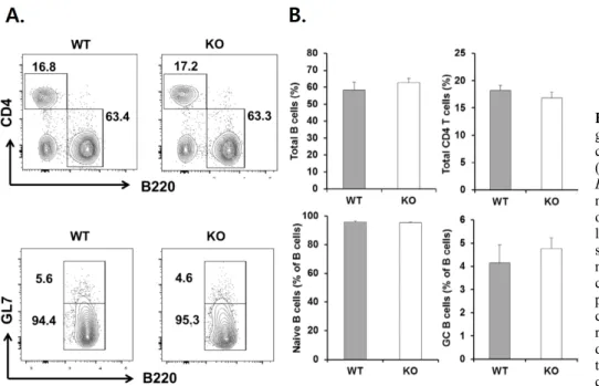
Figure 2. Analysis of naive B, germinal center B, and CD4+ T cell populations in Egr1−/− mice.
(A) Spleen cells from 12 week-old Egr1−/− mice and their WT litter- mates were stained for the presence of GL7, B220 and CD4, and ana- lyzed by FACS. The dot plots show total B (B220+), CD4+ T, naive B (B220+GL7lo), and germinal center (GC) B cell (B220+GL7hi) populations with relevant per- centages. Presented values are representative of six indepen- dent experiments. (B) The propor- tions (mean±SEM) of indicated cell compartments.
Figure 1. A selective lack of Egr-1 expression in Egr1−/− cells.
Splenocytes from WT and Egr1−/− mice were treated with PMA and ionomycin (P/I) for 1 or 3 h, and assayed by semi-quantitative (A) and quantitative RT-PCR (B). Egr1 mRNA expression was normalized to that for β2m. Data are representative of three independent experiments.
RESULTS AND DISCUSSION
Egr-1 expression was selectively abolished in Egr1−/− B cells
We examined whether depletion of Egr1 induces compen- satory expression of other Egr genes. Egr1 mRNA were detected in unstimulated WT B cells, and were more abun- dant 1 h later in response to stimulation with PMA and ionomycin. We failed to detect Egr1 mRNA in Egr1−/−
B cells, regardless of whether cells were stimulated (Fig.
1). We also found that Egr2 and Egr3 mRNA were ex- pressed at similar levels in WT and Egr1−/− B cells. Our findings show that deficient Egr1 expression did not in- duce compensatory expression of Egr2 and Egr3. Thus, our Egr1−/− knockout model appears to be appropriate for investigating the role(s) of Egr-1.
Normal development of naive and germinal center B cells in Egr1−/− mice
Given that a deficiency in Egr-1 affects normal develop- ment of B-lineage cells (7,8), we assessed whether Egr1−/−
mice have an abnormal composition of spleen cells. We observed similar proportions of B220+ and CD4+ cells among WT and Egr1−/− mice (Fig. 2). The proportions of B220+GL7− naive B cells and B220+GL7+ germinal center B cells within the whole B cell population were similar between WT and Egr1−/− mice. Therefore, Egr-1 activity does not appear to be necessary for the develop- ment of naive B cells, CD4+ T cells, or germinal center B cells.
Differentiation of Egr1−/− B cells to plasma cells is defective, while proliferation and apoptosis of Egr1−/− B cells is unaffected
We sought to determine whether a deficiency in Egr-1 af- fects the proliferation and apoptosis of Egr1−/− B cells,
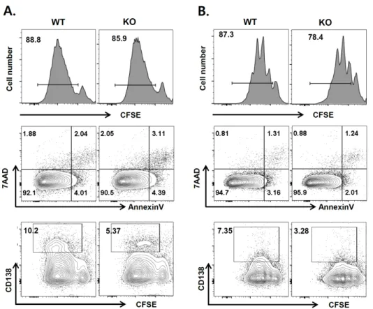
Figure 3. Responses of Egr1−/−
B cells to polyclonal stimulants.
Naive B cells sorted from spleens of WT and Egr-1−/− mice were stained with CFSE and stimulated with LPS/IL-4 (A) or anti-IgM/IL-4 (B). After 4 days, cell proliferation, apoptosis and plasma cell develop- ment were analyzed by FACS. The data presented are representative of results from more than three independent experiments. Values indicate the percentages of cells within indicated areas.
and whether naive Egr1−/− B cells can effectively differ- entiate into plasma cells. Naive B cells sorted from WT and Egr1−/− mice were labeled with CFSE and cultured with LPS plus IL-4 or anti-IgM Ab plus IL-4, and then subjected to flow cytometry. The vast majority of cells proliferated, as determined by the decrease in CFSE in- tensity (CFSEdim cells), when treated with both types of stimulants (Fig. 3). The proportions of CFSEdim cells were not significantly different between WT and Egr1−/− mice, regardless of the stimulants used (88.7±1.2 for LPS/IL-4- treated WT versus 89.5±2.1 for LPS/IL-4-treated KO, p=0.67; 88.2±1.4 for anti-IgM/IL-4-treated WT versus 82.9±4.5 for anti-IgM/IL-4-treated KO, p=0.46). In addi- tion, the proportions of early apoptotic (Annexin V+7-AAD−) and late apoptotic (Annexin V+7-AAD+) cells were equivalent between WT and Egr1−/− mice. However, the proportion of CD138+CFSEdim cells among Egr1−/−
cells was approximately 50% of that seen for WT cells.
These results demonstrate that a deficiency in Egr-1 im- paired the differentiation of B cells into plasma cells, while proliferation and apoptosis of these cells were unaffected.
Thus, Egr-1 appears to play an important role in plasma cell differentiation, and its depletion cannot be adequately compensated for by other Egr family members. Results from previous studies have shown that Egr-1 activity is as- sociated with the proliferation of mature B lymphoma cell lines (2,9). Therefore, it is likely that, rather than not par- ticipating in proliferation, the functions of Egr-1 are re- dundant and compensated for by other Egr family mem- bers, as suggested previously (8).
Egr1−/− mice produce less Abs than their WT litter- mates
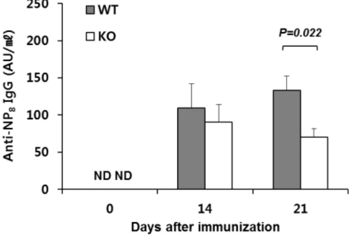
To validate the in vitro effects of Egr-1 deficiency on plas- ma cell development, we immunized Egr1−/− mice and their WT littermates with NP-KLH and alum. We then de- termined serum titers of Abs with high affinity for NP at 14 and 21 days post-immunization (dpi). The anti-NP8 IgG was detected at both dpi; its titer was significantly lower in Egr1−/− mice at 21, but not 14, dpi than that in WT mice (Fig. 4). Our findings reveal that Egr-1 is involved in the production of Abs, possibly by promoting the devel-
Figure 4. Reduced Ab production in Egr1−/− mice. WT and Egr1−/− mice were immunized with NP-KLH/alum, and serum was collected at 14 and 21 days after immunization. Titers of NP-specific IgG were detetmined by ELISA. Values represent the mean±SEM arbitrary unit (AU)/ml, pooled from three in- dependent experiments (n=10∼12 per group). p-values were determined using unpaired two-tailed Student’s t-tests. ND, not detected.
opment of plasma cells through a TD pathway.
Results from several studies have indicated that Egr-1 is possibly associated with plasma cell development. When chronic lymphocytic leukemia B cells were stimulated with phorbol esters, Egr-1 was upregulated, giving rise to cells with a plasmacytic phenotype (15). Egr-1 could bind to the GC-box element of human PRDM1, which encodes Blimp-1, a master regulator of plasma cell differentiation (16). In the light of this result, we speculate that the effect of Egr-1 in the differentiation of B cells into plasma cells may be mediated by the induction of Blimp-1 expression. Trans- genic mice lacking all Egr family members contain lower numbers of Ab-secreting cells than their WT counterparts (8). However, these studies have provided relatively little information regarding the role of Egr-1 during plasma cell differentiation.
In summary, we found that the proliferation and death of B cells was unaffected by the absence of Egr-1, indicat- ing that Egr-1 is not required for these processes. We also showed that Egr1−/− B cells could not efficiently differ- entiate into plasma cells in vitro and in vivo, which was a strong indicator of the crucial and non-redundant role of Egr-1 in the commitment of B cells to a plasma cell fate.
ACKNOWLEDGEMENTS
This work was supported by a NRF grant funded by the Ministry of Science, ICT & Future Planning (NRF-2014 R1A2A1A11052070 awarded to J. Youn).
CONFLICTS OF INTEREST
The authors have no financial conflict of interest.
REFERENCES
1. Tarlinton, D., A. Radbruch, F. Hiepe, and T. Dorner. 2008. Plasma cell differentiation and survival. Curr. Opin. Immunol. 20: 162-169.
2. Gomez-Martin, D., M. Diaz-Zamudio, M. Galindo-Campos, and J. Alcocer-Varela. 2010. Early growth response transcription fac- tors and the modulation of immune response: implications towards autoimmunity. Autoimmun. Rev. 9: 454-458.
3. Adamson, E., I. de Belle, S. Mittal, Y. Wang, J. Hayakawa, K.
Korkmaz, D. O'Hagan, M. McClelland, and D. Mercola. 2003.
Egr1 signaling in prostate cancer. Cancer Biol. Ther. 2: 617-622.
4. Virolle, T., A. Krones-Herzig, V. Baron, G. De Gregorio, E. D.
Adamson, and D. Mercola. 2003. Egr1 promotes growth and sur- vival of prostate cancer cells. Identification of novel Egr1 target genes. J. Biol. Chem. 278: 11802-11810.
5. Adamson, E. D., and D. Mercola. 2002. Egr1 transcription factor:
multiple roles in prostate tumor cell growth and survival. Tumour Biol. 23: 93-102.
6. McMahon, S. B., and J. G. Monroe. 1996. The role of early growth response gene 1 (egr-1) in regulation of the immune response. J. Leukoc. Biol. 60: 159-166.
7. Dinkel, A., K. Warnatz, B. Ledermann, A. Rolink, P. F. Zipfel, K. Burki, and H. Eibel. 1998. The transcription factor early growth response 1 (Egr-1) advances differentiation of pre-B and immature B cells. J. Exp. Med. 188: 2215-2224.
8. Gururajan, M., A. Simmons, T. Dasu, B. T. Spear, C. Calulot, D. A. Robertson, D. L. Wiest, J. G. Monroe, and S. Bondada.
2008. Early growth response genes regulate B cell development, proliferation, and immune response. J. Immunol. 181: 4590-4602.
9. Seyfert, V. L., S. McMahon, W. Glenn, X. M. Cao, V. P. Sukhatme, and J. G. Monroe. 1990. Egr-1 expression in surface Ig-mediated B cell activation. Kinetics and association with protein kinase C activation. J. Immunol. 145: 3647-3653.
10. Ke, J., M. Gururajan, A. Kumar, A. Simmons, L. Turcios, R. L.
Chelvarajan, D. M. Cohen, D. L. Wiest, J. G. Monroe, and S.
Bondada. 2006. The role of MAPKs in B cell receptor-induced down-regulation of Egr-1 in immature B lymphoma cells. J. Biol.
Chem. 281: 39806-39818.
11. Seyfert, V. L., S. B. McMahon, W. D. Glenn, A. J. Yellen, V.
P. Sukhatme, X. M. Cao, and J. G. Monroe. 1990. Methylation of an immediate-early inducible gene as a mechanism for B cell tolerance induction. Science 250: 797-800.
12. Chen, L., S. Wang, Y. Zhou, X. Wu, I. Entin, J. Epstein, S.
Yaccoby, W. Xiong, B. Barlogie, J. D. Shaughnessy, Jr., and F.
Zhan. 2010. Identification of early growth response protein 1 (EGR-1) as a novel target for JUN-induced apoptosis in multiple myeloma. Blood 115: 61-70.
13. Jang, E., W. S. Cho, M. L. Cho, H. J. Park, H. J. Oh, S. M. Kang, D. J. Paik, and J. Youn. 2011. Foxp3+ regulatory T cells control humoral autoimmunity by suppressing the development of long- lived plasma cells. J. Immunol. 186: 1546-1553.
14. Kim, S., K. Park, J. Choi, E. Jang, D. J. Paik, R. H. Seong, and J. Youn. 2015. Foxp3+ regulatory T cells ensure B lymphopoiesis by inhibiting the granulopoietic activity of effector T cells in
mouse bone marrow. Eur. J. Immunol. 45: 167-179.
15. Segel, G. B., T. J. Woodlock, J. Xu, L. Li, R. E. Felgar, D. H.
Ryan, M. A. Lichtman, and N. Wang. 2003. Early gene activation in chronic leukemic B lymphocytes induced toward a plasma cell phenotype. Blood Cells Mol. Dis. 30: 277-287.
16. Mora-Lopez, F., N. Pedreno-Horrillo, L. Delgado-Perez, J. A.
Brieva, and A. Campos-Caro. 2008. Transcription of PRDM1, the master regulator for plasma cell differentiation, depends on an SP1/SP3/EGR-1 GC-box. Eur. J. Immunol. 38: 2316-2324.