28
28 THE EWHA MEDICAL JOURNALTHE EWHA MEDICAL JOURNAL
Solitary Rectal Ulcer Syndrome Mimicking Rectal Cancer
Young Min Choi, Hyun Joo Song, Min Jung Kim
1, Weon Young Chang
1, Bong Soo Kim
2, Chang Lim Hyun
3Departments of Internal Medicine, 1Surgery, 2Radiology, and 3Pathology, Jeju National University School of Medicine, Jeju, Korea
Introduction
Solitary rectal ulcer syndrome (SRUS) is a rare benign and chronic rectal disease that has a wide spectrum of clinical pre- sentations and variable endoscopic findings [1]. Rectal bleed- ing and abdominal pain are known as the main symptoms of SRUS. Usually, it is misdiagnosed through colonoscopy [2];
SRUS mimicking rectal cancer according to endoscopic findings, abdominopelvic computed tomography (CT), positron emission tomography (PET)-CT, and magnetic resonance imaging (MRI) has been very rarely reported [3]. We report the case of a 68-year-old man who presented with an ulcerated mass of the rectum representing an SRUS variant.
Case
A 68-year-old man was referred to Jeju National Univer- sity Hospital with anal pain and difficulty in passing stool. The patient had a history of hypertension and internal hemorrhoids.
His vital signs were normal. There was no history of self-dig- itation, fever, weight loss, or abdominal pain. On initial evalu- ation, the abdomen was soft, bowel sounds were normal, and there was no tenderness in the abdomen. Rectal examination revealed an irregular broad-based ulcerated mass in the rectum.
Initial laboratory findings were normal (hemoglobin level 16.3 g/dL, white blood cell count 8,900/mm3, and platelet count 182,000/mm3). Additionally, liver function test, erythrocyte sedimentation rate, C-reactive protein, and coagulation profile were normal. Carcinoembryonic antigen (CEA) level was 1.72 ng/mL (normal, 0 to 5.0 ng/mL). Stool occult blood test was negative.
Colonoscopy showed a hemorrhagic and circumferential ulcer- ated mass with edema in the anterior rectal wall located 5 cm from the anal verge (Fig. 1A). Abdominopelvic CT exhibited an area of rectal wall thickening with perirectal fatty infiltration and enlargement of multiple small mesocolic lymph nodes, con- sistent with rectal cancer (Fig. 2). However, two repeat biopsies showed ulceration with inflammatory reaction. There was no
Case Report
Ewha Med J 2016;39(1):28-31
http://dx.doi.org/10.12771/emj.2016.39.1.28 pISSN 2234-3180 • eISSN 2234-2591
Solitary rectal ulcer syndrome (SRUS) is a rare benign and chronic rectal disease that has a wide spectrum of clinical presentations and variable endoscopic findings. It is usually diagnosed by histopathological examination through biopsy. A 68-year-old man was referred to our hospital with anal pain and difficulty on bowel movement. Colo- noscopy showed a hemorrhagic ulcerated mass in the rectum. All radiologic findings such as abdominopelvic computed tomography (CT), positron emission tomography- CT and magnetic resonance imaging were suspicious of rectal cancer. Although the patient underwent repeat endoscopic biopsy and one surgical biopsy, the results were not indicative of malignancy. Two months after conservative management, clinical symptoms and colonoscopic findings were markedly improved. Thus, we report this rare case of a 68-year-old man who had a central ulcerated mass that mimicked rectal cancer on gross colonoscopic and radiologic findings, representing an SRUS variant.
(Ewha Med J 2016;39(1):28-31)
Received August 6, 2015 Accepted January 12, 2016 Corresponding author Hyun Joo Song
Department of Internal Medicine, Jeju National University Hospital, 15 Aran 13-gil, Jeju 63241, Korea
Tel: 82-64-754-8142, Fax: 82-64-717-1131 E-mail: songhj@jejunu.ac.kr
Key Words
Solitary; Ulcer; Syndrome; Rectal neoplasms
29
THE EWHA MEDICAL JOURNAL Solitary Rectal Ulcer Syndrome Mimicking Rectal Cancer
evidence of malignancy. However, PET-CT was highly sug- gestive of rectal cancer (maximum standardized uptake value, SUVmax 15.4) with intense fludeoxyglucose uptake in multiple lymph nodes (SUVmax 8.1) (Fig. 3A, B). Furthermore, MRI showed a similar finding, which was irregular wall thickening of
mid-rectum with perirectal infiltration (Fig. 3C). Patient un- derwent repeat biopsy under spinal anesthesia. Incisional biopsy showed a central ulcerated lesion with the surrounding edema invading the muscle layer. A third biopsy also exhibited ulcer- ation with inflammatory reaction (Fig. 4). Two months after
A B
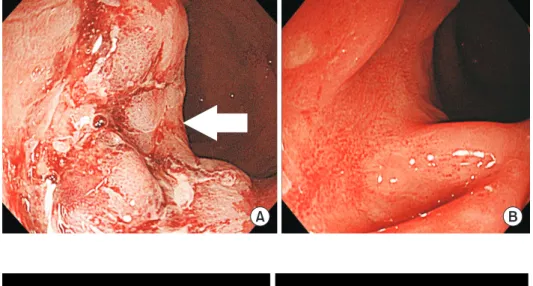
Fig. 1. Endoscopic findings. (A) Colonos- copy shows a broad-based rectal lesion with irregular surface and hemorrhagic ulceration (arrow) (B) Rectal lesion is markedly improved with remnant ulcer- ative scarring two months later.
A B
Fig. 2. Abdominopelvic computed to- mography (CT) findings. (A) Enhanced CT image shows rectal wall thickening (ar- row) and perirectal fatty infiltration. (B) Enlargement of multiple small mesocolic lymph nodes are observed (arrows).
A B C
Fig. 3. Radiologic findings. (A) Positron emission tomography-computed tomography shows intense fludeoxyglucose uptake (arrow) in rectum (maximum standardized uptake value, SUVmax 15.4), and (B) multiple enlarged mesocolic lymph nodes (arrows) with intense FDG uptake (SUV- max 8.1). (C) Magnetic resonance imaging reveals sagittal T2-weighted image of the pelvis showing irregular wall thickening of mid rectum (ar- row).
30 THE EWHA MEDICAL JOURNAL Choi YM, et al
conservative management with stool softener and pain control, clinical symptoms of patient improved. Follow-up colonoscopy showed that the lesion markedly improved with remnant ulcer- ative scarring (Fig. 1B).
Discussion
SRUS is an uncommon disorder of benign and chronic rectal disease with diverse spectrum of clinicopathological abnormali- ties [4-9]. The mean age at presentation is 30s to 40s, with a wide range from 10 to more than 80 years [10]. Although the etiology is presumed to be diverse, the pathogenesis is not entirely understood. First, finger insertion and suppository may cause direct injury [11]. Second, it can be caused by mucosal trauma and ischemia which can be explained by rectal prolapse and paradoxical compression of the pelvic floor [10,12]. There are various symptoms of SRUS [4]. In a single center study, rectal bleeding was present in 82%, abdominal pain in 49%, constipation in 23% and diarrhea in 22%. Histopathological ex- amination is a key to the diagnosis of SRUS. Diagnosis of SRUS is by rule-out of other diseases, ultimately through biopsy. Ra- diologic examination can be done such as abdominopelvic CT or MRI. However, accurate diagnosis is not always possible, and the treatment is still not established. Since SRUS has a benign disease course, it can be managed with conservative treatment.
Patient education and behavioral modification are the first step in the treatment of SRUS, including avoidance of straining and anal digitation, and ingestion of high fiber diet and bulk laxa- tives. If the symptoms do not improve, mucosal prolapse must be suspected. In the selected patient, biofeedback and surgical treatment may be considered. Using a stool softener can reduce straining during defecation and corticosteroid or mesalazine can also be administered. However, their effectiveness has been not proven. If it cannot be treated medically, surgical treatment may be considered [4].
To date, there have been several reports regarding SRUS [3,9,11,13,14]. Colonoscopic findings with suspected malignancy were variable, including tumor, malignant stricture, and ulcer- ative mass. MRI was performed frequently as an additional test.
In some cases, malignancy and inflammation could not be dif- ferentiated. Surgery was performed in several cases. One patient who underwent surgery had intractable symptoms after a short course of ineffective conservative therapy and occult malignancy could not be excluded [15]. In another patient, the ulcer had expanded very quickly to cause a case underwent a second deep biopsy. In most cases, the first biopsy was performed superfi- cially. Some cases underwent a second deep biopsy. Interest- ingly, they revealed rectal cancer on superficial biopsy initially.
However, rectal cancer was ultimately excluded by follow-up circumferential stricture of the rectum [9]. Most cases were
A B
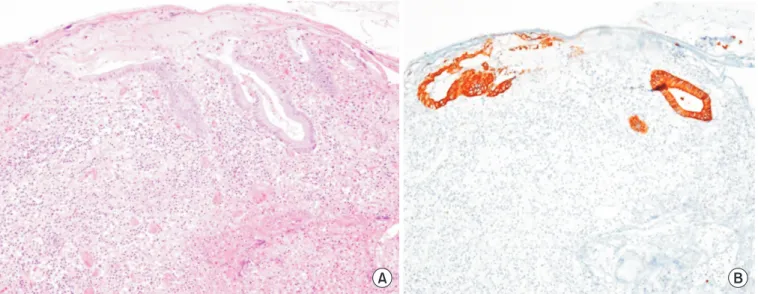
Fig. 4. Pathologic findings. (A) There is surface erosion and ulceration, characterized by discontinuity of the epithelium with acute inflammation and exudates. The ulcerated rectal mucosa and submucosa reveals formation of granulation tissue characterized by an admixture of small ves- sels, numerous inflammatory cells, fibrin and edema (H&E, ×100). (B) Distorted crypts are strongly positive for cytokeratin staining (×100).
31
THE EWHA MEDICAL JOURNAL Solitary Rectal Ulcer Syndrome Mimicking Rectal Cancer
treated conservatively, and surgery was avoided [14].
In this case, rectal cancer was strongly suspected in the be- ginning. On biopsy, only inflammation was revealed. However, confirmation was necessary. The patient did not use finger pen- etration. Pelvic floor dysfunction and rectal prolapse were not visible. On general diagnostic imaging modalities, neither CT nor MRI had distinguished the inflammation from rectal cancer in a previous report [3]. Even PET-CT was not helpful. Fol- lowing a number of tests, SRUS was considered, and ultimately diagnosed. In contrast to this case, other cases were initially di- agnosed as SRUS and later changed to that of malignancy [14].
Therefore, it is necessary to distinguish carefully for malignancy.
Repetitive biopsies are strongly recommended.
In conclusion, SRUS should always be considered in patients with malignant-mimicking rectal cancer. However, we believe it is important not to miss a diagnosis of rectal cancer over the diagnosis of SRUS. We report the case of an SRUS patient who had a central ulcerated mass lesion that mimicked rectal cancer on gross colonoscopic and radiologic findings.
References
1. Abid S, Khawaja A, Bhimani SA, Ahmad Z, Hamid S, Jafri W. The clinical, endoscopic and histological spectrum of the solitary rectal ulcer syndrome: a single-center experience of 116 cases.
BMC Gastroenterol 2012;12:72.
2. Umar SB, Efron JE, Heigh RI. An interesting case of mistaken identity. Case Rep Gastroenterol 2008;2:308-313.
3. Amaechi I, Papagrigoriadis S, Hizbullah S, Ryan SM. Solitary rectal ulcer syndrome mimicking rectal neoplasm on MRI. Br J Radiol 2010;83:e221-224.
4. Zhu QC, Shen RR, Qin HL, Wang Y. Solitary rectal ulcer syn- drome: clinical features, pathophysiology, diagnosis and treat-
ment strategies. World J Gastroenterol 2014;20:738-744.
5. Gordon PH, Nivatvongs S. Solitary rectal ulcer syndrome of the rectum. In: Gordon PH, Nivatvongs S, editors. Principle and practice of surgery for the colon, rectum and anus. 2nd ed. St.
Louis, MO: Quality Medical Publisher; 1999. p.1403-1409.
6. Lawler LP, Fleshman JW. Solitary rectal ulcer, rectocele, hemor- rhoids and pelvic pain. In: Pemberton JH, Swash M, Henry MM, editors. The pelvic floor, its function and disorders. London: WB Saunders; 2002. p.358-362.
7. Chong VH, Jalihal A. Solitary rectal ulcer syndrome: characteris- tics, outcomes and predictive profiles for persistent bleeding per rectum. Singapore Med J 2006;47:1063-1068.
8. Ingle SB, Patle YG, Murdeshwar HG, Hinge Ingle CR. An un- usual case of solitary rectal ulcer syndrome mimicking inflam- matory bowel disease and malignancy. Arab J Gastroenterol 2012;13:102.
9. Perrakis E, Vezakis A, Velimezis G, Filippou D. Solitary rectal ulcer mimicking a malignant stricture. A case report. Rom J Gas- troenterol 2005;14:289-291.
10. Tjandra JJ, Fazio VW, Church JM, Lavery IC, Oakley JR, Milsom JW. Clinical conundrum of solitary rectal ulcer. Dis Colon Rec- tum 1992;35:227-234.
11. Wong WM, Lai KC, Shek TW, Lam SK. Self-inflicted rectal ulcer with exuberant granulation tissue: a lesion that mimics carci- noma. Gastrointest Endosc 2002;55:951-952.
12. Al-Brahim N, Al-Awadhi N, Al-Enezi S, Alsurayei S, Ahmad M.
Solitary rectal ulcer syndrome: a clinicopathological study of 13 cases. Saudi J Gastroenterol 2009;15:188-192.
13. Park HJ, Kim WH, Woo JS, Han KH, Lee SI, Park IS, et al. Solitary rectal ulcer syndrome. Yonsei Med J 1994;35:223-230.
14. Blanco F, Frasson M, Flor-Lorente B, Minguez M, Esclapez P, Garcia-Granero E. Solitary rectal ulcer: ultrasonographic and magnetic resonance imaging patterns mimicking rectal cancer.
Eur J Gastroenterol Hepatol 2011;23:1262-1266.
15. Delgado J, Delgado B, Sztarkier I, Sperber AD, Walfisch S. A soli- tary rectal ulcer mimicking rectal cancer. Gastrointest Endosc 2005;62:309.