NC/Nga 생쥐에서 분리한 T 세포에서 foxp3+ 세포 활성화에 대한 PGA-LM 의 효과
장순남1·김금란1·강상모1,2
*
1건국대학교 대학원 생물공학과, 2건국대학교 미생물공학과
Effects of PGA-LM on CD4+CD25+foxp3+ Treg Cell Activation in Isolated CD4+ T Cells in NC/Nga Mice. Jang, Soon-Nam1, Kum-Lan Kim1, and Sang-Mo Kang1,2*. 1Department of Bioengineering, Gradu- ate School at Konkuk University, Seoul 143-701, Korea, 2Department of Microbial Engineering, Konkuk Univer- sity, Seoul, 143-701, Korea − Poly-γ-glutamic acid (γ-PGA) was mixed natural flora of Bacillus subtilis, contaminated from cooked soybeans. Also, it was performed to find out the antiallergic activity by using NC/
Nga mice, in vitro. The γ-PGA (PGA-HM : PGA-high molecular weight), Molecular weight 300 kDa, was decomposed and made PGA-LM (PGA-low molecular weight) which has molecular weight below 30 kDa by sonication. Therefore, it was same result between PGA-HM and PGA-LM, and reported PGA-LM as basic result. We found that PGA-LM contains antiallergic efficacy that inhibit B cells and Th2 cells activation from isolated CD4+T cells in NC/Nga atopic dermatitis model mice, and not show a cytotoxicity in the hFCs. To investigate the effects of these PGA-LM in vitro, isolation of splenic B cell and CD4+ T cells in atopic derma- titis mice were used. To elucidate the role of PGA-LM in anti-CD40+ interleukin-4 (IL-4)-mediated B-cell activation, showed that the capacity of B cells to expression IL-1β, IL-6, and TNF-α mRNA down-regulated, and IL-10 mRNA up-regulation by PGA-LM treatment, but it had no effect on TGF-β expression. In addition to CD4+IFN-γ+ and CD4+CD25+foxp3+, the functions of PGA-LM in the development of the CD4+
CD25+foxp3+ and CD4+IFN-γ+ cells, the phenotype and functions of PGA-LM induced CD4+CD25+
foxp3+, and CD4+IFN-γ+cells in CD4+ T cells. These results suggested that PGA-LM could change cytokine production and generate CD4+CD25+foxp3+Tregs in NC/Nga mice, and may be effective for immunotherapy in patients with AD.
Key words: γ-PGA, CD4+CD25+foxp3+ Treg, NC/Nga
서 론
기존의 아토피 피부염은 IgE에 감작된 비만세포가 히스타 민(histamine) 등의 화학매체들을 분비시켜 부종, 붉은 반점 및 소양 등과 같은 증상들이 발생되는 것으로 알려져 왔으 나[55], 만성 염증상태를 나타내는 이유와 아토피 피부염환 자에서 흔히 보이는 세포매개성 면역기능의 저하를 충분하 게 설명할 수 없었다[42]. 그 후 세포성 면역에서 중요한 위 치를 차지하는 T 림프절구 아형 또는 그와 관련된 사이토카 인(cytokine)이 면역체계에 관여한다는 사실의 규명으로 이 부분에 대한 설명이 가능해졌고, 세포면역을 담당하는 T 세 포 및 그 아형의 이상, 그로 인해 나타나는 사이토카인의 불 균형이 아토피 피부염을 일으키는 주요 기전이 된다고 설명 하고 있다[35, 36].
최근 CD4+ T 림프구 등의 염증성 세포가 아토피 피부염 의 발생에 중요한 역할을 하는 것으로 보고되고 있는데, T 림프구는 Th1 세포와 Th2 세포로 분화하며[28], Th2 세포 는 interleukin(IL)-4, IL-5, IL-9, IL-13을, Th1 세포는 IL- 2, IFN-γ, TNF-α를 생성하는데, 이 중 Th2 세포에서 분비되 는 사이토카인이 아토피의 발생에 중요한 역할을 한다[1].
Th1 및 Th2 분화에 중요한 역할을 하는 전사인자에는 T-box expressed in T-cells(T-bet), STAT-1, STAT-4, STAT-6, GATA- binding protein-3(GATA-3), c-maf 등이 알려져 있는데 T- bet, STAT-1, STAT-4 등의 전사인자가 활성화되면 Th1 림프 구로의 분화가 촉진되고, STAT-6, GATA-3, c-maf, NFAT 등의 전사인자가 활성화되면 Th2 림프구로의 분화가 촉진 된다[2]. 특히, GATA-3와 T-bet은 신호전달과정에서 유전자 의 전사를 조절하는 상위에 위치하는 전사인자이므로 Th1 및 Th2의 분화에 있어 가장 중요한 전사인자라 할 수 있다 [47, 48]. GATA-3의 활성화로 인해 Th2 세포가 활성화되면 여러 Th2 사이토카인의 생성, 분비가 증가하게 되고 B세포 활성화가 이루어지므로 알레르기 염증반응을 매개하여 아토
*Corresponding author
Tel: 82-2-450-3524, Fax: 82-2-3437-8360 E-mail: [email protected]
피 피부염, 기관지 천식 등의 만성적인 질환을 유발한다[49, 54]. 2003년 Fontenot와 Curotto는 foxp3+ 조절 T 세포 (regulatory T cell, Treg cell)가 직·간접적으로 알레르기 염 증을 조절하여 호산구의 침윤, 염증 IFN-γ 생산 세포 등에 관여함으로써 foxp3+ Treg 세포의 활성화가 자가면역질환 치료에 획기적인 전기가 될 수 있는 결과를 발표하였다[10, 13].
아토피 피부염에 사용되고 있는 치료제들을 살펴보면 연 고 형태로 glucocorticoid와 calcinulin 저해제인 FK-506 (tacrolimus)이 다용되고 있으며, 전신 투여 경우에는 항히스 타민제, 스테로이드 제제, 사이클로스포린, DNA 합성 저해 제(antimetabolite) 등이 사용되고 있으나, 다양한 부작용으 로 인하여 안전하면서도 우수한 효능을 가진 새로운 치료제 의 개발이 요구되고 있다[9, 30, 31]. 이에 본 연구는 기초 자료검색을 통하여 부작용이 없으며 장기간 복용이 가능한 천연물로 폴리감마글루탐산(Poly-γ-glutamic Acid. γ-PGA)의 효과를 검증하고자 하였다. γ-PGA는 글루탐산의 γ-카르복실 기와 글루탐산의 α-아미노기가 아마이드 결합된[6, 37] γ-폴 리펩타이드로 콩 발효식품미생물인 Bacillus sbutilis 등 Bacillus 속이 보유하고 있는 γ-PGA 합성계(γ-PGA synthetase complex, pgsBCA system)에 의해서 생산된다[8]. 우리는 흔 히 청국장 점질물의 주성분으로 γ-PGA를 자주 접하고 있으 며 상업용으로는 Bacillus 속을 탱크 배양하여 대량으로 얻 고 있다. 이러한 γ-PGA는 수용성, 음이온성, 생분해성 원료 및 식용의 아미노산 고분자소재로 고부가가치의 의약품, 화 장품, 기능성 식품, 환경용, 공업용 등으로 적용 범위가 매 우 다양하다[45]. 최근 이 등[29]의 연구에서는 γ-PGA가 대 식세포를 자극하고 TNF-α와 IL-1β의 분비를 유도하여 면역 기능을 증강시키는 역할을 하며, 함 등[17]의 연구에서 γ- PGA는 T 임파구에서 IFN-γ의 분비를 촉진하여 T 세포 및 B 세포, 대식세포, 자연살해세포(NK cell) 등을 활성화시켜 세포 내 세균 및 바이러스 감염에 대한 억제효능 및 암세포 살해에 대한 효능을 가지고 있다는 것이 보고되었다. 이와 같이 γ-PGA가 체내 면역세포를 활성화시켜 면역증강 효과 를 가지고 있다는 것이 확인되었다. 그러나 γ-PGA의 알레 르기 면역질환에 관한 연구에서 아직까지 CD4+CD25+
foxp3+ Treg 세포 활성에 관한 연구는 보고되지 않았다.
본 연구에서는 평균 분자량이 300 kDa의 고분자 γ-PGA(
이하 PGA-HM이라 한다)를 저분자화시켜 저분자 γ-PGA(이 하 PGA-LM이라 한다)를 만들고, in vitro에서 PGA-HM과
PGA-LM의 아토피 억제능을 조사한 결과 각각에서 동일한
실험결과를 얻어 PGA-LM 실험결과 중심으로 보고한다. 실 험내용은 NC/Nga 생쥐에서 B 세포와 T 세포를 순수분리 하고 PGA-LM를 처리함으로 PGA-LM이 염증 유전자 발 현억제 및 CD4+foxp3+ Treg와 IFN-γ+의 활성세포 빈도수 변화에 미치는 영향을 검토하고 아토피 억제 가능성을 조 사하였다.
재료 및 방법 실험재료
실험에 사용된 동물은 수컷 7주령의 SPF(Specific Patho- gen-free) NC/Nga mouse(15~20 g)로 Charles River Japan (Yokohama, Japan)사에서 공급받았다. 실험 당일까지 고형 사료(항생제 무첨가, 삼양사료 Co.)와 물을 충분히 공급하고 온도 22±2oC, 습도 55±15%, 12시간(light-dark cycle)의 환 경에서 1주간 적응시킨 후 in vitro 실험을 위하여 conven- tional한 조건에서 18주령까지 사육하였다. γ-PGA는 바이웰 에서 구입하였으며 평균 분자량은 300 kDa이다.
저분자 γ-PGA (PGA-LM)의 제조
증류수(Distilled water)에 PGA-HM을 10%(w/v)가 되도 록 녹여 sonicator(Branson사)에 60분간 high level(40 khz) 로 sonication하여[39] 분자량 30 kDa 이하 분리 가능한 밀 리포어필터로 여과하여 여과액을 deep freezer에서 24 hr 동 안 냉동시킨 후 freeze dryer(Ilshin사)에 넣어 3~4일 동결건 조하고 결정화시켜 사용하였다.
시약
본 실험에 사용된 시약은 diethyl pyrocarbonate(DEPC), chloroform, trichloroacetic acid, isopropanol, Tris-HCl, KCl, MgCl2, 적혈구 용혈액(ACK lysis solution), DMEM 배양액, dulbecco's phosphate buffered saline(D-PBS), Sulforhodamin B(SRB), 2-isopropanol, sodium dodecyl sulfate(SDS), PMA, ionomycin, FK506, antibiotics는 Sigma사(U.S.A.) 제품을 사 용하였으며, 우태아혈청(fetal bovine serum, FBS)은 Hyclone 사(Logan, U.S.A.) 제품을, anti-CD3-PE(phycoerythrin), anti-CD4-FITC(fluorescein isothiocyanate), anti-CCR3-PE, anti-B220-PE, anti-CD8-FITC, anti-B220-FITC, anti-CD49b- FITC, anti-CD40 mAb, rmIL-4, rmIL-10, BD Cytofix/Cyto- perm plus kit, anti-CD3 mAb, anti-foxp3-PE, anti-IFN-γ- PE, anti-CD28 mAb 등은 Pharmingen사(Torreyana, U.S.A.) 제품을, CD4+ T cell isolation kit와 B cell isolation kit는 Miltenyi Biotec사(Bergisch Gladbach, Germany) 제품을, IL- 4, IFN-γ, IL-5, IL-13 ELISA kit는 BioSource사(California, U.S.A.) 제품을, IgE, IgG1 ELISA kit는 SHIBAYAGI사 (Shibukawa, Japan) 제품을, anti-mouse CCR3 mAb와 anti-mouse CD4 mAb는 Santa-Cruz사(California, U.S.A.) 제품을, LSAB kit는 DAKO사(Glostrup, Denmark) 제품을 사용하였으며, 기타 일반 시약은 특급 시약을 사용하였다.
기기
본 실험에 사용된 기기는 CO2 incubator(Forma scientific Co., U.S.A.), deep-freezer(Sanyo, Japan), real time quanti- tative RT-PCR(Applied Biosystems, U.S.A.), homogenizer
(OMNI, U.S.A.), VarioMACS(Bergisch Gladbach, Germany), FACScalibur(BD, U.S.A.) 및 ELISA leader(Molecular Devices, U.S.A.) 등을 사용하였다.
세포독성 측정
Human fibroblast cells (hFCs) 배양:사람피부조직을 cool D-PBS로 3회 세척한 후 작은 조각으로 절단한 다음, conical tube(15 mL)에 넣어 1,400 rpm에서 5분간 원심분리하고, tube에 DMEM {containing collagenase A(5 mg/mL, BM, Indianapoilis, U.S.A.)와 DNase type(0.15 mg/mL, Sigma), antibiotics(penicillin 104U/mL, streptomycin 10 mg/mL, amphotericin B 25µg/mL)}를 넣고 37oC CO2 배양기에서 2시간 동안 배양하였다. 다음 0.5% trypsin-0.2% EDTA를 첨가한 후 30분간 계속 배양하였다. 배양 후 인산완충용액 으로 약 2회 1500 rpm에서 원심분리한 후 DMEM-10%
FBS에 1주일 동안 배양하였다. 1주일 후 0.5% trypsin-0.2%
EDTA로 hFCs 세포를 분리하여 DMEM-5% FBS 배양액에 105cells/mL 농도로 맞추어 96 well plate에 분주하였다.
SRB assay법: 세포독성측정방법은 SRB assay법[15]을 약 간 변형하여 실험에 사용하였다. hFCs 세포는 37oC, 5%
CO2 배양기에서 1시간 배양한 후 PGA-LM(최종 농도 10 mg/mL, 5 mg/mL, 1 mg/mL, 0.5 mg/mL, 0.25 mg/mL)을 48시간 동안 처리하였다. 배양종료 후에 배양액을 버리고 인 산완충용액으로 2회 세척하였다. 각 well에 50% TCA를 50 µL로 가하고 1시간 동안 4oC에 방치하였다. 증류수로 5회 세척한 다음 well plate를 공기 중에서 건조하였다. SRB (0.4%/1% acetic acid) 용액을 100 µL/well로 가하고 실온에 서 30분간 염색하였다. 그리고 0.1% acetic acid 용액으로 약 45회 세척한 다음 공기 중에서 건조하고 10 mM Tris base로 100 µL/well로 용해시켰다. 이 plate를 plate shaker (Lab-Line, U.S.A.)에서 3.5 speed로 5분간 shaking하고 ELISA reader(molecular devices, U.S.A.) 540 nm에서 흡광 도를 측정하였다.
NC/Nga 생쥐에서 T 및 B세포 분리 및 배양: 일반적인 (Conventional) 상태에서 사육한 18주령의 NC/Nga 생쥐의 눈에서 capillary 관을 이용하여 100 µL의 혈액을 채혈하여 이 중 IgE 함량이 50 µg/mL 이상이고 피부에 발진이 나타 난 NC/Nga 생쥐를 선택하여 실험에 사용하였다. 이를 아토 피피부발진(atopic dermatitis-like skin) NC/Nga 생쥐라고 하였다. 순수한 T 세포와 B 세포를 분리하기 위하여 NC/
Nga 생쥐의 비장을 적출하여 100 mesh에서 분쇄하여 비장 세포를 얻어 2000 rpm에서 5분간 원심분리로 세포를 회수 하였다. 여기에 ACK 용액(8.3 g NH4Cl, 1 g KHCO3, in 1 L of demineralized water + 0.1 mM EDTA)을 실온에서 5분 동안 처리하여 적혈구를 제거하였다. 그리고 비장세포를 2%의 FBS가 함유된 PBS(PBS/FBS)에 1×10⁸/mL로 현탁시 키고 normal rat serum을 5%되게 첨가하여 4oC에서 15분
간 blocking한 후, T 세포 분리는 biotinylated antibody cocktail for lineage {CD8, CD14, CD16, CD19, CD36, CD56, CD123, TCR γ/d, CD235a(glycophorin A)} (CD4+
T cell isolation kit, Miltenyi Biotec)를 가하였고, B 세포 분리는 biotinylated antibody cocktail for lineage {CD43 (Ly-48), CD4(L3T4), Ter-119}(B cell isolation kit, Miltenyi Biotec)를 가하여 각각 4oC에서 15분간 반응시켰다. 각각의 세포를 PBS/FBS로 세척하여 1×10⁷/mL로 현탁하였고, 20 µL의 Anti-Biotin MicroBeads를 가하여 다시 4oC에서 15분 간 반응시켰다. 이 세포를 magnetic column(CS column, Milteny Biotech)을 이용하여 PBS로 세척하여 준비해둔 VarioMACS(Milteny Bio-tech)에 장치하고, magnetic bead 가 표지된 세포를 통과시켰다. PBS로 column을 충분히 세 척한 다음 column을 통과한 부유액을 원심분리하여 lineage 음성인 T 세포와 B 세포를 각각 모았다[34].
NC/Nga 생쥐에서유전자발현분석
1) B 세포에서 RNA 분리 및 cDNA 합성
아토피피부발진 NC/Nga 생쥐에서 분리한 B 세포를 24 well plate에 1×10⁶ cell/well로 분주하고 PGA-LM(100 µg/
mL)을 처리하였고, 약물처리 1시간 후 anti-CD40 mAb (500 ng/mL)와 rmIL-4(recombinant mouse interleukin-4, 500 U/mL, PharMingen)를 넣고 6시간 동시 배양하였다. 그 리고 rmIL-10(recombinant mouse interleukin-10, 50 ng/
mL, Endogen)을 양성대조군으로 사용하였다. 배양 종료 후
배양 상층액을 제거한 후 RNAzolB 500µL를 넣고 용해될 때까지 분쇄하였다. 이 혼합 부유액에 chloroform(CHCl3) 50µL를 첨가한 후 15초간 다시 혼합하였다. 이를 얼음에 15 분간 방치한 후 13,000 rpm에서 원심분리한 후 약 200 µL 의 상층액을 회수하여 2-propanol 200 µL와 동량 혼합 후 천천히 흔들고 얼음에서 15분간 방치하였다. 이를 다시 13,000 rpm에서 원심분리한 후 80% EtOH로 수세하고 3분 간 vaccum pump에서 건조하여 RNA를 추출하였다. 추출한 RNA는 diethyl pyrocarbonate(DEPC)를 처리한 20 µL의 증 류수에 녹여 heating block 75oC에서 불 활성화시킨 후 first strand cDNA합성에 사용하였다[22]. 역전사(reverse trans- cription) 반응은 준비된 total RNA 2 µg을 DNase I(10 U/
mL) 2 U/tube를 37oC heating block에서 30분 동안 반응한 후 75oC에서 10분 동안 변성시켰다. 이에 2.5 µL 10 mM dNTPs mix, 1µL random sequence hexanucleotides(25 pmole/25µL), RNA inhibitor로서 1 µL RNase inhibitor (20 U/µL), 1 µL 100 mM DTT, 4.5 µL 5×RT buffer(250 mM Tris-HCl(pH 8.3), 375 mM KCl, 15 mM MgCl2)를 가 한 후, 1 µL의 M-mLV RT(200 U/µL)를 다시 가하고
DEPC 처리된 증류수로서 최종 부피가 20 µL가 되도록 하
였다. 이 20 µL의 반응 혼합액을 잘 섞은 뒤 2,000 rpm에서 5초 동안 원심침강하여 37oC heating block에서 60분 동안 반응시켜 first-strand cDNA를 합성한 다음, 95oC에서 5분
동안 방치하여 M-mLV RT를 불활성화시킨 후 합성이 완료 된 cDNA를 PCR에 사용하였다[27].
2) Real time quantitative PCR RT-PCR
Real time quantitative PCR은 Applied Biosystems 7500 Real-Time PCR system(Applied Biosystems, U.S.A.)를 이 용하여 수행하였다. 사용된 primers는 아래와 같다.
Cytokine 유전자 발현은 SYBR Green PCR Master mix (ABI)를 사용하였고, internal standard로 G3PDH를 사용하 였고, primer의 최종농도가 200 nM이 되게 반응시켰다.
Quantitative real-time RT-PCR의 RQ(relative quantita- tive) 값은 Witney 등[51]의 다음과 같은 공식에 의하여 측 정하였다.
y = x(1+e)n
(x; starting quantity, y; yield, n; number of cycles, e;
efficiency)
IFN-γ+와 foxp3+ T 세포의 Intracellular Staining 분석: 18 주령의 NC/Nga 생쥐의 비장에서 순수 분리한 CD4+ T 세 포를 미리 α-CD3/α-CD28 Ab(1 µg/mL)가 coating된 24 well plate에 분주하여 자극, 활성화시킨 후 대조군(control, CT)으로 사용하였으며, 분주 후 면역억제제인 cyclosporin A (CsA) 5µg/mL와 PGA-LM 100 µg/mL 등을 가하고 48시 간 동안 동시 배양하여 각각 양성대조군(positive Control, PC)과 실험군으로 사용하였다. 또한 α-CD3/α-CD28 Ab(1 µg/mL)로 자극하지 않은 only CD4+ T 세포는 정상군으로 사용하였다. 배양 종료 후 BD Cytofix/Cytoperm Plus kit (with BD GolgiPlug, 555028)와 mouse CD4+CD25+foxp3+
flow cytometry kit(from BioLegend)를 이용하여 각각 세포 내 염색을 통하여 CD4+IFN-γ+ 세포와 CD4+CD25+foxp3+
Treg cell의 형광염색을 실시하였다. 반응 후 3회 이상 인산
완충용액으로 수세한 후 형광 유세포분석기(Cytometry, BD, U.S.A.)로 분석하였다. 분석프로그램은 CellQuest 프로그램 으로 활성세포(%) 비율을 산출하였다.
통계처리
다양한 실험으로부터 얻은 결과는 mean ± standard error 로 기록하였고, 유의성 검증은 Student's t-test분석방법을 이 용하여 결정하였다.
결과 및 고찰
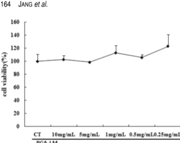
PGA-LM의 in vitro 세포독성
세포독성 물질은 세포의 증식·분화·기능을 저해하며, 세포분열을 할 때 염색체 복제 이전 단계에서 염색체의 활 성을 방해한다. Sonication하여[39] 분자량 30 kDa 이하의
PGA-LM을 얻은 후 PGA-LM이 정상세포에 미치는 영향을
SRB법으로 측정하여 안정성 여부를 관찰하였다[33]. 본 연 구에서 사용된 사람섬유아세포(hFCs)는 정상세포로 세포 성 장, 분열, 그리고 약물에 대한 반응이 최소한으로 조절되는 데, 보편적으로 세포독성 판정에 있어서 기준이 되는 세포 로 알려져 있다[16]. 그러나 본 실험에 사용된 NC/Nga 생쥐 의 T와 B 세포는 약물에 대한 반응이 세포분열, 사이토카인 의 자가분비 및 타가분비 등이 다양하게 나타나 세포독성 측 정에 적합하지 않아 보통 hFCs로 대체하여 실험을 한다.
PGA-LM이 hFCs에 나타내는 세포독성을 실험하였다.
hFCs을 96 well plate에 5×10⁴ 세포로 분주하여 subculture 한 후 PGA-LM을 10 mg/mL, 5 mg/mL, 1 mg/mL, 0.5 mg/
mL, 0.25 mg/mL 농도로 48시간 동시 배양하였다. 그 후 핵 내 단백질인 sulforhodamine B protein량을 측정하는 방법으 로 1차 검색방법인 비색법 중 가장 민감하고 안정한 방법으 로 알려져 있는 SRB방법[46]으로 세포독성을 실험한 결과,
PGA-LM를 처리하지 않은 상태인 대조군의 세포생존율은
100.0±5.8(%)로 나타났다. 그리고 PGA-LM를 10 mg/mL, 5 mg/mL, 1 mg/mL, 0.5 mg/mL, 0.25 mg/mL 농도로 처리한 실험군도 각각 대조군에 비하여 처리한 모든 농도에서 세포 독성을 나타내지 않았다(Fig. 1). Fig. 1에 의하면 오히려 PGA-LM 0.25 mg/mL 첨가에서는 약 25%의 세포생존율을 증가시키는 것으로 나타났다. 따라서 γ-PGA은 현재도 식이 가 가능하여 섭취하고 있는 식품으로 PGA-LM도 안전성이 매우 우수할 것으로 사료되었다.
Gracials 등의 보고에 의하면[39] 20 kHz로 γ-PGA를 초음 파 처리 시 분자량이 1/30 이하로 작아지는 것을 알 수 있 다. 이러한 초음파처리 특성원리를 이용하여 본 논문에서는 분자량 300 kDa인 PGA-HM을 40 kHz로 1시간 초음파처리 하고 밀리포어필터로 여과하여 분자량이 1/10이하 즉, 30 kDa 이하 PGA-LM을 얻었다. 이 PGA-LM은 hFCs의 세포 독성 실험에서 독성이 없는 것으로 나타났는데 PGA-HM도 Primers used for real time PCR.
Gene Primer Sequence
G3PDH Forward 5' TGAAGCAGGCATCTGAGGG 3' Reverse 5' CGAAGGTGGAAGAGTGGGAG 3' IL-
1 beta
Forward 5' CAACCAACAAGTGATATTCTCCATG 3' Reverse 5' GATCCACACTCTCCAGCTGCA 3'
TNF- alpha
Forward 5' TTCTGTCTACTGAACTTCGGGGT- GATCGGTCC 3'
Reverse 5' GTATGAGATAGCAAATCGGCTGACG- GTGTGGG 3'
IL-6 Forward 5' TCCAGTTGCCTTCTTGGGAC 3' Reverse 5' GTGTAATTAAGCCTCCGACTTG 3' IL-10 Forward 5' AAGCAGCCTTGCAGAAAAGA 3'
Reverse 5' TGGAAGTGGGTGCAGTTAT 3' TGF-
beta 1
Forward 5' TGGAGCAACATGTGGAACTC 3' Reverse 5' CTGCCGTACAACTCCAGTGA 3'
같은 결과를 보여, 고분자와 저분자의 γ-PGA 모두 세포독 성이 없는 것을 알 수 있었다.
PGA-LM에 의한 아토피피부발진 NC/Nga 생쥐 B 세포의 염증 유전자 발현 분석
NC/Nga 생쥐의 B세포에서 IL-1β, IL-6, 그리고 TNF-α mRNA 유전자발현분석: 생쥐의 B 세포는 anti-CD40/rmIL- 4로 자극하면 전사인자인 NF-κB의 활성화로 IL-1β, IL-6, 그리고 TNF-α mRNA의 발현이 증가되고, B 세포의 증식 과 분화 및 IgE 생산이 유도되는 것으로 알려져 있다[23].
아토피피부발진 NC/Nga 생쥐의 비장세포를 anti-CD40/
rmIL-4로 자극하여 활성화시킨 후 PGA-LM이 IL-1β, IL-6 그리고 TNF-α mRNA 유전자 발현을 억제하는지를 관찰하 였다. 결과는 Fig. 2에 B 세포에 anti-CD40/rmIL-4를 처리 한 대조군에 대한 PGA-LM 처리군의 상대정량(RQ)값으로 나타내었다. Fig. 2와 같이 무처리군(정상군)의 IL-1β, IL-6, 그리고 TNF-α mRNA 유전자 발현은 대조군의 RQ값이 1 일 때 각각 0.795, 0.109, 0.491이었고, anti-CD40/rmIL-4와
PGA-LM을 동시 배양한 실험군은 대조군의 RQ값이 1일 때
IL-1β, IL-6, 그리고 TNF-α mRNA 유전자 발현이 0.715, 0.608, 그리고 0.557로 유전자 발현은 현저하게 억제되었고, TNF-α mRNA 유전자 발현은 46% 이상 감소를 나타내었다.
그리고 PGA-HM에서도 같은 현상을 보였다. 또한 rmIL-10 을 처리하고 anti-CD40/rmIL-4로 자극한 양성대조군(PC)의 유전자 발현은 대조군의 RQ값이 1일 때 IL-1β, IL-6, 그리 고 TNF-α mRNA 유전자 발현이 0.233, 0.546, 그리고 0.375로 IL-1β, IL-6 그리고 TNF-α mRNA 유전자 발현은
55~75%이상 현저하게 억제되었다.
Kanda 등[21]은 최근 아토피 연구에서 ketoconazole이 사
람혈액에서 분리한 B 세포에서 anti-CD40/rmIL-4에 의한 IgE 생산과 IgG1 및 IL-1과 IL-6의 생산량을 억제하여 항아 토피 효과를 나타내었고, 그 기전이 cAMP signal의 억제에 기인한다고 보고하였다. 따라서 본 연구에서 PGA-LM은 Kanda 등의 보고와 같이 염증 cytokine인 IL-1β, IL-6, 그리 고 TNF-α를 만드는 유전자 발현을 억제한다고 생각되었다.
아토피피부발진 NC/Nga 생쥐의 비장 B 세포에서 IL-10와 TGF-β mRNA 유전자발현분석: 아토피피부발진 NC/Nga 생 쥐의 비장 B 세포를 anti-CD40/rmIL-4로 자극하여 활성화 시킨 후 PGA-LM이 IL-10과 TGF-β mRNA 유전자 발현에 관여하는지를 관찰하였다. 결과는 Fig. 3에 대조군에 대한
PGA-LM 처리군의 상대정량(RQ) 값으로 나타내었다. Fig.
3과 같이 무처리군(정상군)의 IL-10과 TGF-β mRNA 유전 자 발현은 대조군의 RQ값이 1일 때 각각 0.593과 0.816이 었고, anti-CD40/rmIL-4와 PGA-LM을 동시 배양한 실험군 은 대조군의 RQ값이 1일 때 IL-10과 TGF-β mRNA 유전 자 발현이 1.885와 0.798이었고, IL-10 mRNA 유전자 발현 은 대조군(RQ)보다 189% 증가를 하였고, TGF-β 유전자 발 현은 20%이상 억제되었다. 그리고 PGA-HM에서도 같은 현 상을 보였다. 또한 양성대조군인 rmIL-10을 처리하고 anti- Fig. 1. Cytotoxicity effects of PGA-LM on human fibroblast
cells (hFCs). Human fibroblast cells (hFCs) were cultured with various concentration of PGA-LM for 48 hr and the cell viability was measured by SRB method. The results are expressed the mean± S.E (N=6). Statistically significant value compared with control group data by T test.
Fig. 2. Effects of PGA-LM on IL-1β, IL-6 and TNF-α mRNA expression by PGA-LM plus anti-CD40/rmIL-4-stimulated murine NC/Nga B cells. Isolated B cells from atopy dermatitis- like skin NC/Nga mice were either stimulated with anti-CD40/
rmIL-4 (10 µg/mL) or treated with PGA-LM (100 µg/mL). B cells were not treated (normal; only B cells), co-cultured with anti- CD40/rmIL-4 (CT), anti-CD40/rmIL-4 plus rmIL-10 (500 U/mL) (PC), and anti-CD40/rmIL-4 plus PGA-LM (100 µg/mL) for 4hrs.
IL-1β, IL-6, and TNF-α mRNA synthesized by real-time PCR was analyzed. IL-1β, IL-6, and TNF-α mRNA express were measured real-time PCR. The amount of SYBR Green was measured at the end of each cycle. The cycle number at which the emission inten- sity of the sample rises above the baseline is referred as to the RQ (relative quantitative) and is proportional to the target concentra- tion. Real time PCR was performed in duplicate and analyzed by a Applied Biosystems 7500 Real-Time PCR system.
CD40/rmIL-4로 자극한 실험군의 유전자 발현은 대조군의 RQ값이 1일 때 IL-10 그리고 TGF-β mRNA 유전자 발현이 0.815와 0.473으로 대조군에 비하여 모두 억제를 나타내었다.
Punnonen 등[40]은 IL-10에 대한 연구에서 사람의 PBMC 에서 B 세포를 순수 분리하여 anti-CD40 mAb와 rIL-4로 자극하면 IL-4에 의해 유도되는 IgG4와 IgE가 증가되는데
IL-10과 동시 배양하면 거의 억제된다는 것을 확인하고 IL-
10이 IgE의 항알레르기 효과를 나타낸다는 것을 보고하였 다. Komai-Koma와 Wilkinson[26]에 의하면 TGF-β가 B 세포에 anti-CD40 mAb와 rIL-4를 처리하여 배양할 경우 B 세포의 분화와 IgE 생성을 억제한다고 보고하였다. 그러나 본 연구 결과에서는 B 세포에서 TGF-β의 유전자 발현이
PGA-LM 처리군에서 대조군과 차이를 나타내지 않았다. 이
러한 결과는 TGF-β가 T 세포나 대식세포 등에서 분비되어 B 세포의 분화나 IgE 생성을 억제하는데 반하여, 우리가 B 세포에서 TGF-β의 유전자 발현을 분석한 것이 그 원인이라 생각된다.
생쥐의 B 세포에 anti-CD40+IL-4로 자극하면 IgE 생산과 B 세포 증식이 일어나고, 또한 신호전달기전인 NK-B와 표 면분자인 CD23이 증가하게 된다[20]. Guido 등[18]은 최근 연구에서 in vitro에서 vitamin D(1α,25-dihydroxyvitamin D3)를 처리하여 B 세포의 분화와 IgE 생산을 감소시키는 결 과를 얻어 비타민 D가 NF-B의 활성을 억제하는 기전을 보 고하였다. 또한 Worm 등[53]은 in vitro에서 사람의 PBMC 와 B 세포에 retinoic acid를 처리한 후 anti-CD40+IL-4로 자극하면 IgE 생산이 억제된다는 결과를 발표하였다. 또한 Kimata, Kiniwa, Vercelli 등[24, 25, 50]은 B 세포에 anti- CD40+IL-4의 자극으로 IgE 생산의 억제는 cytokine으로 IFN-γ, IL-8, IL-10, IL-12 등을 보고하였다. 그리고 Kang 등[23]은 생쥐의 B 세포에 NF-B를 활성화시키는 anti-CD40 과 LPS 또는 rHRF를 처리한 후 RT-PCR로 IL-1, IL-2, IL-6, IL-10, TNF-α, 그리고 TGF-β mRNA 유전자 분석을 관찰한 결과 IL-1, IL-6, TNF-α, 그리고 TGF-β가 유도되는 결과를 얻었다.
본 결과에서 대조군에 비해 PGA-LM군에서 Fig. 2와 같 이 IL-1β, IL-6, TNF-α, 그리고 Fig. 3과 같이 TNF-β
mRNA 유전자 발현이 억제되었고, IL-10 mRNA 발현은 증
가를 나타내었다. 그리고 PGA-HM에서도 같은 현상을 보였 다. 이러한 결과로 PGA-HM과 PGA-LM이 B 세포에서 NF-B의 활성조절 기전에 관여하는 것으로 생각된다.
NC/Nga 생쥐의 CD4+ T세포에서 CD4+IFN-γ+의 세포내염 색 분석: 1980년대 후반 Mosmann 등[36]이 생쥐에서 면역 반응을 일으키는 조력 T 세포(naiver T cell)에는 IL-2, IFN- γ 등을 분비하는 Th1 세포와 IL-4, IL-5를 분비하는 Th2 세 포가 존재한다고 보고하였다. 이 후 많은 연구에서 Th1 반 응과 Th2 반응은 서로 길항작용을 하면서 균형을 유지하고 있고[12], 아토피 및 천식 같은 알레르기 질환의 면역학적
병인과 관련하여 이러한 Th1/Th2 반응의 불균형이 핵심적 인 요인임을 알아내었다. 알레르기 질환의 면역학적 발생기 전과 관련하여 Th2 가설을 기반으로 하여 아토피 및 천식 발생과 관련하여 대표적인 Th1 사이토카인인 IFN-γ의 역할 에 대한 많은 연구가 진행되었다[41]. 이들 연구결과를 바탕 으로 많은 연구자들이 IFN-γ가 알레르기 반응을 억제한다는 가설에 동의하고 있다. 그러나 최근 연구에서 기존의 Th2 가설과는 정반대로 아토피 및 천식환자의 면역기관에서 오 히려 IFN-γ가 감염에 의하여 증가되었다는 연구결과들이 발 표되었다[32].
본 연구는 아토피피부발진 NC/Nga 생쥐의 비장에서
CD4+ T 세포를 VarioMACS를 사용하여 순수 분리하여 미
리 coating해 놓은 α-CD3/α-CD28(1 µg/mL)로 자극하여 활 성화시킨 후 72시간 배양하여 IFN-γ+를 세포내 형광염색으 로 분석하였다. 그 결과 Fig. 4에서 보듯 α-CD3/α-CD28(1 µg/mL)로 자극하지 않은 only CD4+ T 세포군의 CD4+IFN- γ+는 2.9%이었고, anti-CD3/anti-CD28(1 µg/mL)로 자극한 대조군의 CD4+IFN-γ+는 10.8%이었다. 양성대조군으로 cyclosporin A(1µg/mL)를 α-CD3/α-CD28(1 µg/mL)와 동 시 배양한 CsA 처리군의 CD4+IFN-γ+는 6.8%이었고, 실험 Fig. 3. Effects of PGA-LM on IL-10, and TGF-β mRNA expression by PGA-LM plus anti-CD40/rmIL-4-stimulated murine NC/Nga B cells. Isolated B cells from atopy dermatitis- like skin NC/Nga mice were either stimulated with anti-CD40/
rmIL-4 (10 µg/mL) or treated with PGA-LM (100 µg/mL). B cells were not treated (normal; only B cells), co-cultured with anti- CD40/rmIL-4 (CT), anti-CD40/rmIL-4 plus rmIL-10 (500 U/mL) (PC), and anti-CD40/rmIL-4 plus PGA-LM (100 µg/mL) for 4hrs.
IL-10, and TGF-β mRNA synthesized by real-time PCR was ana- lyzed. IL-10, and TGF-β mRNA express were measured real-time PCR. The amount of SYBR Green was measured at the end of each cycle. The cycle number at which the emission intensity of the sample rises above the baseline is referred as to the RQ (rela- tive quantitative) and is proportional to the target concentration.
Real time PCR was performed in duplicate and analyzed by a Applied Biosystems 7500 Real-Time PCR system.
군인 PGA-LM(100 µg/mL)를 α-CD3/α-CD28(1 µg/mL)와 동시 배양한 PGA-LM 처리군의 CD4+IFN-γ+는 15.4%로 대조군에 비하여 1.5배 이상 증가하였다. 그리고 PGA-HM 에서도 PGA-LM과 같은 결과를 보였다.
Bellinghausen 등[5]의 연구에서 insect venom이 T 세포 의 IL-10과 IFN-γ의 증가를 촉진시켜 알레르기반응에 대한 Th2 세포에서 Th1 세포로 전환시키는 면역억제제 효능을 보 고하였고, 이와 같이 본 연구에서도 PGA-LM이 알레르기 반응을 억제하는 IFN-γ를 증가시켜 알레르기 반응에 우위 한 Th2 세포에서 Th1 세포로 shift시킬 수 있을 것으로 생 각된다.
NC/Nga 생쥐의 CD4+ T 세포에서 CD4+CD25+foxp3+
Treg의 세포내염색분석: 면역 체계는 자가항원을 인식하는 면역세포는 선택적으로 제거하고, 면역 반응의 조건이 충족 되지 않았을 때는 알레르기를 유도하여 숙주를 보호한다. 면 역반응을 직접적으로 억제할 수 있는 세포가 있을 것이라는 의견이 오래 전부터 제기되어 왔으나 이 세포의 존재는 오 랫동안 논쟁거리가 되어왔고, 최근의 연구 결과에 의해서야 비로소 조절 T 세포(regulatory T cells, Treg cells)의 존재 가 드러났다[43]. 조절 T 세포는 흉선(thymus)으로부터 직접 유래한 자연(natural) Treg와 말초(periphery)에서 미성숙 (naive) T cell이 항원의 자극을 받아 유도되는 유도(induci- ble) Treg이 존재한다고 알려져 있다. 이러한 Treg의 특징은 세포 표면에 CD25(IL-2Rα)를 지속적으로 발현하고 있고
foxp3+라는 전사인자를 특이적으로 발현한다. 따라서 CD25
와 foxp3+를 marker로써 CD4+CD25+foxp3+ natural Treg 을 식별할 수 있고 Treg가 직접적으로 다양한 면역 반응을 억제하는 역할을 한다는 연구 결과들이 발표되었다[14].
foxp3+ Treg은 흉선에서 만들어지고 이들은 naive T 세포 의 활성화와 성장 및 다양한 면역 반응을 주관하는 Th1, Th2, Th17을 포함한 effector T 세포로의 분화를 억제한다 [44]. Periphery에서는 비록 그 기전은 명확하지 않지만 naive
T 세포가 foxp3+ Treg으로 분화하기도 한다. 최근의 연구 결과에 따르면 Treg은 대식세포, Th17 세포 분화, 과잉 Th2 세포의 GATA3, 그리고 B 세포와 같은 다양한 면역세포들 에 대하여 Treg의 세포 표면에 발현하는 CTLA4, GITR,
CD25 그리고 LAG3 등을 통하여 면역억제 신호를 전달하
거나 면역 세포들 사이의 상호작용을 저해한다고 한다[11].
본 연구는 아토피피부발진 NC/Nga 생쥐의 비장에서
CD4+ T 세포를 VarioMACS를 사용하여 순수 분리하여 미
리 coating해 놓은 α-CD3/α-CD28(1 µg/mL)로 자극하여 활 성화시킨 후 72시간 동안 PGA-LM과 동시 배양하여 CD4+CD25+foxp3+ Treg 세포를 세포내 형광염색으로 분석 하였다. 그 결과 Fig. 5에서 보듯이 α-CD3/α-CD28(1 µg/
mL)로 자극하지 않은 only CD4+ T 세포군의 CD4+CD25+
foxp3+는 0.8±0.25%이었고, α-CD3/α-CD28(1 µg/mL)로 자 극한 대조군의 CD4+CD25+foxp3+는 5.2±0.28%이었다. 실 험군인 PGA-LM(100 µg/mL)를 α-CD3/α-CD28(1 µg/mL) 와 동시 배양한 PGA-LM 처리군의 CD4+CD25+foxp3+는
13.7±3.2%로 대조군에 비하여 1.65배 이상의 유의성 있는
증가를 나타내었다(p<0.01). 그리고 PGA-HM도 PGA-LM과 같은 결과를 보였다.
Bellanti 등[4]의 연구에서는 인체의 방어 기전인 면역체계 는 T 세포의 활성화를 중심으로 이루어진다고 보고하였다.
Th1/Th2 세포들은 서로 상호 견제를 통하여 균형을 이루는
데 어떤 이유에서 Th2 세포 쪽으로 기울게 되면 알레르기 반응이 일어난다. 그래서 PGA-LM 처리군의 CD4+CD25+
foxp3+ Treg 세포의 유도로 알레르기 반응에서 IL-4를 분비 하는 Th2 세포의 GATA-3 전사인자를 억제하는 결과로 우 위한 Th2 세포에서 Th1 세포로 shift시킬 수 있을 것으로 생 각된다. 이러한 결과는 최근 Peter(2008)의 보고에서 활성화 된 CD4+CD25+foxp3+ Treg 세포의 Th2 세포 억제기전에 의 내용과 일치하는 연구결과이다[3].
CD4+CD25+ T 림프구(Treg cells)는 다른 T 세포의 아형 Fig. 4. Effects of PGA-LM on IFN-γ+ intracellular staining analysis by α-CD3/α-CD28 stimulated murine CD4+ T cells. Isolated CD4+ T cells from atopy dermatitis-like skin NC/Nga mice were either stimulated with α-CD3/α-CD28(1µg/mL) or treated with PGA-LM (100 µg/mL). CD4+ T cells were not treated(normal; only cells), co-cultured with α-CD3/α-CD28 (1 µg/mL) (Control), and with α-CD3/
α-CD28 (1 µg/mL) plus PGA-LM (100 µg/mL), and with α-CD3/α-CD28 (1 µg/mL) plus cyclosporin A (CsA, 5 µg/mL) for 48hours.
After a 48-hours stimulation, 0.6 µL/mL monensin (GolgiStop; BD Pharmingen) was added and included in subsequent incubations until fixation. Cells were stained with FITC-labeled anti-CD4. After fixation in Cytofix/Cytoperm (BD Pharmingen), cells were washed twice and permeabilized in Perm/Wash buffer (BD PharMingen), stained with PE-labeled anti-IFN-γ mAb (BD Pharmingen) and immediately analyzed on a FACScalibur (BD Biosciences) using CellQuest software (BD Biosciences).
들처럼 흉선에서 발생되고 전사인자인 foxp3+을 발현하여 다양한 T 림프구의 활동들을 제어하는 능력을 갖고 있다. 그 러나 Treg 세포가 T 림프구의 활동을 억제하는 기작에 대해 서는 몇 가지 모델이 제시되었지만 각 모델마다 논쟁의 여 지가 있고 그 메커니즘이 명확하지 않다[13, 19]. ‘Nature Immunology` 최신호에는 Treg 세포가 IL-2를 선택적으로 사 용하여 결과적으로 목표 T 림프구의 사멸을 유도하는 새로 운 모델을 제시하고 있다[38]. Brunkow 등[7]의 연구에서는 조절 T 세포(Treg cells)에서 발현되는 foxp3+ 전사인자는 자가 면역 질환들을 조절하는데 필수적이라고 보고하였다.
따라서 세포독성이 없는 PGA-HM(data not shown)과 PGA-LM은 IL-1β, IL-6, 그리고 TNF-α mRNA 유전자 발 현을 억제하고 IL-10, mRNA 유전자 발현을 증가시켜 염증 유전자 발현을 억제하였다. 그리고 CD4+ T 세포에서 CD4+IFN-γ+ 및 CD4+CD25+foxp3+ Treg의 세포내 형광염 색으로 분석한 결과 대조군에 비해 그 값이 유의적으로 증 가하였다. PGA-HM과 PGA-LM에 의한 이러한 염증유전자 발현억제와 CD4+IFN-γ+ 및 CD4+CD25+foxp3+ 증가로 아 토피 억제 가능성이 있을 것으로 사료된다.
요 약
γ-PGA는 우리 전통 콩 발효식품인 청국장의 끈적끈적한 점액성의 성분으로, 매우 다양한 기능을 가지고 있는 천연 소재이다. 이러한 γ-PGA가 아토피발진 억제 가능성을 알아 보기 위해 NC/Nga 생쥐를 사용하여 in vitro 실험을 실시하 였다. γ-PGA(PGA-HM, 분자량 300 kDa)를 초음파처리로 저분자화시킨 30 kDa 이하의 저분자 PGA-LM를 만들고, 고 분자 PGA-HM과 PGA-LM을 사용하여 실험하였는데 동일 한 결과를 얻어 PGA-LM 실험결과 중심으로 보고한 것이 다. 아토피 피부발진 NC/Nga 생쥐의 비장에서 B 세포와 T
세포를 순수 분리하여 항알레르기 작용에 대한 in vitro 실 험을 실시하였다. PGA-LM은 hFCs에 대한 세포독성 실험 에서 모든 농도에서 세포독성을 나타내지 않았다. PGA-LM 이 B 세포 분화 및 활성화에 미치는 영향을 관찰하기 위하 여, NC/Nga 생쥐의 비장에서 순수 분리한 B 세포에 anti- CD40/rmIL-4로 자극한 결과, 대조군은 전사인자인 NF-κB 의 활성화로 IL-1β, IL-6, 그리고 TNF-α mRNA의 발현이 증가되었다. 그러나 PGA-LM과 양성대조군인 rmIL-10 투여 군은 염증사이토카인 IL-1β, IL-6 그리고 TNF-α mRNA 유 전자 발현이 감소하였고, IL-10 mRNA 유전자 발현은 증가 하였으나 TGF-β mRNA의 유전자 발현은 대조군과 큰 차이 가 나타나질 않았다. 또한 CD4+ T 세포에 PGA-LM 100 µg/ml를 처리한 후 4일간 동시 배양하여 CD4+IFN-γ+와 CD4+CD25+foxp3+ Treg 세포를 세포내 염색으로 분석한 결과에서는 CD4+IFN-γ+인 Th1 세포의 증가와 CD4+
CD25+foxp3+ Treg 세포를 증가시켜 알레르기반응에서 우
위한 Th2 세포에서 Th1 세포로 전환시키는 면역조절 역할 을 나타내었다. 이상의 결과로 NC/Nga 생쥐에서 PGA-LM 은 염증유전자 발현을 억제시키고 IFN-γ+의 증가 및 조절 T 세포의 유도로 아토피피부염의 피부발진을 치료하는 면역 조절제로 사용될 수 있을 것으로 생각된다.
R
EFERENCES1. Abbas, A. K., K. M. Murphy, and A. Sher. 1996. Functional diversity of helper T lymphocytes. Nature. 383: 787-793.
2. Agnello, D., C. S. Lankford, J. Bream, A. Morinobu, M.
Gadina, J. J. O'Shea, and D. M. Frucht. 2003. Cytokines and transcription factors that regulate T helper cell differ- entiation: new players and new insights. J. Clin. Immunol.
23: 147-161.
3. Barnes, P. J. 2008. Immunology of asthma and chronic Fig. 5. Effects of PGA-LM on CD4+CD25+foxp3+ intracellular staining analysis by α-CD3/α-CD28 stimulate dmurine NC/Nga CD4+T cells. Isolated CD4+T cells from atopy dermatitis-like skin NC/Nga mice were either stimulated with α-CD3/α-CD28 (1 µg/mL) or treated with PGA-LM (100 µg/mL). CD4+T cells were not treated (normal; only cells), co-cultured with α-CD3/α-CD28 (1 µg/mL) (Control), and with α-CD3/α-CD28 (1 µg/mL) plus PGA-LM (100 µg/mL, 10 µg/mL,) for 48 hrs. After a 48-hours stimulation, 0.6 µL/mL monensin (GolgiStop; BD Pharmingen) was added and included in subsequent incubations until fixation. Cells were stained with FITC- labeled anti-CD4,and Cychrom5.5-labeled anti-CD25. After fixation in Cytofix/Cytoperm (BD Pharmingen), cells were washed twice and permeabilized in Perm/Wash buffer (BD PharMingen), stained with PE-labeled anti-foxp3 mAb (BD Pharmingen) and immediately ana- lyzed on a FACScalibur (BD Biosciences) using CellQuest software (BD Biosciences). Statistically significant value compared with NC/
Nga mice group data by student's t-test (**p<0.01).
obstructive pulmonary disease. Nat. Rev. Immunol. 8: 183- 192.
4. Bellanti, J. A. 1998. Cytokines and allergic diseases: clinical aspects. Allergy Asthma Proc. 19: 337-341.
5. Bellinghausen, I., G. Metz, A. H. Enk, S. Christmann, J.
Knop, and J. Saloga. 1997. Insect venom immunotherapy induces interleukin-10 production and a Th2-to-Th1 shift, and changes surface marker expression in venom-allergic subjects. Eur. J. Immunol. 27: 1131-1139.
6. Bordi, F., C. Cametti, and G. Paradossi. 1996. A com- parative study of the high-frequency dielectric properties of poly (alpha-glutamate) and poly (gamma-glutamate) aqueous solutions. Biopolymers 40: 485-494.
7. Brunkow, M. E., E. W. Jeffery, K. A. Hjerrild, B. Paeper, L.
B. Clark, S. A. Yasayko, J. E. Wilkinson, D. Galas, S. F.
Ziegler, and F. Ramsdell. 2001. Disruption of a new forkhead/winged-helix protein, scurfin, results in the fatal lymphoproliferative disorder of the scurfy mouse. Nat.
Genet. 27: 68-73.
8. Choi, H. J. and M. Kunioka. 1995. Preparation conditions and swelling equilibria of hydrogel prepared by [gamma]- irradiation from microbial poly([gamma]-glutamic acid).
Radiation Physics and Chemistry 46: 175-179.
9. Cooper, K. D. 1994. Atopic dermatitis: recent trends in pathogenesis and therapy. J. Invest. Dermatol. 102: 128-137.
10. Curotto de Lafaille, M. A., N. Kutchukhidze, S. Shen, Y.
Ding, H. Yee, and J. J. Lafaille. 2008. Adaptive Foxp3+
regulatory T cell-dependent and -independent control of allergic inflammation. Immunity. 29: 114-126.
11. Elias, K. M., A. Laurence, T. S. Davidson, G. Stephens, Y.
Kanno, E. M. Shevach, and J. J. O'Shea. 2008. Retinoic acid inhibits Th17 polarization and enhances FoxP3 expression through a Stat-3/Stat-5 independent signaling pathway.
Blood. 111: 1013-1020.
12. Fiorentino, D. F., M. W. Bond, and T. R. Mosmann. 1989.
Two types of mouse T helper cell. IV. Th2 clones secrete a factor that inhibits cytokine production by Th1 clones. J.
Exp. Med. 170: 2081-2095.
13. Fontenot, J. D., M. A. Gavin, and A. Y. Rudensky. 2003.
Foxp3 programs the development and function of CD4+
CD25+ regulatory T cells. Nat. Immunol. 4: 330-336.
14. Fontenot, J. D. and A. Y. Rudensky. 2005. A well adapted regulatory contrivance: regulatory T cell development and the forkhead family transcription factor Foxp3. Nat. Immunol.
6: 331-337.
15. Gross, S. S., E. A. Jaffe, R. Levi, and R. G. Kilbourn. 1991.
Cytokine-activated endothelial cells express an isotype of nitric oxide synthase which is tetrahydrobiopterin-depen- dent, calmodulin-independent and inhibited by arginine analogs with a rank-order of potency characteristic of activated macrophages. Biochem. Biophys. Res. Commun.
178: 823-829.
16. Gurbay, A., C. Garrel, M. Osman, M. J. Richard, A. Favier, and F. Hincal. 2002. Cytotoxicity in ciprofloxacin-treated human fibroblast cells and protection by vitamin E. Hum
Exp Toxicol 21: 635-641.
17. Hahm, J. H., T. Y. Lee, J. S. Lee, C. Park, M. H. Sung, and H. Poo. 2004. Antitumor effect of Poly-glutamic acid by modulating cytokine production and NK cell activity.
International Meeting of the Federation of Korean Microbiological Societies p. 21-22.
18. Heine, G., K. Anton, B. M. Henz, and M. Worm. 2002.
1alpha,25-dihydroxyvitamin D3 inhibits anti-CD40 plus IL- 4-mediated IgE production in vitro. Eur. J. Immunol. 32:
3395-3404.
19. Hori, S., T. Nomura, and S. Sakaguchi. 2003. Control of regulatory T cell development by the transcription factor Foxp3. Science. 299: 1057-1061.
20. Jabara, H. H., S. M. Fu, R. S. Geha, and D. Vercelli. 1990.
CD40 and IgE: synergism between anti-CD40 monoclonal antibody and interleukin 4 in the induction of IgE synthesis by highly purified human B cells. J. Exp. Med. 172: 1861- 1864.
21. Kanda, N. and Watanabe, S. 2002. Ketoconazole suppresses interleukin-4 plus anti-CD40-induced IgE class switching in surface IgE negative B cells from patients with atopic dermatitis. J. Invest. Dermatol. 119: 590-599.
22. Kaneda, N., K. Kobayashi, H. Ichinose, F. Kishi, A.
Nakazawa, Y. Kurosawa, K. Fujita, and T. Nagatsu. 1987.
Isolation of a novel cDNA clone for human tyrosine hydroxylase: alternative RNA splicing produces four kinds of mRNA from a single gene. Biochem. Biophys. Res.
Commun. 146: 971-975.
23. Kang, H. S., M. J. Lee, H. Song, S. H. Han, Y. M. Kim, J. Y.
Im, and I. Choi. 2001. Molecular identification of IgE- dependent histamine-releasing factor as a B cell growth factor. J. Immunol. 166: 6545-6554.
24. Kimata, H., A. Yoshida, C. Ishioka, I. Lindley, and H.
Mikawa. 1992. Interleukin 8 (IL-8) selectively inhibits immunoglobulin E production induced by IL-4 in human B cells. J. Exp. Med. 176: 1227-1231.
25. Kiniwa, M., M. Gately, U. Gubler, R. Chizzonite, C. Fargeas, and G. Delespesse. 1992. Recombinant interleukin-12 sup- presses the synthesis of immunoglobulin E by interleukin-4 stimulated human lymphocytes. J. Clin. Invest. 90: 262-266.
26. Komai-Koma, M. and P. C. Wilkinson. 1997. TGF-beta stimulates but IFN-gamma inhibits growth-related activation of locomotion of human B cells. J. Immunol. 158: 3125- 3129.
27. Kuhn, J. and S. Binder. 2002. RT-PCR analysis of 5' to 3'- end-ligated mRNAs identifies the extremities of cox2 transcripts in pea mitochondria. Nucleic Acids Res. 30: 439- 446.
28. Larche, M., D. S. Robinson, and A. B. Kay. 2003. The role of T lymphocytes in the pathogenesis of asthma. J. Allergy Clin. Immunol. 111: 450-463.
29. Lee, J. S., H. Poo, C. J. Kim, Y. H. Choi, C. Park, T. Y. Lee, and M. H. Sung. 2004. Effect of poly-gamma-glutamic acid as an adjuvant and immune response stimulatory factor in vitro and in vivo. The 16th Annual Meeting of the Korean
Society for Molecular and Cellular Biology. pp. 14-15.
30. Leung, D. Y. 2000. Atopic dermatitis: new insights and opportunities for therapeutic intervention. J. Allergy Clin.
Immunol. 105: 860-876.
31. Leung, D. Y. and T. Bieber. 2003. Atopic dermatitis. Lancet.
361: 151-160.
32. Magnan, A. O., L. G. Mely, C. A. Camilla, M. M. Badier, F.
A. Montero-Julian, C. M. Guillot, B. B. Casano, S. J. Prato, V. Fert, P. Bongrand, and D. Vervloet. 2000. Assessment of the Th1/Th2 paradigm in whole blood in atopy and asthma.
Increased IFN-gamma-producing CD8(+) T cells in asthma.
Am. J. Respir. Crit. Care Med. 161: 1790-1796.
33. Martin, A. and M. Clynes. 1993. Comparison of 5 microplate colorimetric assays for in vitro cytotoxicity testing and cell proliferation assays. Cytotechnology. 11: 49-58.
34. Miltenyi, S., W. Muller, W. Weichel, and A. Radbruch. 1990.
High gradient magnetic cell separation with MACS.
Cytometry 11: 231-238.
35. Mosmann, T. R., H. Cherwinski, M. W. Bond, M. A.
Giedlin, and R. L. Coffman. 1986. Two types of murine helper T cell clone. I. Definition according to profiles of lymphokine activities and secreted proteins. J. Immunol.
136: 2348-2357.
36. Mosmann, T. R. and R. L. Coffman. 1989. TH1 and TH2 cells: different patterns of lymphokine secretion lead to different functional properties. Annu. Rev. Immunol. 7: 145- 173.
37. Obst, M. and A. Steinbuchel. 2004. Microbial Degradation of Poly(amino acid)s. Biomacromolecules 5: 1166-1176.
38. Pandiyan, P., L. Zheng, S. Ishihara, J. Reed, and M. J.
Lenardo. 2007. CD4+CD25+Foxp3+ regulatory T cells induce cytokine deprivation-mediated apoptosis of effector CD4+ T cells. Nat. Immunol. 8: 1353-1362.
39. Perez-Camero, G., F. Congregado, J. J. Bou, and S. Munoz- Guerra. 1999. Biosynthesis and ultrasonic degradation of bacterial poly(gamma-glutamic acid). Biotechnol. Bioeng.
63: 110-115.
40. Punnonen, J., R. de Waal Malefyt, P. van Vlasselaer, J. F.
Gauchat, and J. E. de Vries. 1993. IL-10 and viral IL-10 prevent IL-4-induced IgE synthesis by inhibiting the accessory cell function of monocytes. J. Immunol. 151:
1280-1289.
41. Robinson, D. S., Q. Hamid, S. Ying, A. Tsicopoulos, J.
Barkans, A. M. Bentley, C. Corrigan, S. R. Durham, and A.
B. Kay. 1992. Predominant TH2-like bronchoalveolar T- lymphocyte population in atopic asthma. N. Engl. J. Med.
326: 298-304.
42. Ruiz, R. G., J. F. Price, D. Richards, and D. M. Kemeny.
1990. Lack of relation between IgE in neonatal period and later atopy. Lancet. 336: 808.
43. Sakaguchi, S. 2005. Naturally arising Foxp3-expressing CD25+CD4+ regulatory T cells in immunological tolerance to self and non-self. Nat. Immunol. 6: 345-352.
44. Sakaguchi, S. and F. Powrie. 2007. Emerging challenges in regulatory T cell function and biology. Science 317: 627- 629.
45. Shih, I. L. and Y. T. Van. 2001. The production of poly- (gamma-glutamic acid) from microorganisms and its various applications. Bioresour. Technol. 79: 207-225.
46. Skehan, P., R. Storeng, D. Scudiero, A. Monks, J. McMahon, D. Vistica, J. T. Warren, H. Bokesch, S. Kenney, and M. R.
Boyd. 1990. New colorimetric cytotoxicity assay for anti- cancer-drug screening. J. Natl. Cancer Inst. 82: 1107-1112.
47. Szabo, S. J., S. T. Kim, G. L. Costa, X. Zhang, C. G.
Fathman, and L. H. Glimcher. 2000. A novel transcription factor, T-bet, directs Th1 lineage commitment. Cell. 100:
655-669.
48. Szabo, S. J., B. M. Sullivan, C. Stemmann, A. R. Satoskar, B. P. Sleckman, and L. H. Glimcher. 2002. Distinct effects of T-bet in TH1 lineage commitment and IFN-gamma production in CD4 and CD8 T cells. Science. 295: 338-342.
49. Ting, C. N., M. C. Olson, K. P. Barton, and J. M. Leiden.
1996. Transcription factor GATA-3 is required for develop- ment of the T-cell lineage. Nature. 384: 474-478.
50. Vercelli, D., H. H. Jabara, K. Arai, T. Yokota, and R. S.
Geha. 1989. Endogenous interleukin 6 plays an obligatory role in interleukin 4-dependent human IgE synthesis. Eur. J.
Immunol. 19: 1419-1424.
51. Witney, A. A., D. L. Doolan, R. M. Anthony, W. R. Weiss, S.
L. Hoffman, and D. J. Carucci. 2001. Determining liver stage parasite burden by real time quantitative PCR as a method for evaluating pre-erythrocytic malaria vaccine efficacy. Mol. Biochem. Parasitol. 118: 233-245.
52. Wittig, H. J., J. Belloit, I. De Fillippi, and G. Royal. 1980.
Age-related serum immunoglobulin E levels in healthy subjects and in patients with allergic disease. J. Allergy Clin.
Immunol. 66: 305-313.
53. Worm, M., J. M. Krah, R. A. Manz, and B. M. Henz. 1998.
Retinoic acid inhibits CD40 + interleukin-4-mediated IgE production in vitro. Blood. 92: 1713-1720.
54. Zheng, W. and R. A. Flavell. 1997. The transcription factor GATA-3 is necessary and sufficient for Th2 cytokine gene expression in CD4 T cells. Cell 89: 587-596.
(Received March 4, 2009/Accepted May 13, 2009)