Introduction
Parosteal osteosarcoma (POS), like periosteal, intracortical, and high- grade surface osteosarcomas, is a type of surface osteosarcoma.1-3) Of these, POS accounts for 65% of juxtacortical osteosarcomas and is frequently encountered as a low-grade lesion with a low propen- sity to metastasize and 5- and 10-year survival rates of 80-90%.4-6) Treatment with a wide operative margin and reconstruction using a prosthesis has been advocated.7,8) Furthermore, lobulated parosteal lesions may be of higher-grade, and radiological evidence of inva- sion into the medullary canal or the presence of a non-mineralized soft-tissue mass of larger than 1 cm3 may suggest a poor progno- sis.9-12)
Although previous reports have addressed the importance of ob- taining a wide surgical margin in the treatment of parosteal osteosar- coma, in the clinical setting, intralesional resection is possible. Two
Clinical Outcome of Parosteal Osteosarcoma
Won Seok Song, Dae-Geun Jeon, Wan Hyeong Cho, Chang Bae Kong, Sang Hyun Cho, Kwang Ryul Lee, and Soo-Yong Lee
Department of Orthopedic Surgery, Korea Cancer Center Hospital, Seoul, Korea
factors appear to be associated with under-treatment; misdiagnosis due to its radiologic and pathologic similarities with other benign tumors7,8,13), and deliberate compromise of its surgical margin, due to its reported excellent survival and indolent growth after intralesional resection.
In the largest series of POS conducted to date, the risk of recur- rence after intralesional or marginal resection was found to be sig- nificant.6,7,13) However, the surgical management of locally recurrent POS and its clinical course after tumor recurrence has not been well- defined.
Our primary study goal was to evaluate correlations between clinicopathologic findings and oncologic outcomes, and our second- ary goal was to ascertain the fate of patients after treatment for local recurrence.
Materials and Methods
We retrospectively reviewed the records of 30 parosteal osteosar- coma (POS) patients treated between 1990 and 2010. However, we excluded 8 of the 30 for; incomplete data (2 patients), no surgery (2 patients), and a follow-up period of less than 2 years (4 patients).
Therefore, the final study population consisted of 22 patients (Table Received March 25, 2013 Revised May 14, 2013 Accepted May 20, 2013
Correspondence to: Dae-Geun Jeon
Department of Orthopedic Surgery, Korea Cancer Center Hospital, 215-4, Gongneung- dong, Nowon-gu, Seoul 139-706, Korea
TEL: +82-2-970-1242 FAX: +82-2-970-2403 E-mail: dgjeon@kcch.re.kr
Purpose: The purpose of this study was to evaluate the oncologic outcomes of parosteal osteosarcoma (POS) and to ascertain the fates of patients after local recurrence (LR).
Materials and Methods: The authors retrospectively reviewed 22 POS patients with an average follow-up of 114 months (range: 36-235 months). Seven of the 22 patients were referred after LR. There were 17 Stage IB and 5 Stage IIB (G2, 2; dedifferentiation, 3). Tumors were located in the femur (11) and in other locations (11). Initial surgical margins were wide in 10, marginal in 5, and intralesional in 7. Correla- tions between clinico-pathologic variables and LR and clinical courses after LR were evaluated.
Results: The 10-year overall survival rate was 85.7%. Three (14%) patients developed distant metastasis and all of them succumbed to the disease. Nine (41%) patients developed LR. Tumor location, resection type, and surgical margin were found to be correlated with LR. At final follow-up, 7 of the 9 patients that experienced local failure achieved no evidence of disease.
Conclusion: A substantial risk of misdiagnosis exists, especially for POS in other than a femoral location. Recurrent tumor re-excision is possible in most cases; however, patients with an aggressive recurrence pattern deserve special attention.
Key words: parosteal osteosarcoma, local recur
Copyrights © 2013 by The Korean Bone and Joint Tumor Society
“This is an Open Access article distributed under the terms of the Creative Commons Attribution Non-Commercial License (http://creativecommons.org/licenses/by-nc/3.0/) which permits unrestricted non-commercial use, distribution, and reproduction in any medium, provided the original work is properly cited.”
대한골관절종양학회지:제19권 제1호 2013
1). There were 8 males and 14 females. Patients ages ranged from 6 to 68 years (mean, 28 years). Seven of the 22 patients were referred for more than one local recurrence after surgery by other hospitals.
Clinical data were obtained from the patient charts and medical
records, preoperative roentgenograms, and pathology slides of con- sulting surgeon and pathologists. Radiographic imaging studies were available for all patients. Specific radiographic findings, including location, size, and the presences of medullary invasion and of a non-
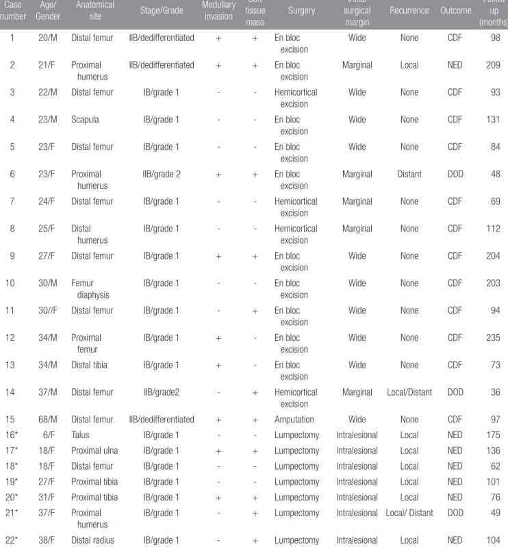
Table 1. Patient Demographics and Treatment Outcomes Case
number Age/
Gender
Anatomical
site Stage/Grade Medullary invasion
Soft tissue mass
Surgery
Initial surgical
margin
Recurrence Outcome
Follow up (months) 1 20/M Distal femur IIB/dedifferentiated + + En bloc
excision
Wide None CDF 98
2 21/F Proximal humerus
IIB/dedifferentiated + + En bloc excision
Marginal Local NED 209
3 22/M Distal femur IB/grade 1 - - Hemicortical
excision
Wide None CDF 93
4 23/M Scapula IB/grade 1 - - En bloc
excision
Wide None CDF 131
5 23/F Distal femur IB/grade 1 - - En bloc
excision
Wide None CDF 84
6 23/F Proximal humerus
IIB/grade 2 + + En bloc
excision
Marginal Distant DOD 48
7 24/F Distal femur IB/grade 1 - - Hemicortical
excision
Marginal None CDF 69
8 25/F Distal humerus
IB/grade 1 - - Hemicortical
excision
Marginal None CDF 112
9 27/F Distal femur IB/grade 1 + + En bloc
excision
Wide None CDF 204
10 30/M Femur
diaphysis
IB/grade 1 - - En bloc
excision
Wide None CDF 203
11 30//F Distal femur IB/grade 1 - + En bloc
excision
Wide None CDF 94
12 34/M Proximal femur
IB/grade 1 + - En bloc
excision
Wide None CDF 235
13 34/M Distal tibia IB/grade 1 + - En bloc
excision
Wide None CDF 73
14 37/M Distal femur IIB/grade2 - + Hemicortical
excision
Marginal Local/Distant DOD 36
15 68/M Distal femur IIB/dedifferentiated + + Amputation Wide None CDF 97
16* 6/F Talus IB/grade 1 - - Lumpectomy Intralesional Local NED 175
17* 18/F Proximal ulna IB/grade 1 + + Lumpectomy Intralesional Local NED 136
18* 18/F Distal femur IB/grade 1 - - Lumpectomy Intralesional Local NED 62
19* 27/F Proximal tibia IB/grade 1 - - Lumpectomy Intralesional Local NED 101
20* 31/F Proximal tibia IB/grade 1 + + Lumpectomy Intralesional Local NED 76
21* 37/F Proximal humerus
IB/grade 1 - + Lumpectomy Intralesional Local/ Distant DOD 49
22* 38/F Distal radius IB/grade 1 - + Lumpectomy Intralesional Local NED 104
*Referred patients.
CDF, continous disease free; NED, no evidence of disease; DOD, dead of disease; AWD, alive with disease.
mineralized soft tissue mass, were noted.11) Locations of primary tumors were; femur (11), humerus (4), tibia (3), and one case each at talus, radius, ulna, and scapula. Tumor sizes ranged from 3 to 23 cm in maximum diameter (mean 7.1 cm). Nine (40.9%) patients had intramedullary tumor extension, and a non-mineralized soft tissue mass was observed in 11 (50%) patients by CT or MRI. Pathologic materials were analyzed to confirm the diagnoses. Five of the 7 referred patients were pathologically confirmed to have POS after intralesional excision and the other 2 were diagnosed to have a be- nign bone tumor at referral centers. Five of the 15 patients managed at our institute did not undergo biopsy and the remaining 10 patients underwent open biopsy. No patient showed metastasis at presenta- tion, and no patient underwent initial chemo- or radiotherapy. Ex- tent of surgery was decided by MRI or CT. Two types of resection methods were used; compartmental (en-bloc) resection and more conservative hemicortical resection. The indications for en-bloc re- section were a large tumor, the presence of intramedullary invasion, and a local recurrence. Hemicortical resection was performed for small-to-moderate sized tumors with no intramedullary invasion.14) However, conservative resection was performed in two patients that underwent intralesional excision at another hospital. After surgery, surgical margins were evaluated using pathologic specimens; both bone and soft tissue margins were evaluated. A wide margin was de- fined as one with more than 3 millimeters of normal soft tissue and more than 2 centimeters of normal bone. Initial surgical margin was wide in 10, marginal in 5, and intralesional in 7 patients. Pathologic specimens were evaluated to determine the presence of high grade or dedifferentiated regions. The Musculoskeletal Tumor Society
staging system was used to assess stage; Grade 1 lesions were as- signed to Stage I and Grades 2 and 3 to Stage II.15) Dedifferentiation was defined as limited areas of high-grade tumor in a lesion that was predominantly low-grade. There were 17 Stage IB lesions and 5 Stage IIB lesions. Plain anteroposterior and lateral radiographic examinations were performed three monthly until 2 years, and bian- nually thereafter. Computed tomography of the chest and a whole body bone scan were performed biannually. For patients with lung metastasis, adjuvant chemotherapy was carried out using a modified T10 protocol, which included methotrexate (8-12 g/m2), adriamycin (60 mg/m2), and cisplatin (100 mg/m2). Follow-up duration was at least 36 months (average: 114 months, range: 36-235 months), and follow-up duration was defined as the time between the date of index operation to date of death or last visit. Patient survivals were
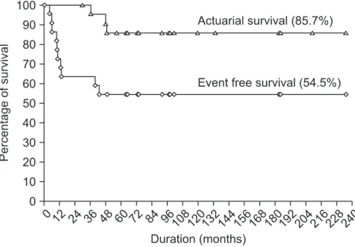
Figure 1. The 10-year overall and event free survival rates were determined using the Kaplan-Meier plot.
Figure 2. Patient 21 was a 37-year- old woman who was misdiagnosed as Nora’s lesion. (A) Initial anteroposterior radiograph shows an ossified mass on the posterolateral aspect of humerus.
(B) This anteroposterior radiograph was taken after 4 episodes of intralesional procedure at referral hospital. Note ill- defined calcified nodules which were located around proximal humerus. (C) The patient underwent segmental excision and reconstruction with recycled autograft.
Local recurrence was noted on this anteroposterior radiograph taken after 3 month later. Concomitant metastasis was also identified.
plotted using the Kaplan-Meier method, and correlations between clinical variables and outcomes were evaluated using the chi-square test.
Results
1. Clinical outcomes of all study subjects
The 10-year overall and event free survival rates for the 22 study subjects determined using the Kaplan-Meier method were 85.7±
5.1% and 54.6±10.6%, respectively (Fig. 1). Nineteen (86%) of the 22 patients were alive at a mean follow-up of 100 months. One pa- tient (case 6) died of pulmonary metastasis at 48 months after index surgery. This patient developed pulmonary metastasis at 21 months after index surgery without evidence of local relapse, but despite metastasectomy and adjuvant chemotherapy, died 27 months later.
The other patient (case 14) with marginal resection and high-grade POS developed local recurrence at 13 months after index surgery, and despite re-excision with wide margin, succumbed to another local recurrence and concomitant pulmonary metastasis. Remaining one patient (case 21) was initially misdiagnosed as Nora’s lesion and underwent four episodes of intralesional excision over 35 months.
After referral, this patient received en-bloc resection of humerus, nevertheless, local recurrence and fulminant metastasis (lung, thigh, and lower leg) developed and eventually expired 49 months from initial intralesional procedure. A pathologic examination of the en- bloc resected specimen in this patient showed dedifferentiated POS (Fig. 2).
Nine (41%) of the 22 patients developed local relapse, and median time to first local recurrence was 22 months (range, 4-43 months).
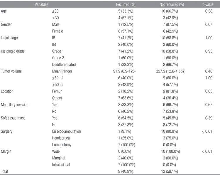
The clinico-pathological variables found to be correlated with lo-
Table 2. Patient and Tumor Characteristics of 9 Patients with Local Recurrence and 13 Patients without Local Recurrence
Variables Recurred (%) Not recurred (%) p-value
Age ≤30 5 (33.3%) 10 (66.7%) 0.38
>30 4 (57.1%) 3 (42.9%)
Gender Male 1 (12.5%) 7 (87.5%) 0.07
Female 8 (57.1%) 6 (42.9%)
Initial stage IB 7 (41.2%) 10 (58.8%) 1.00
IIB 2 (40.0%) 3 (60.0%)
Histologic grade Grade 1 7 (41.2%) 10 (58.8%) 0.93
Grade 2 1 (50.0%) 1 (50.0%)
Dedifferentiated 1 (33.3%) 2 (66.7%)
Tumor volume Mean (range) 91.9 (0.9-125) 397.9 (12.6-4,552) 0.48
≤50 ml 6 (40.0%) 9 (60.0%) 1.00
>50 ml 3 (42.9%) 4 (57.1%)
Location Femur 2 (18.2%) 9 (81.8%) 0.03
Others 7 (63.6%) 4 (36.4%)
Medullary invasion Yes 3 (33.3%) 6 (66.7%) 0.67
No 6 (46.2%) 7 (53.8%)
Soft tissue mass Yes 6 (54.5%) 5 (45.5%) 0.39
No 3 (27.3%) 8 (72.7%)
Surgery En bloc/amputation 1 (9.1%) 10 (90.9%) < 0.01
Hemicortical 1 (25.0%) 3 (75.0%)
Lumpectomy 7 (100.0%) 0 (0.0%)
Margin Wide 0 (0.0%) 10 (100.0%) < 0.01
Marginal 2 (40.0%) 3 (60.0%)
Intralesional 7 (100.0%) 0 (0.0%)
Total 9 (40.9%) 13 (59.1%)
cal recurrence were primary tumor location, type of resection, and surgical margin (Table 2). With respect to primary tumor location, local recurrence occurred in the femur in 2 (18%) of the 11 and in other locations in 7 (64%). The majority of patients that experienced local recurrence had undergone incomplete surgical resection (7 of 9 patient underwent lumpectomy). According to margin status, all the 7 patients with an intralesional mar- gin and 2 of 5 patients with a marginal margin developed local recurrence. None of 10 patients with wide margin showed local recurrence.
2. Treatment and clinical outcomes of locally recurrent patients (Table 3)
The 7 referred patients experienced an average of two lo- cal recurrences (range; 1-4). The majority of recurrences required the removal of an entire segment of bone to achieve a wide operative margin, but no patient under- went amputation. After surgery to obtain wide margin for local failure, 7 (78%) of the 9 patients with local recur- rence developed at least one further recurrence. Average time to subsequent recurrence was 24.1 months (range 3-40 months). At final follow-up, 7 of the 9 patients that experienced local failure did not show further recurrence.
However, the other 2 patients had developed concomi- tant lung metastasis.
Discussion
POS has better survival than classic high-grade intra- medullary osteosarcoma, and often behaves in an indo- lent manner, even after inadvertent procedures. However, with the exception of POS of the femur, lack of famil- iarity with POS with knowledge of the aforementioned characteristics can cause surgeons to underestimate the risk of POS, which could result in a patient missing the opportunity of surgical cure.
Although the conclusions that can be drawn from this small series are limited, this study reconfirms the impor- tance of a sound surgical margin, and demonstrates that there is ample opportunity to under-treat POS, especially in a non-femoral location. Furthermore, it shows that re- excision to overcome further recurrence after an incom- plete procedure is difficult to achieve.
This study is limited by its size and the use of hetero- Table 3. Treatment and Outcome of Patients with Local Recurrence Time to 1st Type of initial ic thologCase Initial pa diagnosisnumberoperationLR (months) No. of intralesional or marginal procedure
Operation to achieve wide margin Subsequent recurrence (months) No. of subsequent recurrence
Metastasis† (site)OutcomeFollow up (months) 2Dedifferentiated POSEn-bloc resection101Re-excision361-NED209 14POS G2Hemicortocal excision131En-bloc excision & tumor prosthesis 8221 (lung)DOD36 16*OsteochondromaLumpectomy184Talectomy391-NED175 17*POS G1Lumpectomy152En-bloc excision & recycled autograft 51-NED136 18*POS G1Lumpectomy 71Hemicortocal excisionNoneNone-NED32 19*POS G1Lumpectomy171En-bloc excision & recycled autograft401-NED81 20*POS G1Lumpectomy101En-bloc excision & recycled autograft381-NED56 21*Nora's lesionLumpectomy213Segmental resection & recycled autograft 3138 (lung, soft tissue)DOD49 22*POS G1Lumpectomy202En-bloc excision & recycled autograftNoneNone-NED84 *Referred case,† Months after initial treatment. LR, local recurrence; POS, parosteal osteosarcoma; NED, no evidence of disease; AWD, alive with disease.
geneous surgical techniques. We acknowledge the heterogeneities caused by the different resection methods used and the relatively large proportion of referred patients. Nevertheless, our objective was to analyze the outcomes of various surgical conditions.
The overall survival achieved is similar to those of numerous pre- vious studies (Table 4). The clinico-pathologic variables previously reported to be correlated with the oncologic results of POS include intramedullary invasion, histologic grade, soft tissue mass, and sur- gical margin.7,11,13,15,16) However, we were unable to determine the prognostic significances of these variables with the exception of sur- gical margin. All 9 local recurrences were associated with marginal or intracapsular procedures, which reconfirms that a wide surgical procedure should be viewed as the gold standard when treating POS.
The local recurrence rate of 41% found in our study compares with that of the Mayo clinic report, in which referred patients constituted 41% (28/67) of the cohort. Interestingly, as compared with other re- ports, the proportion with a location other than the femur (50%) was high in the present study, and these sites were found to be associated with a significantly higher rate of local recurrence. Seven of the 9 patients that experienced local recurrence were referred due to an inadvertent procedure, and of these, 6 had a non-femoral primary site. Furthermore, although the diagnosis of POS is often believed to be relatively straightforward using plain radiographs, reported series suggest that risk of underdiagnosis is substantial.7,13) In those two studies, of the 21 patients that experienced local recurrence, 13 (72%) were initially misdiagnosed as having exostosis, myositis os- sificans, osteoma, or osteitis, which suggests that primary physicians are unfamiliar with POS presenting with an aberrant location and radiologic pattern (case 21).
No matter what clinical situations lead to recurrence, in cases of local failure, the surgeon should decide on a type of surgery that re- sults in a wide margin. Outcomes after local recurrence differ from those of classic high-grade osteosarcoma. Grimer et al. reported that 31 (41%) of 96 patients with local recurrence developed lung metas- tasis either before or at the time of recurrence, and 68 patients either aborted surgery (24 patients) or required amputation (44 patients).17) On the other hand, although there is a risk of dedifferentiation or an increase in histologic grade after repeated recurrence, the majority of patients can be successfully controlled.7,13,18) Therefore, in cases of local recurrence, resection with a wide margin, by whatever method, should be respected. In a meta-analysis of 21 locally recurrent pa- tients, although a half of them underwent amputation, 90% of pa- tients were free of disease at last follow up (Fig. 3).7,13) However, be- cause these two studies were reported around 20 years ago, it is likely
that the abilities of imaging modalities to define the extent of local Table 4. Summary of Publications Concerning Parosteal Osteosarcoma Patient Author/Year number Enneking stage (IB/IIB/III) Histologic gradeLocationSurgical margin (1st OP)Local recurrence (%)
Metastasis (%)Overall survivalMean FU duration (yr)G1G2DedifferentiatedFemur (%)OthersIntralesionalMarginalWide Temple et al. 20003825/12/1 2611 129 (76%) 9 219174/38 (11%)1/37 (3%)38/38 (100%)6.75 (0.5-19) Okada et al. 1994 226* (67)NA/NA/01573237142 (63%)84 6253533/67 (49%)14/67 (21%)56/67 (84%)13 (2-41) Ritschl et al. 199133NA 23 9 123 (70%)1010 32011/33 (33%)5/33 (15%)29/33 (88%)8 (2-23) Han et al. 2008217/14/0 711 316 (76%) 5 2 6132/21 (10%)1/21 (5%)20/21 (95%)9.1 (2.5-22) Current study2217/5/0 17 2 31111 7 5109/22 (41%)3/22 (14%)3/22 (86%)9.5 (3-19.5) *226 patients were registered and outcomes of 67 (managed at that center) patients were presented. OP, operation; FU, follow up; G, grade; NA, not assessed.
recurrence would have been limited. Nowadays, surgical planning in recurrent patients is supported by the accuracy of MRI, which translates into a high rate of limb salvage. Nevertheless, patients with repeated recurrence after procedures that were presumed to achieve a sound margin should not be spared amputation.
In conclusion, we reconfirm the importance of achieving a sound surgical margin when treating POS, and emphasize that the risk of under-treatment not be ignored, especially for cases with a non- femoral location and without typical plain radiologic characteristics.
Furthermore, we found that by using advanced imaging modalities, re-excision without a mutilating procedure was possible in the ma- jority of cases with the exception of those with an aggressive disease pattern after repeated recurrences.
References
1. Raymond AK. Surface osteosarcoma. Clin Orthop Relat Res.
1991;(270):140-8.
2. Unni KK, Dahlin DC, Beabout JW. Periosteal osteogenic sar- coma. Cancer. 1976;37:2476-85.
3. van der Heul RO, von Ronnen JR. Juxtacortical osteosarcoma.
Diagnosis, differential diagnosis, treatment, and an analysis of eighty cases. J Bone Joint Surg Am. 1967;49:415-39.
4. Campanacci M, Picci P, Gherlinzoni F, Guerra A, Bertoni F, Neff JR. Parosteal osteosarcoma. J Bone Joint Surg Br.
1984;66:313-21.
5. Han I, Oh JH, Na YG, Moon KC, Kim HS. Clinical outcome of Figure 3. A diagram shows the outcome in a meta-analysis of 21 locally recurrent patients. Nearly a half (10/21) of them underwent amputation to manage the local recurrence (LR, local recurrence; NED, no evidence of disease; DOD, dead of disease).
parosteal osteosarcoma. J Surg Oncol. 2008;97:146-9.
6. Okada K, Frassica FJ, Sim FH, Beabout JW, Bond JR, Unni KK. Parosteal osteosarcoma. A clinicopathological study. J Bone Joint Surg Am. 1994;76:366-78.
7. Enneking WF, Springfield D, Gross M. The surgical treatment of parosteal osteosarcoma in long bones. J Bone Joint Surg Am. 1985;67:125-35.
8. Kavanagh TG, Cannon SR, Pringle J, Stoker DJ, Kemp HB.
Parosteal osteosarcoma. Treatment by wide resection and prosthetic replacement. J Bone Joint Surg Br. 1990;72:959-65.
9. Ahuja SC, Villacin AB, Smith J, Bullough PG, Huvos AG, Marcove RC. Juxtacortical (parosteal) osteogenic sarcoma:
histological grading and prognosis. J Bone Joint Surg Am.
1977;59:632-47.
10. Bertoni F, Bacchini P, Staals EL, Davidovitz P. Dedifferentiated parosteal osteosarcoma: the experience of the Rizzoli Institute.
Cancer. 2005;103:2373-82.
11. Jelinek JS, Murphey MD, Kransdorf MJ, Shmookler BM, Malawer MM, Hur RC. Parosteal osteosarcoma: value of MR imaging and CT in the prediction of histologic grade. Radiol- ogy. 1996;201:837-42.
12. Wold LE, Unni KK, Beabout JW, Sim FH, Dahlin DC. Dedif- ferentiated parosteal osteosarcoma. J Bone Joint Surg Am.
1984;66:53-9.
13. Ritschl P, Wurnig C, Lechner G, Roessner A. Parosteal os- teosarcoma. 2-23-year follow-up of 33 patients. Acta Orthop Scand. 1991;62:195-200.
14. Lewis VO, Gebhardt MC, Springfield DS. Parosteal osteosar- coma of the posterior aspect of the distal part of the femur.
Oncological and functional results following a new resection technique. J Bone Joint Surg Am. 2000;82A:1083-8.
15. Temple HT, Scully SP, O'Keefe RJ, Katapurum S, Mankin HJ.
Clinical outcome of 38 patients with juxtacortical osteosar- coma. Clin Orthop Relat Res. 2000;(373):208-17.
16. Sheth DS, Yasko AW, Raymond AK, et al. Conventional and dedifferentiated parosteal osteosarcoma. Diagnosis, treatment, and outcome. Cancer. 1996;78:2136-45.
17. Grimer RJ, Sommerville S, Warnock D, et al. Management and outcome after local recurrence of osteosarcoma. Eur J Cancer.
2005;41:578-83.
18. Luck JV Jr, Luck JV, Schwinn CP. Parosteal osteosar- coma: a treatment-oriented study. Clin Orthop Relat Res.
1980;(153):92-105.
방골성 골육종의 임상결과
송원석 • 전대근 • 조완형 • 공창배 • 조상현 • 이광열 • 이수용
원자력병원 정형외과
목적: 방골성 골육종 환자의 치료 결과와 국소 재발 후의 결과에 대해서 알아보고자 하였다.
대상 및 방법: 22명의 방골성 골육종 환자의 치료 결과를 후향적으로 분석하였다. 평균 추시기간은 114개월(범위; 36-235개월)이었
다. 22명 중 7명은 국소 재발 후에 전원 되었다. 병기는 17명에서 IB였고, 5명은 IIB (G2, 2명; 역분화, 3명)이었다. 종양의 위치는 대퇴 골(11명), 기타 부위(11명)이었다. 최초 절제연은 광범위 절제연 10명, 변연 절제연 5명, 병소내 절제가 7명이었다. 여러 임상 및 병리인 자와 국소 재발과의 연관성, 그리고 국소 재발 후의 임상 경과를 조사하였다.
결과: 10년 생존율은 85.7%이었다. 3명(14%)에서 원격 전이를 보였고 이들은 모두 사망하였다. 9명(41%)에서 국소 재발이 있었다. 종
양의 위치, 절제 방법 및 절제연이 국소재발과 관련이 있었다. 국소 재발 후 수술 한 환자 9명 중 최종 추시 시 7명에서는 무병 상태였 다.
결론: 방골성 골육종의 오진의 가능성이 높으며 특히 대퇴골 이외에 발생한 경우 오진이 많았다. 대부분의 재발성 종양에 대한 재 절제
는 가능하나 공격적 성향을 보이며 재발한 경우에는 주의가 필요할 것으로 생각된다.
색인단어: 방골성 골육종, 국소 재발
접수일 2013년 3월 25일 심사수정일 2013년 5월 14일 게재확정일 2013년 5월 20일 교신저자 전대근
서울시 노원구 공릉동 215-4, 원자력병원 정형외과
TEL 02-970-1242, FAX 02-970-2403, E-mail dgjeon@kcch.re.kr
Copyrights © 2013 by The Korean Bone and Joint Tumor Society
“This is an Open Access article distributed under the terms of the Creative Commons Attribution Non-Commercial License (http://creativecommons.org/licenses/by-nc/3.0/) which permits unrestricted non-commercial use, distribution, and reproduction in any medium, provided the original work is properly cited.”
The Journal of the Korean Bone and Joint Tumor Society Vol. 19, No. 1 (June 2013)