Korean J Gastroenterol Vol. 63 No. 3, 183-186 http://dx.doi.org/10.4166/kjg.2014.63.3.183 pISSN 1598-9992 eISSN 2233-6869
CASE REPORT
Korean J Gastroenterol, Vol. 63 No. 3, March 2014 www.kjg.or.kr
특발성 정맥경화성 대장염: 만성 허혈성 장염의 드문 예
최종민, 이강녕, 김해수, 이상기, 이정규, 이성원, 이오영, 최호순
한양대학교 의과대학 내과학교실
Idiopathic Phlebosclerotic Colitis: A Rare Entity of Chronic Ischemic Colitis
Jong Min Choi, Kang Nyeong Lee, Hae Su Kim, Sang Ki Lee, Jung Gyu Lee, Sung Won Lee, Oh Young Lee and Ho Soon Choi Department of Internal Medicine, Hanyang University College of Medicine, Seoul, Korea
Colonic wall thickening is frequently encountered in various conditions, from acute or chronic inflammatory disease to colorectal carcinoma. Colonic wall thickening may be accompanied by calcifications in mucinous adenocarcinoma of the colon, leiomyosarco- ma of the colon, schistosomiasis japonica, and phlebosclerotic colitis. Phlebosclerotic colitis is a rare entity of chronic ischemic colitis associated with sclerosis and fibrosis of mesenteric veins. Although its development is usually insidious, and, thus its diagnosis can be delayed, characteristic findings in phlebosclerotic colitis are calcifications of mesenteric veins as well as colonic wall thickening with calcifications. We report on a 71-year-old woman who presented with chronic diarrhea and intermittent hematochezia, who was first misdiagnosed as mucinous adenocarcinoma of the colon, but finally diagnosed as a rare entity of chronic ischemic colitis, phlebosclerotic colitis. Differential points of phlebosclerotic colitis from other diseases, including leiomyosarcoma and schistosomiasis japonica, are also described. (Korean J Gastroenterol 2014;63:183-186) Key Words: Ischemic colitis; Mesenteric veins; Sclerosis; Vascular calcification
Received June 13, 2013. Revised August 9, 2013. Accepted August 13, 2013.
CC This is an open access article distributed under the terms of the Creative Commons Attribution Non-Commercial License (http://creativecommons.org/licenses/
by-nc/3.0) which permits unrestricted non-commercial use, distribution, and reproduction in any medium, provided the original work is properly cited.
교신저자: 이강녕, 133-791, 서울시 성동구 왕십리로 222, 한양대학교 의과대학 내과학교실
Correspondence to: Kang Nyeong Lee, Department of Internal Medicine, Hanyang University College of Medicine, 222 Wangsimni-ro, Seongdong-gu, Seoul 133-791, Korea. Tel: +82-2-2290-8339, Fax: +82-2-2290-8314, E-mail: [email protected]
Financial support: None. Conflict of interest: None.
INTRODUCTION
Colonic wall thickening is frequently encountered in clin- ical practice. It is related to a variety of entities, including nor- mal variants, inflammatory conditions, and colonic neo- plasms.1 Development of colonic wall thickenings may be not only primary, but also secondary in many clinical settings as- sociated with body fluid retention due to heart failure, chronic liver disease, and renal insufficiency, and in other infiltrative diseases such as amyloidosis. Colonic wall thickenings can be accompanied by calcifications in mucinous adenocarci- noma of the colorectum,1,2 leiomyosarcoma of the colon,3 schistosomiasis japonica,4 and phlebosclerotic colitis (PC), a rare entity of ischemic colitis.
Ischemic colitis may develop by occlusion or stenosis in mesenteric arteries or veins due to atherosclerotic, throm- botic, or embolic causes, or by insufficient tissue perfusion without occlusive lesions of mesenteric vessels. Ischemic colitis by non-thrombotic occlusion of mesenteric veins is rarely reported; however, it may be associated with systemic disease, such as amyloidosis, lupus, or rheumatoid arthritis.
Ischemic colitis due to disturbances in mesenteric blood flow due to sclerotic changes and calcifications of the venous wall is defined as PC. However, the etiology and pathogenesis of this rare entity of chronic ischemic colitis has not yet been clearly defined. Herein, we report on a case of colonic wall thickening with calcifications in PC, which was first mis- diagnosed as a mucinous adenocarcinoma of the colon.
184 최종민 등. 특발성 정맥경화성 대장염
The Korean Journal of Gastroenterology Fig. 1. Simple radiograph shows linear calcifications in the right
abdominal area (arrow).
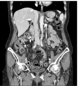
Fig. 2. Abdominal CT shows colonic wall thickenings accompanied by mural and mesenteric venous calcifications in the ascending colon (arrows).
CASE REPORT
A 71-year-old woman was admitted because of chronic di- arrhea and hematochezia for the past seven months. One year ago, she had found that her stool caliber became narrow.
However, no other alarming symptoms or signs suggestive of malignancy, including weight loss and fever, were noted. She had been living in America since immigrating 10 years ago.
Concern for medical costs urged her to return to her home- town city, Seoul, Korea for further evaluation. Just before ad- mittance to our hospital, she had undergone abdominal CT at a local clinic and was informed that colonic cancer was suspected. She does not drink alcohol and had not taken any herbal medications, and she had no past medical history of chronic liver or kidney disease.
At admission, vital signs were body temperature of 36.8oC, pulse rate of 82/min, respiratory rate of 18/min, and blood pressure of 120/70 mmHg. Abdomen was soft and non-ten- der, but bowel sounds were slightly increased. Digital rectal examination found no rectal bleeding. Laboratory findings were white blood cells of 3,800/mm3, hemoglobin of 11.8 g/dL, platelets of 251,000/mm3, total protein of 6.8 g/dL, al- bumin of 3.9 g/dL, BUN 10.6 mg/dL, creatinine 0.72 mg/dL, total bilirubin 0.43 mg/dL, AST 15 U/L, ALT 6 U/L, and CEA 2.9 ng/mL. Chest X-ray showed no enlargement of the heart.
Electrocardiogram showed non-specific ST-T wave change.
No ova or cyst suggesting infection of parasite was detected in stool examination.
We found multiple linear radiopacities in the right abdomi- nal area on simple X-ray (Fig. 1). Abdominal CT showed dif- fuse thickening of bowel walls with mural calcifications from the cecum to splenic flexure; surrounding mesenteric veins were also calcified (Fig. 2). Wall thickening with calcifications of the colonic wall and mesenteric vein was not observed on the distal colonic wall. On the other hand, abdominal CT showed no findings suggestive of portal hypertension, such as contrast enhancement of the paraumbilical vein, dilated portal or mesenteric veins, splenomegaly, or ascites. In addi- tion, no varices or portal hypertensive gastropathy suggest- ing portal hypertension were found on upper gastrointestinal endoscopy. Colonoscopy showed dark purple edematous mucosa and areas of luminal narrowing with multiple ero- sions or ulcerations (Fig. 3). The colonic mucosal lesions were distributed from the cecum to the sigmoid colon, but be- came less severe toward the distal colon with only mild dark edematous mucosa in the sigmoid colon. Microscopic exami- nations of colonic tissues obtained from the ascending and transverse colon showed marked thickening and calcifica- tions of the colonic mucosal and submucosal layers with ul- cerations and a large focus of dystrophic calcification.
Mesenteric veins also showed an abnormally thickened wall with hyalinization (Fig. 4). After conservative management, she was discharged and returned to the Unites States.
Choi JM, et al. Idiopathic Phlebosclerotic Colitis 185
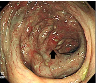
Vol. 63 No. 3, March 2014 Fig. 3. Colonoscopy shows dark purple edematous mucosa with
areas of luminal narrowing in the ascending colon. Multiple erosions or ulcerations with surrounding erythema are also noted (arrow).
Fig. 4. Microscopic examinations. Marked thickening and calcifications of the colonic mucosal and submucosal layers are noted. Mesenteric veins show an abnormally thickened wall with hyalinization (arrow) (H&E, ×200).
DISCUSSION
Colonic wall thickenings are frequently found because co- lonic responses are nonspecific to various stimuli, including infection, inflammation, or ischemia. This case of colonic wall thickening represents a rare entity of chronic ischemic colitis, PC. Although the suggested pathogenesis was that mesen- teric veins gradually become sclerotic and fibrotic resulting in colonic wall thickening with calcifications, currently, its eti- ology and pathogenesis are not clearly defined.
Whatever the etiology and pathogenesis of PC are, its char-
acteristic finding of colonic wall thickening with calcifications on CT should be distinguished from mucinous adenocarci- noma, leiomyosarcoma, and schistosomiasis japonica. In mucinous adenocarcinoma of the colon, colonic wall thicken- ing with mural calcifications is irregular and asymmetric with short-segment involvement, compared with regular and sym- metric thickening with long segment involvement in PC.1 Wall thickening in mucinous adenocarcinoma also tends to spread abruptly to the unaffected colonic wall and to cause obstruction of the bowel due to severe luminal narrowing.
However, in our case, asymmetric thickening and severe nar- rowing in certain areas of the colonic wall might have caused confusion in differentiation of PC from mucinous adenocar- cinoma, although the involvement of the long segment of the colon was strongly suggestive of PC. In leiomyosarcoma, a mesenchymal cancer of the colon, colonic wall thickenings are bulky mural masses with calcifications, heterogeneous hyper-enhancement, exophytic growth, and cystic degenera- tion.3 In schistosomiasis japonica, in which schistosoma ja- ponicum, a kind of parasite, causes granulomatous disease in the intestine, colonic wall thickening with calcifications in- dicates calcified eggs originating from larvae matured in the portal vein, and are frequently shown in the distal colon com- pared with proximal involvement in PC.4 Taken together, the presence of mesenteric venous calcifications around the af- fected colon on abdominal CT is a differential point of PC from these diseases.
Colonoscopy of our patient showed dark purple dis- colorations of the mucosa with mucosal edema and eryth- ema, erosions or ulcerations, and luminal narrowing, which are characteristic findings in PC. The dark purple discoloration of the mucosa seems to be caused by chronic congestion with ischemia associated with sclerotic and fibrotic changes of the mesenteric veins. However, certain toxic agents that can show staining in the colonic mucosa after being absorbed through the colonic wall were also suggested as a cause. In addition, the reason that PC usually involves the right-sided colon was also suggested to be due to the fact that certain toxic agents metabolized by colonic bacteria in the proximal colon were absorbed in the region.5 However, in our patient, mucosal lesions of varying severity were distributed through- out the colon, ranging from the cecum to the sigmoid colon with no skipped lesions. One report suggested that it may gradually progress to the entire colon.6 In addition, our pa-
186 최종민 등. 특발성 정맥경화성 대장염
The Korean Journal of Gastroenterology
tient denied use of any herbal medication and had no predis- posing conditions that might increase mesenteric venous pressure. Therefore, the cause of PC in our patient seems to be idiopathic.
Microscopic findings of PC are variable. Colonic wall thick- enings consist mainly of marked submucosal fibrosis; how- ever, they may contain a variable degree of vascular con- gestion, hemosiderin-laden macrophages, erythrocyte ex- travasation, and brown to black-pigmented macrophages.
Venous wall thickenings around the involved colon and mes- entery are also shown, and are accompanied by fibrosis, scle- rosis, and hyalinization as well as calcifications, which are suggested to result from coagulative necrosis in the muscle layer and subsequent myointimal hyperplasia of mesenteric veins.7 Then, the hyperplastic myointima are damaged re- peatedly, leading to gradual occlusion of the mesenteric veins. These phlebosclerotic changes may be caused by long-term elevated pressure to the veins in conditions such as right-sided heart failure, liver cirrhosis, and portal hyper- tension.2,8 In addition, the suggested predisposing con- ditions include diabetes mellitus, dyslipidemia, and hemo- dialysis.5,9 On the other hand, all of these changes of the mes- enteric veins are supposed to be attributable to certain toxic agents that are absorbed into venous return of the proximal colonic wall. Similarly, association of chronic use of herbal medicine and predilection of East Asians of female gender with PC has been reported.5
As in our patient, symptoms of PC include abdominal dis- comfort, diarrhea, constipation, hematochezia, and weight loss with chronic and insidious development.2 The chronic and insidious development of symptoms may be attributable to the chronic congestion of mesenteric veins, thus resulting in delayed diagnosis of PC. For diagnosis of PC, other modal- ities except abdominal CT or colonoscopy are complemen- tary. In simple abdominal radiography, multiple threadlike calcifications may be noted along the right hemicolon.
Barium study of the colon shows disappearance of semilunar folds, luminal irregularities, and overall stenosis with rigidity.
Angiography in PC may show narrowing of the marginal ar- teries with dilatation and tortuosity of veins along the vasa recta. In terms of treatment, reports have suggested that con- servative management with bowel rest and hydration are suf- ficient in PC.2 However, when conservative management fails to result in improvement of persistent abdominal pain or
ileus, surgery may be required.2 PC may be complicated by dehydration, hemorrhage, perforation, and death.10
This report indicates that the combination of chronic ab- dominal discomfort with diarrhea, right-sided multiple calci- fications on a simple abdominal radiography, dark purple and edematous mucosa with erosions and luminal narrowing on colonoscopy, and wall thickening with calcifications both in colon and in mesenteric veins is suggestive of PC, a very rare entity of chronic ischemic colitis. In addition, other dis- eases of colonic wall thickening with calcifications should be differentiated, particularly mucinous adenocarcinoma of the colon, although the differentiation might not be difficult be- cause mucinous adenocarcinoma shows focal thickening of the colonic wall instead of long segment involvement.
Therefore, awareness of this disease entity is needed be- cause despite insidious and rare presentation of PC, its endo- scopic and radiologic findings are characteristic.
REFERENCES
1. Macari M, Balthazar EJ. CT of bowel wall thickening: significance and pitfalls of interpretation. AJR Am J Roentgenol 2001;176:
1105-1116.
2. Iwashita A, Yao T, Schlemper RJ, et al. Mesenteric phlebo- sclerosis: a new disease entity causing ischemic colitis. Dis Colon Rectum 2003;46:209-220.
3. Lee SH, Ha HK, Byun JY, et al. Radiological features of leiomyom- atous tumors of the colon and rectum. J Comput Assist Tomogr 2000;24:407-412.
4. Lee RC, Chiang JH, Chou YH, et al. Intestinal schistosomiasis ja- ponica: CT-pathologic correlation. Radiology 1994;193:539-542.
5. Hiramatsu K, Sakata H, Horita Y, et al. Mesenteric phlebo- sclerosis associated with long-term oral intake of geniposide, an ingredient of herbal medicine. Aliment Pharmacol Ther 2012;
36:575-586.
6. Chen MT, Yu SL, Yang TH. A case of phlebosclerotic colitis with involvement of the entire colon. Chang Gung Med J 2010;33:
581-585.
7. Chang KM. New histologic findings in idiopathic mesenteric phlebosclerosis: clues to its pathogenesis and etiology--prob- ably ingested toxic agent-related. J Chin Med Assoc 2007;70:
227-235.
8. Kang HY, Noh R, Kim SM, Shin HD, Yun SY, Song IH. Phlebosclerotic colitis in a cirrhotic patient with portal hypertension: the first case in Korea. J Korean Med Sci 2009;24:1195-1199.
9. Song JH, Kim JI, Jung JH, et al. A case of phlebosclerotic colitis in a hemodialysis patient. Korean J Gastroenterol 2012;59:40-43.
10. Kato T, Miyazaki K, Nakamura T, Tan KY, Chiba T, Konishi F.
Perforated phlebosclerotic colitis--description of a case and re- view of this condition. Colorectal Dis 2010;12:149-151.