Copyright © 2019 The Korean Society for Bone and Mineral Research
This is an Open Access article distributed under the terms of the Creative Commons Attribution Non-Commercial Li- cense (http://creativecommons.org/licenses/by-nc/4.0/) which permits unrestricted non-commercial use, distribu- tion, and reproduction in any medium, provided the original work is properly cited.
Effects of Thyrotropin Suppression on Bone Health in Menopausal Women with Total Thyroidectomy
Eun Heui Kim1, Yun Kyung Jeon1, Kyoungjune Pak2, In-Joo Kim2, Seong-Jang Kim3, Seunghyeon Shin2, Bo Hyun Kim1, Sang Soo Kim1, Byung-Joo Lee4, Jeong-Gyu Lee5, Tae Sik Goh6, Keunyoung Kim2
1Division of Endocrinology and Metabolism, Department of Internal Medicine and Biomedical Research Institute, Pusan National University Hospital, Busan;
2Department of Nuclear Medicine and Biomedical Research Institute, Pusan National University Hospital, Busan;
3Department of Nuclear Medicine and Research Institute for Convergence of Biomedical Science and Technology, Yangsan Pusan National University Hospital, Yangsan;
4Department of Otorhinolaryngology-Head and Neck Surgery, Pusan National University School of Medicine and Biomedical Research Institute, Pusan National University Hospital, Busan;
5Department of Family Medicine, Pusan National University School of Medicine, Busan;
6Department of Orthopaedic Surgery and Biomedical Research Institute, Pusan National University Hospital, Busan, Korea
Background: This study examined the change in the trabecular bone score (TBS), areal bone mineral density (aBMD), and osteoporosis in postmenopausal women who under- went thyrotropin (TSH)-suppressive therapy for treating papillary thyroid cancer after a total thyroidectomy procedure. Methods: We evaluated 36 postmenopausal women who received a total thyroidectomy for papillary thyroid cancer and were undergoing TSH suppressive therapy with levothyroxine. Postmenopausal women (n=94) matched for age and body mass index were recruited as healthy controls. The aBMD and TBS of the lumbar spine were compared between dual energy X-ray absorptiometry (DXA) at baseline and at follow-up after an average of 4.92 years. Results: There was no signifi- cant difference in the rate of diagnoses of osteoporosis, osteopenia, or normal bone sta- tus between the 2 groups during the baseline DXA evaluation. However, the TBS was significantly lower whereas aBMD did not show significant difference at the time of baseline DXA measurement (1st DXA, 1.343±0.098 vs. 1.372±0.06317, P<0.001; 2nd DXA, 1.342±0.095 vs. 1.370±0.062, P<0.001). The TBS and aBMD did not differ signifi- cantly between the initial and follow-up DXA images in both groups of TSH suppressive patients and controls. Conclusions: The average value of TBS and aBMD did not signifi- cantly change during the follow-up period. The TSH suppressive therapy was revealed as not a significant factor for the progressive deterioration of bone status during long term follow-up.
Key Words: Absorptiometry, photon · Bone density · Postmenopause · Thyroid neoplasms
INTRODUCTION
Among many endocrinological causes of secondary osteoporosis (e.g., Cush- ing’s syndrome, hyperparathyroidism, hypogonadism), conditions of excess or de- ficient thyrotropin (thyroid-stimulating hormone [TSH]) and thyroid hormones are potentially damaging to bone status.[1,2] Although endogenous hyperthy- roidism is associated with an increased risk of osteoporosis due to increased os- Corresponding author
Keunyoung Kim
Department of Nuclear Medicine and Biomedical Research Institute, Pusan National University Hospital, 179 Gudeok-ro, Seo-gu, Busan 49241, Korea
Tel: +82-51-240-7389 Fax: +82-51-241-5570 E-mail: [email protected] Received: January 7, 2019 Revised: January 28, 2019 Accepted: February 4, 2019
No potential conflict of interest relevant to this article was reported.
Original Article
pISSN 2287-6375 eISSN 2287-7029
teoclastic resorption and decreased bone formation,[3,4]
the effect of suppressive thyroxine therapy on osteoporo- sis remains unclear. Recent concerns have emerged that lifelong TSH-suppressive therapy may have the potentially harmful effects on the heart and skeleton induced by sub- clinical hyperthyroidism [5] differentiated thyroid cancer (DTC) is the most common endocrine malignancy and is generally an indolent tumor with a 10-year overall survival rate of 80% to 95%.[6]
Osteoporosis is the recent concern regarding lifelong TSH-suppressive therapy.[5,7-11] Osteoporosis is a major healthcare challenge worldwide; the number of newly di- agnosed cases and associated costs of osteoporotic frac- ture are increasing with age. Both menopause and hyper- thyroidism are the risk factor of osteoporosis,[12] and wom- en with hyperthyroidism experience their first fractures earlier during life than do those without.[13] Recent stud- ies reported that, suppressed TSH level is significantly cor- related with the risk of osteoporosis after total thyroidec- tomy.[14,15] Osteoporosis is characterized by a low bone mass and microarchitectural deterioration. The World Health Organization’s standard criterion for diagnosing osteopo- rosis uses densitometry tools, such as dual energy X-ray absorptiometry (DXA) for measuring areal bone mineral density (aBMD; mg/cm2) and calculating a T-score. The tra- becular bone score (TBS) is a new textural index obtained from DXA, that evaluates pixel gray-level variations of the lumbar spines has recently been proposed as an represen- tative parameter for the trabecular bone microarchitecture independent of BMD.[16] According to recent study, Recu- ded TBS has association with increased osteoporotic verte- bral fractures risk in patients with osteopenia, as well as in those with normal BMD.[17] In healthy euthyroid post-meno- pausal women, the TBS is negatively correlated with free thyroxine (fT4) levels within range for high-to-normal.[18]
In postmenopausal women with DTC, a longer duration of TSH suppression was independently associated with a de- creased lumbar spine TBS.[19] From a clinical perspective, the additional information about bone quality provided by the TBS could aid the management of patients with osteo- penia, as well as those with normal BMD who have risk fac- tors for osteoporosis.
We designed this retrospective study to determine whether suppressive L-thyroxine (LT4) therapy after a total thyroid- ectomy influences the TBS, and compare the incidence of
osteoporosis with a healthy control group.
METHODS
1. Study subjectsThis retrospective study included postmenopausal wom- en with DTC who underwent a total thyroidectomy and began follow-up testing between October 2013 and Janu- ary 2017 at Hospital. The strict selection criteria for the TSH- suppression group were as follows: (1) no distant metasta- sis or invasion of locoregional structures; (2) postmenopaus- al status with a baseline DXA evaluation; (3) treatment with LT4 at suppressive doses to maintain a serum TSH level <0.3 mU/I; (4) fT4 level within the normal range; (5) free of thy- roid cancer on clinical and laboratory examinations during follow-up (negative neck ultrasound and undetectable se- rum thyroglobulin); and (6) regular follow-up visits. Because menopause is a risk factor for osteoporosis, all patients un- derwent repeat DXA for evaluation of bone mineral densi- ty. We enrolled postmenopausal women (n=36) with pap- illary thyroid cancer who underwent total thyroidectomy and TSH suppression.
2. Healthy control group
To identify the healthy controls, we enrolled postmeno- pausal women (n=94) who visited our hospital for a health check-up and matched in age and body mass index (BMI) to the treatment group. They had annual checkup for lum- bar spine DXA and thyroid function tests (serum TSH, fT4, and T3). The participants in the healthy control group had serum TSH levels within the normal reference range and did not have hypo- or hyperthyroidism or a history of ma- lignant disease. Exclusion criteria for all participants includ- ed renal or hepatic impairment and exposure to any medi- cation that affects bone metabolism such as glucocorticoids or anti-osteoporotic drugs.
TSH and fT4 levels were measured using the convention- al BRAHMS manual immunoradiometric assay (Thermo Fisher Scientific BRAHMS GmbH, Hennigsdorf, Germany).
Laboratory analyses of calcium, phosphate, and alkaline phosphatase levels were performed on the same day as DXA.
This retrospective study was approved by our Institution- al Review Board (IRB), which waived the requirement for written consent (IRB No. H-1711-008-061).
3. DXA
The aBMD of the lumbar vertebrae (L1-4) was measured on DXA scans (Lunar Prodigy; GE Medical Systems, Milwau- kee, WI, USA) and analyzed using Encore software (version 13.0; GE Healthcare, Madison, WI, USA). Calibration and quality assurance tests were performed daily. The coeffi- cient of variation for precise measurement of the aBMD of the lumbar spine was 0.34%. T-scores were defined accord- ing to the number of standard deviations (SDs) from the mean BMD of a reference group from the general popula- tion aged 25 to 35 years and matched for sex, as follows: l normal, lowest T-score≥-1.0; osteopenia, -2.5<lowest T- score<-1.0; and osteoporosis, ≤-2.5. The TBS of the lum- bar spine was extrapolated from the lumbar spine DXA file using iNsight software (version 3.0.2.0; Medimaps, Meri- gnac, France). A TBS was classified by a cutoff of >1.350 for a normal TBS value according to an international working group of TBS users for postmenopausal women.[20] The ratios of TBS and aBMD deterioration were calculated ac- cording to the baseline and final follow-up values.
4. Statistical analyses
All normally distributed variables were expressed as means
±SD, while variables with non-normal distributions are ex- pressed as medians and interquartile ranges (IQRs; 25-75%).
To compare categorical data between the treatment and control groups, the χ2 test was used. A paired t-test was used to compare the follow-up DXA scans. The Mann–Whitney U test was used to compare the median values of non-nor- mally distributed variables between the treatment and con- trol groups. P-values <0.05 were considered statistically significant. The statistical analyses were performed using both MedCalc® for Windows software (version 16.4.3; Med- Calc, Mariakerke, Belgium) and GraphPad Prism® (version 6;
GraphPad Software, La Jolla, CA, USA).
RESULTS
1. Patient characteristics
There was no difference in the median follow-up period from the baseline DXA evaluation between the TSH sup- pression group (4.0±1.7 years) and the control group (4.4±
1.7 years; P=0.923). The median age (60.5±5.5 years vs.
60.8±5.5 years, P=0.127) and BMI (23.31±2.77 vs. 23.20±
2.93, P=0.356) also did not differ between the TSH suppres-
sion and control groups. The TSH-suppressive therapy was maintained for 4.66±1.52 years after total thyroidectomy at the baseline DXA study. The daily dose of LT4 for the pa- tient group was 200 to 750 µg/day or 2.500±0.774 µg/kg.
Of the patients in the TSH suppression group, 32 (88.89%) received calcium/vitamin D supplementation. Of the par- ticipants in the control group, 34 (36.17%) received calci- um/vitamin D supplementation. The TSH levels of all pa- tients in the TSH suppression group were nearly non-de- tectable (median, 0.000; IQR, 0.000-0.254), whereas those in the control group were within the normal range (medi- an, 1.975; IQR, 1.141-1.644; P<0.001). The fT4 levels of all participants were within the normal range; however, the mean level differed significantly between the TSH suppres- sion group (1.706±0.319) and control group (1.240±0.165;
P<0.001). The baseline clinical characteristics of the partic- ipants in both groups are shown in Tables 1 and 2.
2. Comparison of DXA results between the TSH suppression and control groups during follow-up
There was no significant difference in the rate of diagno- Table 1. Baseline characteristics of patients with papillary thyroid cancer
Patient characteristics Value (n=36)
1st DXA after surgery (year) 4.657±1.520
Tumor size (cm) 0.98 (0.5-1.0)
Microcarcinoma 22 (61.1%)
Extrathyroidal extension
Yes 17 (47.2%)
No 18 (50.0%)
NA 1 (2.8%)
N stage
N0 20 (55.6%)
N1a 15 (41.7%)
NA 1 (2.8%)
Radioiodine therapy
None 6 (16.7%)
30 mCi 3 (8.3%)
100 mCi 9 (25.0%)
150 mCi 28 (77.8%)
Dose of levothyroxine
µg/day 135±3.06
mg/kg 2.214±0.031
The data is presented as mean±standard deviation, median (interquar- tile range), or number (%).
DXA, dual energy X-ray absorptiometry; NA, not applicable.
sis of osteoporosis, osteopenia, or normal bone status be- tween the 2 groups during the baseline DXA evaluation (Fig. 1). The average aBMD of the patients who received TSH-suppressive therapy was not significantly different from that of the control group (1st DXA, 0.997±0.149 vs.
1.081±0.148, P=0.415; 2nd DXA, 0.986±0.145 vs. 1.074±
0.147, P=0.670; Table 2) and did not change significantly during follow-up (Fig. 2A). The mean TBS at the 1st DXA and follow-up DXA evaluations was significantly lower in patients with TSH suppression than in the control group (1st DXA, 1.344±0.098 vs. 1.372±0.063, P<0.001; 2nd DXA, 1.342±0.095 vs. 1.370±0.062, P<0.001; Table 2) where- as TBS was not significantly changed comparing at base- line and follow-up regardless of TSH suppressive treatment (Fig. 2B). According to the TBS threshold of 1.350, the sta- tus of the microarchitecture was not significantly different between the 2 groups on the follow-up scans.
DISCUSSION
After a total thyroidectomy, exogenous LT4 therapy for suppression of TSH and radio-iodine ablation therapy have been considered the standard treatments for DTC.[21-23]
These treatments reduce the risk of tumor recurrence and improve patient survival.[6,24] A meta-analysis by McGriff et al.[25] reported that TSH-suppressive therapy decreased the risk of major adverse clinical events, including disease progression, recurrence, and death. Despite studies show- ing that TSH suppressive therapy improves patient survival, there is increasing concern about the adverse effects of lifelong therapy, because DTC patients have a long life ex- pectancy after surgery.
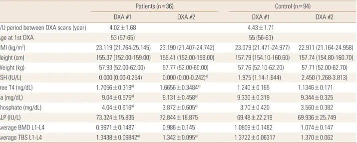
The critical clinical perspective on bone status has emerged Table 2. Clinical and laboratory values during baseline and follow-up dual energy X-ray absorptiometry evaluations
Patients (n=36) Control (n=94)
DXA #1 DXA #2 DXA #1 DXA #2
F/U period between DXA scans (year) 4.02±1.68 4.43±1.71
Age at 1st DXA 53 (57-65) 55 (56-63)
BMI (kg/m2) 23.119 (21.764-25.145) 23.190 (21.407-24.742) 23.079 (21.471-24.977) 22.911 (21.164-24.958) Height (cm) 155.37 (152.00-159.00) 155.41 (152.00-159.00) 157.79 (154.10-160.60) 157.74 (154.80-160.70) Weight (kg) 57.93 (52.00-62.00) 57.77 (52.00-60.00) 57.76 (52.10-62.20) 57.71 (52.00-62.70) TSH (IU/L) 0.000 (0.00-0.254) 0.000 (0.00-0.242)a) 1.975 (1.14-1.644) 2.450 (1.268-3.813)
Free T4 (ng/dL) 1.7056±0.319a) 1.6656±0.3484a) 1.240±0.165 1.1346±0.171
Ca (mg/dL) 9.04±0.575a) 9.131±0.458a) 9.330±0.319 9.344±0.325
Phosphate (mg/dL) 4.04±0.616a) 3.872±0.605a) 3.70±0.420 3.560±0.382
ALP (IU/L) 73.324±15.835 72.844±18.875 69.48±22.219 69.936±25.749
Average BMD L1-L4 0.9971±0.1487 0.986±0.145 1.0809±0.1482 1.074±0.147
Average TBS L1-L4 1.3438±0.09842a) 1.342±0.095a) 1.3722±0.06317 1.370±0.062
The data is presented as the mean±standard deviation, or median (interquartile range).
a)P<0.01 vs. the control group.
F/U, follow-up; DXA, dual energy X-ray absorptiometry; BMI, body mass index; TSH, thyroid-stimulating hormone; Ca, calcium; ALP, alkaline phospha- tase; BMD, bone mineral density; TBS, trabecular bone score.
Fig. 1. Diagnosis of dual energy X-ray absorptiometry (DXA) during follow-up. For DXA, T-scores ≥-1.0 standard deviations (SDs) from the reference mean were defined as normal; T-scores between -2.5 and -1.0 SDs from the reference mean were defined as osteopenia;
T-scores ≤-2.5 SDs from the reference mean were defined as osteo- porosis. This follows the World Health Organization criteria. TSH, thyroid-stimulating hormone.
100 90 80 70 60 50 40 30 20 10 0
Proportion of the participants (%)
TSH suppression
group TSH suppression
group Control
group Control
group
Baseline Follow up
Normal Osteopenia Osteoporosis
22
32 74
10
10 10
10
46
38
4 22
largely because of the extended survival and resulted long- term subclinical hyperthyroid state. Since Ross et al.[11] re- ported that long-term exposure to exogenous thyroid hor- mone was associated with bone loss, studies had reported that TSH-suppressive therapy may cause a significant dete- rioration of BMD in older patients.[14,26-28] However, the effect of exogenous TSH-suppressive therapy remains un- der debate. Heemstra et al.[28] suggested that postmeno- pausal women undergoing long-term TSH-suppressive therapy suffer from a lower BMD and higher risk of osteo- porosis compared with controls. In other studies, the change in the BMD of postmenopausal patients with DTC on sup- pressive therapy was unclear,[29] or there were no signifi- cant effect of long-term TSH-suppressive therapy on BMD or bone turnover markers.[30,31]
The result of this study revealed the average aBMD of the patients who received TSH-suppressive therapy was
not significantly decreased than that of the control group during follow-up. It coincided with report of Kim et al.[32], the study of 100 postmenopausal women and 24 premeno- pausal women on TSH suppression LT4 therapy, there was no significant change of BMD and bone turnover marker.
The TBS decreased during the follow-up DXA evaluation over a mean of 4.02 years without statistical significance.
Although there was no significant change of TBS, there was significant difference between patients and control at the time of baseline DXA measurement. One study report- ed that the TBS of the lumbar spine was independently and negatively correlated with the duration of TSH-sup- pressive therapy in DTC patients.[19] Our data were adjust- ed for age, BMI, and follow-up period, but we could not find the correlation between the TBS and TSH suppression duration. We expect that patients on TSH suppression will have a worse TBS at earlier period of TSH suppression, and Fig. 2. Changes of mean areal bone mineral density (aBMD) and trabecular bone score (TBS) at the 1st dual energy X-ray absorptiometry scan and 2nd follow-up scan. Both (A) aBMD and (B) TBS did not change significantly during follow-up using paired t-test. TSH, thyroid-stimulating hormone.
1.5 1.4 1.3 1.2 1.1 1.0 0.9 0.8 0.7 0.6 Average aBMD (g/cm2)
Baseline Follow up
TSH suppression group
P=0.1336
1.6
1.5
1.4
1.3
1.2
1.1
Average TBS
Baseline Follow up
TSH suppression group
P=0.0977
1.6
1.5
1.4
1.3
1.2
Average TBS
Baseline Follow up
Control group
P=0.6808 1.5
1.4 1.3 1.2 1.1 1.0 0.9 0.8 0.7 Average aBMD (g/cm2)
Baseline Follow up
Control group
P=0.3459
A
B
no more aggravation after that, compared with healthy menopausal women, as described by Moon et al.[19].
DXA considered as the reference standard method for determining bone density and commonly sued way of mon- itoring treatment efficacy in clinical settings;[33] however, DXA has limited ability to evaluate BMD or bone quality.
The TBS provides indirect indices of the trabecular microar- chitecture [34] and changes prior to the T-score of DXA.
TBS additive value BMDs in terms of the properties of a de- graded microarchitecture of the lumbar spine than are tra- ditional density measurements.[16] A low TBS significantly increases the risk of fragility fractures independent of BMD.
[35] In particular, the TBS has been shown to be better in identifying in subjects with osteoporotic whose BMDs were normal or osteopenia on DXA with degraded microarchi- tecture and vertebral fractures.[17]
TSH is hypothesized to promote osteoblast differentia- tion by enhancing alkaline phosphatase levels and prevent- ing bone loss, leading to skeletal remodeling and improved microarchitecture according to preclinical studies.[36] Ad- ditionally, a high level of fT4 causes skeletal toxicity, while triiodothyronine stimulates osteoclasts that increase bone turnover.[37] However, the significant difference only ap- peared at the early DXA measurement and there was no significant change during long term follow up period of median 4.66 years. We can infer that TSH suppression us- ing an excessive dose of LT4 may have a negative effect on skeletal remodeling immediately after total thyroidectomy.
Moreover, menopause is a major risk factor for osteoporo- sis. Therefore, post-menopausal women with a high-to-nor- mal fT4 level undergoing TSH suppression with LT4 thera- py experience more severe distress from a decrease in BMD in early stage of TSH suppressive therapy.
Based on our analysis, we suggest that concerns about osteoporosis in DTC patients can identify low TBS, reduce fracture risk and improve quality of life. Furthermore, we strongly advise an in-depth assessment of bone density with TBS in postmenopausal patients with DTC undergo- ing TSH-suppressive LT4 therapy, especially in the early stage of TSH suppressive therapy. This management pro- cess is essential because long-term TSH-suppressive LT4 therapy in postmenopausal period will unavoidably affect to further decrease in BMD.[18,19,28,38]
Our study had several limitations, including a relatively small patient population. The retrospective study design
did not allow us to confirm the laboratory findings of bone turnover markers that could explain the participants’ bone metabolism or to review the radiologic images for evalua- tion of fragility fractures during follow-up or to identify oth- er medical history affecting bone mass and quality. Because of the critical period of bone loss starting at menopause, the unknown menopausal status during total thyroidecto- my is another potential limitation.
CONCLUSIONS
In conclusion, the results of our study have clinical impli- cations for and evaluating TBS in menopausal patients on long-term TSH-suppressive therapy. More prospective stud- ies of TBS and its relationship with the risk of fracture, and multivariate analysis for the identification of risk factors for progression to osteoporosis, are needed to confirm our find- ings.
ACKNOWLEDGMENTS
Keunyoung Kim was supported by the Bio & Medical Tech- nology Development Program of the NRF funded by the Korean government, MSIP (No. 2018M3A9E8066252).
AUTHOR CONTRIBUTION
Conceptualization: Kim IJ, Jeon YK, Kim K. Data curation:
Goh TS, Shin S, Lee JG, Park K. Formal analysis: Kim K, Kim EH. Investigation: Goh TS, Shin S, Lee JG, Park K. Methodol- ogy: Kim SJ, Kim BH, Kim SS, Lee BJ. Software: Park K. Vali- dation: Kim IJ, Jeon YK, Kim SJ, Kim BH, Kim SS, Lee BJ. Writ- ing - original draft: Kim K, Kim EH. Writing - review & edit- ing: Jeon YK, Kim K.
REFERENCES
1. Abe E, Marians RC, Yu W, et al. TSH is a negative regulator of skeletal remodeling. Cell 2003;115:151-62.
2. Sendak RA, Sampath TK, McPherson JM. Newly reported roles of thyroid-stimulating hormone and follicle-stimu- lating hormone in bone remodelling. Int Orthop 2007;31:
753-7.
3. Eriksen EF, Mosekilde L, Melsen F. Trabecular bone remod- eling and bone balance in hyperthyroidism. Bone 1985;
6:421-8.
4. Mosekilde L, Melsen F, Bagger JP, et al. Bone changes in hyperthyroidism: interrelationships between bone mor- phometry, thyroid function and calcium-phosphorus me- tabolism. Acta Endocrinol (Copenh) 1977;85:515-25.
5. Biondi B, Cooper DS. Benefits of thyrotropin suppression versus the risks of adverse effects in differentiated thyroid cancer. Thyroid 2010;20:135-46.
6. Cady B, Cohn K, Rossi RL, et al. The effect of thyroid hor- mone administration upon survival in patients with dif- ferentiated thyroid carcinoma. Surgery 1983;94:978-83.
7. Klein Hesselink EN, Klein Hesselink MS, de Bock GH, et al.
Long-term cardiovascular mortality in patients with dif- ferentiated thyroid carcinoma: an observational study. J Clin Oncol 2013;31:4046-53.
8. Flynn RW, Bonellie SR, Jung RT, et al. Serum thyroid-stim- ulating hormone concentration and morbidity from car- diovascular disease and fractures in patients on long-term thyroxine therapy. J Clin Endocrinol Metab 2010;95:186- 93.
9. de Melo TG, da Assumpção LV, Santos Ade O, et al. Low BMI and low TSH value as risk factors related to lower bone mineral density in postmenospausal women under levo- thyroxine therapy for differentiated thyroid carcinoma.
Thyroid Res 2015;8:7.
10. Bauer DC, Ettinger B, Nevitt MC, et al. Risk for fracture in women with low serum levels of thyroid-stimulating hor- mone. Ann Intern Med 2001;134:561-8.
11. Ross DS, Neer RM, Ridgway EC, et al. Subclinical hyperthy- roidism and reduced bone density as a possible result of prolonged suppression of the pituitary-thyroid axis with L-thyroxine. Am J Med 1987;82:1167-70.
12. Ross DS. Hyperthyroidism, thyroid hormone therapy, and bone. Thyroid 1994;4:319-26.
13. Solomon BL, Wartofsky L, Burman KD. Prevalence of frac- tures in postmenopausal women with thyroid disease.
Thyroid 1993;3:17-23.
14. Grimnes G, Emaus N, Joakimsen RM, et al. The relationship between serum TSH and bone mineral density in men and postmenopausal women: the Tromso study. Thyroid 2008;
18:1147-55.
15. Wang LY, Smith AW, Palmer FL, et al. Thyrotropin suppres- sion increases the risk of osteoporosis without decreasing recurrence in ATA low- and intermediate-risk patients with differentiated thyroid carcinoma. Thyroid 2015;25:300-7.
16. Pothuaud L, Carceller P, Hans D. Correlations between grey- level variations in 2D projection images (TBS) and 3D mi- croarchitecture: applications in the study of human trabe- cular bone microarchitecture. Bone 2008;42:775-87.
17. Lee JE, Kim KM, Kim LK, et al. Comparisons of TBS and lum- bar spine BMD in the associations with vertebral fractures according to the T-scores: a cross-sectional observation.
Bone 2017;105:269-75.
18. Hwangbo Y, Kim JH, Kim SW, et al. High-normal free thy- roxine levels are associated with low trabecular bone scores in euthyroid postmenopausal women. Osteoporos Int 2016;
27:457-62.
19. Moon JH, Kim KM, Oh TJ, et al. The effect of TSH suppres- sion on vertebral trabecular bone scores in patients with dif- ferentiated thyroid carcinoma. J Clin Endocrinol Metab 2017;102:78-85.
20. Silva BC, Bilezikian JP. Trabecular bone score: perspectives of an imaging technology coming of age. Arq Bras Endo- crinol Metabol 2014;58:493-503.
21. Cooper DS, Doherty GM, Haugen BR, et al. Revised Ameri- can Thyroid Association management guidelines for pa- tients with thyroid nodules and differentiated thyroid can- cer. Thyroid 2009;19:1167-214.
22. Biondi B, Filetti S, Schlumberger M. Thyroid-hormone ther- apy and thyroid cancer: a reassessment. Nat Clin Pract En- docrinol Metab 2005;1:32-40.
23. Pacini F, Schlumberger M, Dralle H, et al. European con- sensus for the management of patients with differentiat- ed thyroid carcinoma of the follicular epithelium. Eur J En- docrinol 2006;154:787-803.
24. Balme HW. Metastatic carcinoma of the thyroid success- fully treated with thyroxine. Lancet 1954;266:812-3.
25. McGriff NJ, Csako G, Gourgiotis L, et al. Effects of thyroid hormone suppression therapy on adverse clinical outcomes in thyroid cancer. Ann Med 2002;34:554-64.
26. Sugitani I, Fujimoto Y. Effect of postoperative thyrotropin suppressive therapy on bone mineral density in patients with papillary thyroid carcinoma: a prospective controlled study. Surgery 2011;150:1250-7.
27. Kung AW, Yeung SS. Prevention of bone loss induced by thyroxine suppressive therapy in postmenopausal wom- en: the effect of calcium and calcitonin. J Clin Endocrinol Metab 1996;81:1232-6.
28. Heemstra KA, Hamdy NA, Romijn JA, et al. The effects of thyrotropin-suppressive therapy on bone metabolism in
patients with well-differentiated thyroid carcinoma. Thy- roid 2006;16:583-91.
29. Quan ML, Pasieka JL, Rorstad O. Bone mineral density in well-differentiated thyroid cancer patients treated with suppressive thyroxine: a systematic overview of the litera- ture. J Surg Oncol 2002;79:62-9.
30. Lee MY, Park JH, Bae KS, et al. Bone mineral density and bone turnover markers in patients on long-term suppres- sive levothyroxine therapy for differentiated thyroid can- cer. Ann Surg Treat Res 2014;86:55-60.
31. Franklyn JA, Betteridge J, Daykin J, et al. Long-term thy- roxine treatment and bone mineral density. Lancet 1992;
340:9-13.
32. Kim CW, Hong S, Oh SH, et al. Change of bone mineral density and biochemical markers of bone turnover in pa- tients on suppressive levothyroxine therapy for differenti- ated thyroid carcinoma. J Bone Metab 2015;22:135-41.
33. WHO Study Group. Assessment of fracture risk and its ap- plication to screening for postmenopausal osteoporosis.
Report of a WHO Study Group. World Health Organ Tech Rep Ser 1994;843:1-129.
34. Leslie WD, Johansson H, Kanis JA, et al. Lumbar spine tex- ture enhances 10-year fracture probability assessment.
Osteoporos Int 2014;25:2271-7.
35. Bousson V, Bergot C, Sutter B, et al. Trabecular bone score (TBS): available knowledge, clinical relevance, and future prospects. Osteoporos Int 2012;23:1489-501.
36. Sampath TK, Simic P, Sendak R, et al. Thyroid-stimulating hormone restores bone volume, microarchitecture, and strength in aged ovariectomized rats. J Bone Miner Res 2007;22:849-59.
37. Kim HY, Mohan S. Role and mechanisms of actions of thy- roid hormone on the skeletal development. Bone Res 2013;
1:146-61.
38. Kim MK, Yun KJ, Kim MH, et al. The effects of thyrotropin- suppressing therapy on bone metabolism in patients with well-differentiated thyroid carcinoma. Bone 2015;71:101-5.