Fitz-Hugh-Curtis Syndrome in A 15-year-old Adolescent with Right Upper Quadrant Abdominal Pain : Case Report
Kyuwhan Jung, M.D., Taejin Park, M.D., Sung-Eun Jung, M.D., Kwi-Won Park, M.D., Hyun-Young Kim1, M.D.
Division of Pediatric Surgery, Seoul National University Children’s Hospital, Seoul, Korea
Submission : 11 / 10 / 24 Acceptance : 12 / 2 / 20 Correspondence:Hyun-Young Kim, M.D.
Division of Pediatric Surgery, Seoul National University Children’s Hospital101 Daehang-ro, Jongno-gu, Seoul 110-744, Korea
Tel : 02)2072-2478, Fax : 02)747-5130 E-mail: [email protected]
INTRODUCTION
Fitz-Hugh-Curtis syndrome (FHCS) is a perihepatic infectious disease, first described by Thomas Fitz Hugh and Arthur Curtis in 1930, that occurs in patients with pelvic inflammatory disease. FHCS is characterized by liver capsular inflammation without direct hepatic invasion, most commonly caused by Neisseria gonorrhea and Chlamydia trachomatis. The pathogens can be directly identified from exudates obtained by laparotomy or laparoscopy during the acute stage. Finding of a distinctive violin string-like pattern of perihepatic adhesions during the chronic stage aids in the diagnosis.
Recently, the use of arterial-phase computed
tomography (CT) scanning has enabled management of FHCS with antibiotics without the need of invasive procedures.
Here, a 15-year-old adolescent presenting with right upper quadrant abdominal pain and FHCS is reported.
CASE
A 15-year-old female visited the emergency room (ER) for relief of right upper quadrant abdominal pain that began one month previously. The right upper quadrant pain extended to the entire abdominal area and had progressively worsened in the 9 days preceding the ER visit. The patient had a history of 10 incidents of sexual intercourse, the last being 5 days prior to presenting to the ER.
The medical history was significant for well-controlled type I diabetes treated with insulin. The patient reported a history of
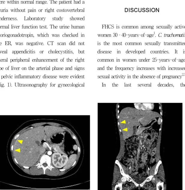
Fig. 1. CT scan showing lateral peripheral enhancement of the right liver and signs of PID (arrowhead). Panel A and B depict axial image and coronal image, respectively.
alcohol use that typically involved ingestion of a bottle of Korean whisky one to two times a month. The patient was a freshman in high school. The menstrual cycle was reported to be regular from 28-30 days with six to seven days of bleeding. The last menstrual period prior to the ER visit was 29 days ago. The patient did not experience increased vaginal discharge. Vital signs were within normal range. The patient had a pyuria without pain or right costovertebral tenderness. Laboratory study showed normal liver function test. The urine human choriogonadotropin, which was checked in the ER, was negative. CT scan did not reveal appendicitis or cholecystitis, but lateral peripheral enhancement of the right lobe of liver on the arterial phase and signs of pelvic inflammatory disease were evident (Fig. 1). Ultrasonography for gynecological
examination revealed a 3 cm left ovarian cyst and a vaginal swab was positive for C.
trachomatis. N. gonorrhea grew in the urine culture. No other pathogens of sexually transmitted diseases (STDs) were recovered. The patient was diagnosed with FHCS and was treated with oral antibiotics, doxycycline and metronidazole, for 4 weeks.
The patient improved with medication.
DISCUSSION
FHCS is common among sexually active women 30–40-years-of-age1. C. trachomatis is the most common sexually transmitted disease in developed countries. It is common in women under 25-years-of-age, and the frequency increases with increased sexual activity in the absence of pregnancy2,3. In the last several decades, the
confirmative diagnosis of FHCS has been the demonstration of a typical “violin-string appearance” in laparotomy or laparoscopical examination or the isolation of causative organisms in the peritoneal fluid or a specimen obtained during procedures.
Nowadays, CT is sufficient in diagnosis of FHCS, ruling out acute appendicitis or cholecystitis if added to clinical information.
Dynamic CT is a highly sensitive imaging modality for the early detection of FHCS, which shows marked hepatic capsular enhancement at the arterial phase because of increased blood flow at the inflamed hepatic capsule4. Ultrasonography may help rule out diseases of the gallbladder and liver4.
In the acute phase of FHCS, mild exudative inflammation of the hepatic capsule is apparent in the presence or absence of capsular congestion, punctuate hemorrhage, and fibrinous exudation. In the chronic phase, a violin-string appearance that reflects the adhesion between the hepatic surface and peritoneum develops.
Pathophysiologically, enhancement in the arterial phase reflects an increased blood flow at the inflamed hepatic capsule, whereas enhancement in the delayed phase reflects the early capsular fibrosis4-6.
There have been few reports of FHCS in adolescents. About 4 % of cases with pelvic inflammatory disease (PID) and Chlamydia infection develop into FHCS in adolescents,
which is a lower than in adults4,7. Factors such as poor treatment compliance8, unfavorable socio-medical services, and low economic mobility might contribute to the lower frequency detected in adolescents9. However, adolescents are more vulnerable to PID and STD due to eversion of the cervix and exstrophy of the transformation zone of uterine columnar cells8. In addition, the loss of the cervical mucus plug and blood creates a favorable environment for bacterial proliferation, which enhances the risk for an ascending infection with menstruation or intercourse10. Recurrence of Chlamydia infection may be very frequent, up to 30-50 %, in teenagers11. These recurrent infections can interfere with reproduction, leading to infertility due to fibrosis and scarring of reproductive system12.
Sexually active adolescents should be educated about protection against STDs and evaluated for such problems once a year13. Abdominal pain is the most common symptom of patients presenting to the ER.
When patients complain of right upper quadrant pain, diseases such as acute cholecystitis, duodenal ulcer, and varicella zoster are typically considered in the differential diagnosis. However, when sexually active female patients complain of right upper quadrant pain and lower abdominal pain simultaneously, an ultrasound should be promptly performed to rule out diseases
of the gallbladder and liver, and a CT scan should be performed to confirm the diagnosis. FHCS should be considered as well as a variety of STDs1.
Conventional treatment for FHCS is similar to therapy for PID. The administration of appropriate antibiotics such as tetracycline, doxycycline, erythromycin, ofloxacin, and azithromycin should be started as soon as FHCS is suspected. The treatment period may have to be extended according to the pain, which subsides with antibiotic therapy4.
REFERENCES
1. Woo SY, Kim JI, Cheung DY, Cho SH, Park SH, Han JY, Kim JK: Clinical outcome of Fitz-Hugh-Curtis syndrome mimicking acute biliary disease. World J Gastroenterol 14:6975-6980, 2008
2. Land JA, Evers JLH: Chlamydia infection and subfertility. Best Pract & Res Clinic Obstet Gynecol 16:901-912, 2002
3. Risser WL, Risser JM, Benjamins LJ, Feldmann JM: Incidence of Fitz-Hugh- Curtis syndrome in adolescents who have pelvic inflammatory disease. J Pediatr Adolesc Gynecol 20:179-180, 2007
4. Kim JH, Oh SH: Two adolescent cases of Fitz-Hugh-Curtis syndrome. Korean J Pediatr 52:1038-1043, 2009
5. Lopez-Zeno JA, Keith LG, Berger GS: The Fitz-Hugh-Curtis syndrome revisited.
Changing perspectives after half a century.
J Reprod Med 30:567-582, 1985
6. Nishie A, Yoshimitsu K, Irie H, Yoshitake T, Aibe H, Tajima T, Shinozaki K, Kakihara D, Matsuura T, Takahashi M, Kamochi N, Onitsuka H, Honda H:
Fitz-Hugh-Curtis syndrome. Radiologic manifestation. J Comput Assist Tomogr 23:786-791, 2003
7. Wang SP, Eschenbach DA, Holmes KK, Wager G, Grayston JT: Chlamydia trachomatis infection in Fitz-Hugh-Curtis syndrome. Am J Obstet Gynecol 138:1034, 1980
8. Pletcher JR, Slap GB: Pelvic inflammatory disease. Pediatr Rev 19:363-367, 1998 9. Hills SD, Joesoef R, Marchbanks PA,
Wasserheit JN, Cates W Jr, Westrom L:
Delayed care of pelvic inflammatory disease as a risk factor for impaired fertility. Am J Obstet Gynecol 16:95, 2003 10. Rice PA, Schachter J: Pathogenesis of
pelvic inflammatory disease: what are the questions? JAMA 266:2587-2593, 1991 11. Hillis SD, Nakashima A, Marchbanks PA,
Addiss DG, Davis JP: Risk factors for recurrent Chlamydia tranchomatis infec- tions in women. Am J Obstet Gynecol 170:801-806, 1994
12. Patton DL, Sweeney YT, Kuo CC:
Demonstration of delayed hypersensitivity in Chlamydia trachomatis salpingitis in monkeys: a pathogenic mechanism of tubal damage. J Inf Dis 169:680-683, 1994 13. Centers for Disease Control and Prevention.
Recommendations for the prevention and management of Chlamydia trachomatis infections. MMWR Recomm Rep 4(RR- 12):1-37, 1993
우상복부 통증을 호소하는 15세 청소년에서 발생한 Fitz-Hugh-Curtis 증후군 1예
서울대학교어린이병원 소아외과
정규환 · 박태진 · 정성은 · 박귀원 · 김현영1
Fitz-Hugh-Curtis 증후군은 골반 내 염증성 질환을 가진 환자의 직접적 간 실질 침범이 없는 간피막 염증에 의한 간주위염으로, 1930년대에 Thomas Fitz-Hugh와 Arthur Curtis에 의해 보고 되었다. Neisseria gonorrhea나 Chlamydia trachomatis에 의해 발병하며, 항생제로 치료되는 양성 성교전파질환이다. 대부분 가임기의 젊은 여성에서 발견되지만, 15세의 청소년에서 진단된 증례가 있어 보고하는 바이다. 15세 여자 환자가 1개월 전부터 발생한 간헐적인 우상복부와 하복부의 통 증을 주소로 내원하였다. 환자는 한 달 전 남자친구와 첫 성교를 한 이후, 5일 전까지 10여 차례 정도 성교를 하였다. 사회력 상 고등학교 1학년 생이고, 월경 주기는 28-30일로 규칙적이었다. 내 원 당일 시행한 임신 반응 검사는 음성이었다. 시행한 복부전산화단층촬영에서 우측 간엽의 가쪽 부분이 동맥기 조영증강을 보이며 골반내감염을 동반하고 있었다. 부인과 검진 상 질경부 면봉 검 사에서 Chlamydia trachomatis 양성소견을 보였으며, 소변배양검사에서 Neisseria gonorrhea가 동 정되었다. Fitz-Hugh-Curtis 증후군 진단 하에 4주간 doxycycline과 metronidazole의 경구용 항생 제 복용 후 호전되었다.
(J Kor Assoc Pediatr Surg 17(2):188~192), 2011.
Index Words:Fitz-Hugh-Curtis syndrome, Chlamydia trachomatis, Neisseria gonorrhea, Adolescent
본 논문의 요지는 2010년도 6월 대구에서 개최된 제26회 대한소아외과학회 춘계학술대회에서 구연되었음.
교신저자:김현영
서울특별시 종로구 대학로 101, 110-744 서울대학교 어린이병원 소아외과 Tel : 02)2072-2478, Fax : 02)747-5130
E-mail : [email protected]