ISSN: 2233-601X (Print) ISSN: 2093-6516 (Online)
Received: September 17, 2018, Revised: October 17, 2018, Accepted: October 22, 2018, Published online: April 5, 2019
Corresponding author: Hyo Jun Jang, Department of Thoracic and Cardiovascular Surgery, Hanyang University Seoul Hospital, Hanyang University College of Medicine, 222-1 Wangsimni-ro, Seongdong-gu, Seoul 04763, Korea
(Tel) 82-2-2290-8463 (Fax) 82-2-2290-8466 (E-mail) [email protected]
© The Korean Society for Thoracic and Cardiovascular Surgery. 2019. All right reserved.
This is an open access article distributed under the terms of the Creative Commons Attribution Non-Commercial License (http://creativecommons.org/
licenses/by-nc/4.0) which permits unrestricted non-commercial use, distribution, and reproduction in any medium, provided the original work is properly cited.
Do Blebs or Bullae on High-Resolution Computed
Tomography Predict Ipsilateral Recurrence in Young Patients at the First Episode of Primary Spontaneous Pneumothorax?
Sungjoon Park, M.D. 1 , Hyo Jun Jang, M.D. 2 , Ju Hoon Song, R.N. 2 , So Young Bae, M.D. 2 , Hyuck Kim, M.D., Ph.D. 2,3 , Seung Hyuk Nam, M.D. 4 , Jun Ho Lee, M.D., Ph.D. 2
1
Department of Thoracic and Cardiovascular Surgery, Inje University Sanggye Paik Hospital, Inje University College of Medicine, Seoul, Korea;
2Department of Thoracic and Cardiovascular Surgery, Hanyang University Seoul Hospital, Seoul,
Korea;
3Department of Thoracic and Cardiovascular Surgery, Hanyang University College of Medicine, Seoul, Korea;
4
Department of Thoracic and Cardiovascular Surgery, Hanyang University Guri Hospital, Guri, Korea
Background: The relationship between the size of bullae and pneumothorax recurrence is controversial. The aim of this study was to retrospectively evaluate the role of blebs or bullae in predicting ipsilateral re- currence in young patients experiencing their first episode of primary spontaneous pneumothorax (PSP) who underwent conservative treatment. Methods: A total of 299 cases of first-episode PSP were analyzed. The sta- tus of blebs or bullae was reviewed on high-resolution computed tomography (HRCT). The dystrophic severity score (DSS; range, 0 to 6 points) was calculated based on HRCT. Results: The 5-year recurrence rate was 38.2%. In univariate analysis, age (<20 years), body mass index (<20 kg/m
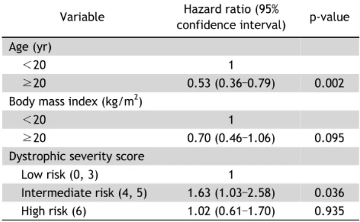
2), a unilateral lesion, and inter- mediate risk (DSS 4 and 5) were associated with recurrence. Sex; smoking history; and the presence, number, and maximal size of blebs or bullae were not related to recurrence. In Cox regression, age and intermediate risk were independent risk factors for recurrence. High risk (DDS 6) was not an independent risk factor.
Conclusion: The presence, number, and size of blebs or bullae did not affect ipsilateral recurrence. DSS failed to show a positive correlation between severity and recurrence. The decision to perform surgery in patients experiencing their first episode of PSP should not be determined by the severity of blebs and bullae.
Key words: 1. Pneumothorax 2. Recurrence
3. Computed tomography 4. Bullae
Introduction
Primary spontaneous pneumothorax (PSP) com- monly occurs in young patients [1]. Its recurrence rate ranges from 22.8% to 42% in patients who re- ceive conservative treatment after the first episode [2]. The recurrence rate of patients treated with vid-
eo-assisted thoracoscopic surgery (VATS) was re- ported to range from 0% to 13% [2]. The British Thoracic Society (BTS) and American College of Chest Physicians (ACCP) guidelines recommend con- servative treatment including observation, aspiration, and chest tube drainage as the first-line treatment of first-episode PSP [3,4]. Still, there is no glob-
https://doi.org/10.5090/kjtcs.2019.52.2.91
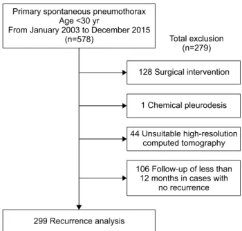
Fig. 1. Flow diagram of study design.
ally-accepted risk stratification system for PSP to de- termine high-risk patients who would benefit from VATS rather than conservative treatment at initial presentation [5]. Recently, a randomized controlled trial (RCT) showed a positive correlation between the risk of recurrence and the size of bullae based on high-resolution computed tomography (HRCT) [6].
However, the data remain insufficient. Moreover, an- other study showed no correlation between the pres- ence of bullae and recurrence [7]. The aim of this study was to evaluate the role of blebs or bullae de- tected using HRCT in predicting ipsilateral recurrence in young patients experiencing their first episode of PSP who underwent conservative treatment.
Methods
1) Patients
This study enrolled 524 patients younger than 30 years who visited the hospital for their first episode of PSP at a single institution from January 2003 to December 2015. PSP was defined as pneumothorax without underlying lung disease described in the medical record at the initial diagnosis. To exclude uncertain diagnoses of PSP in medical records, pa- tients were limited to those younger than 30 years.
This study was reviewed and approved by the Institutional Review Board and informed consent was waived (IRB approval no., 2019-02-018-001).
Fifty-four patients with simultaneous or sequential bilateral pneumothorax were counted as 2 cases for both thoraces. A total of 578 cases were extracted from outpatient and inpatient clinics, including the emergency department. Age, sex, body mass index (BMI), distribution of pneumothorax, smoking history, final management for first-episode PSP, and HRCT findings were collected from the patients’ medical records. Of the 578 cases, 128 underwent VATS, and 1 underwent chemical pleurodesis. After excluding the cases of VATS and chemical pleurodesis, cases with unsuitable HRCT findings (n=44) and those with less than 12 months of follow-up without recurrence (n=106) were also excluded. Finally, 299 cases were enrolled to evaluate ipsilateral recurrence (Fig. 1).
2) High-resolution computed tomography ex- aminations
HRCT without contrast media was generally con-
ducted within 1–5 days after hospitalization. In our practice, if a patient had sequential bilateral pneumo- thorax and was evaluated using HRCT for pneumo- thorax on 1 side, HRCT was not conducted for the contralateral pneumothorax within 1 year. In this study design, HRCT used to assess pneumothorax on 1 side was assumed to be suitable to evaluate the status of blebs or bullae for a contralateral pneumo- thorax if it occurred within 1 month. If HRCT was assessed more than 1 month before the event, it was regarded as unsuitable for evaluation of the con- tralateral status. Such cases were excluded from the study.
HRCT scans were performed using a 64-channel multi-slice CT scanner (Philips Brilliance 64; Philips Medical Systems, Amsterdam, The Netherlands) with the following parameters: 120 kV, 250 mA (planned mA with D-Dom dose modulation), sharp filter, 5-mm slice thickness, 5-mm slice increment, and a pitch of 0674 units. Coronal reconstruction was applied with 2-mm slice thickness and 2-mm slice increments.
The presence, maximal size, and distribution of
blebs or bullae were reviewed. Blebs were defined as
gas-containing spaces smaller than 1 cm within the
visceral pleura, and bullae were defined as gas-con-
taining spaces 1 cm or more in diameter within the
visceral pleura [8]. Some authors have defined blebs
as being smaller than 2 cm [9-11], and a recent RCT
showed that bullae larger than 2 cm were a risk fac- tor for recurrence [6]. Therefore, bullae were divided into smaller (<2 cm) and larger bullae (≥2 cm).
The dystrophic severity score (DSS) was proposed by Ouanes-Besbes et al. [7] and modified by Casali et al.
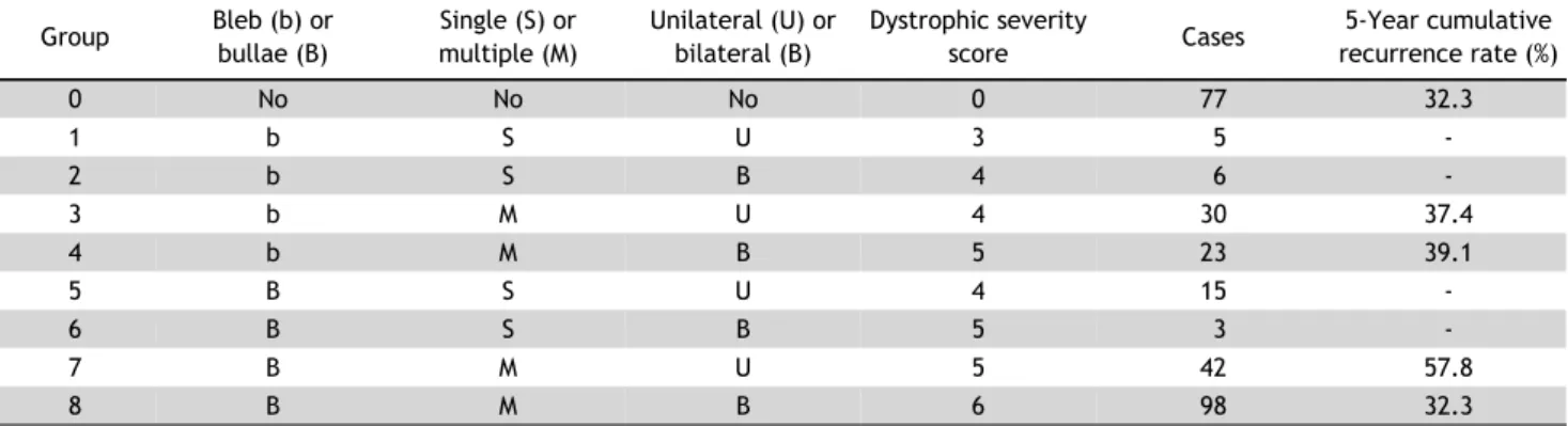
[12]. The modified formula is the sum of scores for type (1 or 2 points for blebs or bullae), number (1 or 2 points for single or multiple), and distribution (1 or 2 points for unilateral or bilateral). If blebs or bullae were seen on HRCT, the DSS ranged from 3 to 6 points. If there were no blebs or bullae in HRCT, the DSS was recorded as 0 points. Cases were div- ided into 3 risk groups according to DSS: low risk (0 or 3 points), intermediate risk (4 or 5 points), and high risk (6 points). Two thoracic surgeons (SB and HJ) reviewed the HRCT scans. SB reviewed all HRCT scans, and HJ reviewed only the cases without blebs or bullae that were confirmed by SB.
3) Management
The indications for chest tube drainage were dysp- nea, a distance of more than 1 cm between the later- al lung margin and chest wall, and gradually increas- ing size of the pneumothorax. Observation, including bed rest or outpatient clinic follow-up, was indicated in patients with no symptoms and a small pneumo- thorax, with less than 1 cm between the lateral lung margin and chest wall.
VATS was performed in 128 cases for persistent air leaks (>5 days, 53 cases), a history of con- tralateral pneumothorax (23 cases), complete lung collapse or tension pneumothorax (18 cases), simul- taneous bilateral pneumothorax (8 cases), army serv- ice or studying abroad (8 cases), hemothorax (2 cas- es), and unknown reasons (16 cases). In a single case, chemical pleurodesis was performed in a pa- tient who refused VATS for persistent air leaks.
4) Follow-up
Telephone interviews were performed in July 2018 by an investigator (JS). The incidence of recurrence was assessed through a telephone interview in 228 cases (39.5%), while the remaining 350 cases (60.5%) were reviewed (by JS) using medical records. The last outpatient clinic record of the thoracic department or the last inpatient admission record from other departments was accepted for review. The last outpatient clinic record from other
departments was regarded as an incomplete record for evaluating the patient’s history of pneumothorax.
Cases with less than 12 months of follow-up and no recurrence were regarded as having been lost to fol- low-up and were excluded from the recurrence anal- ysis, because most cases of recurrence develop dur- ing the first year [13]. The follow-up period was de- fined as extending from the date of diagnosis of the first PSP episode to the date of the last medical re- cord or telephone interview. The recurrence-free pe- riod was defined as extending from the date of diag- nosis of the first PSP episode to the date of ipsi- lateral recurrence in cases with ipsilateral recurrence and from the date of diagnosis of first PSP episode to the last follow-up date in cases without recurrence. The cumulative recurrence rate (cRR) was calculated as the rate of experiencing at least 1 recurrence.
5) Statistical analysis
All descriptive data were expressed as the fre- quency and mean with standard deviation. Frequency was compared using the chi-square and Fisher exact tests for dichotomous variables, and continuous vari- ables were compared using the t-test and analysis of variance. The primary endpoint was ipsilateral cRR calculated by time to event using Kaplan-Meier analysis. The ipsilateral cRR according to each varia- ble was calculated, and cRR curves were compared using the log-rank test. All p-values <0.05 were con- sidered to indicate statistical significance. Cox re- gression was performed to identify variables in- dependently associated with recurrence. All variables with p-values <0.05 were included in the analysis.
Statistical analysis was performed using IBM SPSS ver. 24.0 (IBM Corp., Armonk, NY, USA).
Results
1) Characteristics of the patients
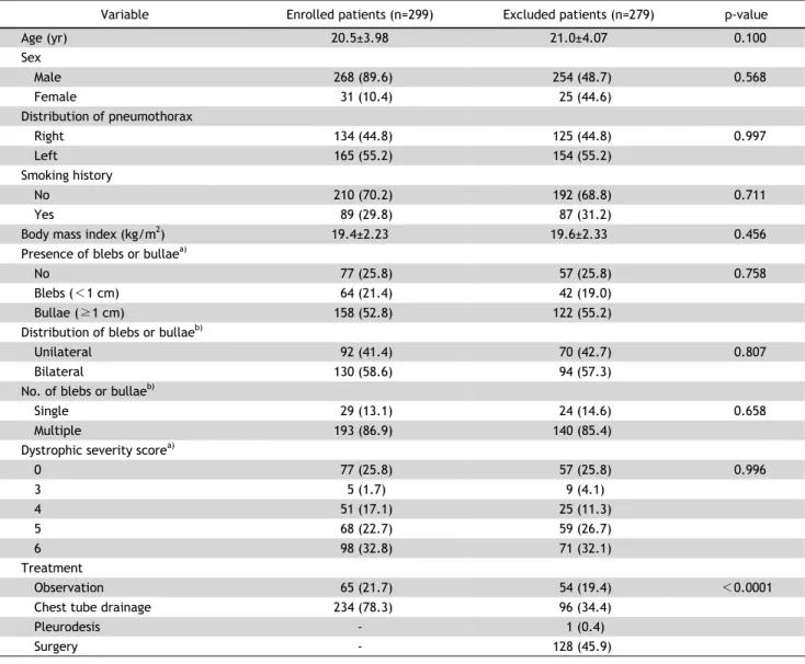
The characteristics of the enrolled and excluded
groups were compared for each variable. Except for
treatment modality, there was no difference between
the groups (Table 1). A total of 299 cases were en-
rolled for the recurrence analysis. The mean age of
the enrolled group was 20.5±3.98 years. Males were
predominant (89.6%). The mean BMI was 19.4±2.23
kg/m
2. In 77 cases (25.8%), there were no visible
Table 1. Demographics and high-resolution computed tomography findings in enrolled and excluded patients
Variable Enrolled patients (n=299) Excluded patients (n=279) p-value
Age (yr) 20.5±3.98 21.0±4.07 0.100
Sex
Male 268 (89.6) 254 (48.7) 0.568
Female 31 (10.4) 25 (44.6)
Distribution of pneumothorax
Right 134 (44.8) 125 (44.8) 0.997
Left 165 (55.2) 154 (55.2)
Smoking history
No 210 (70.2) 192 (68.8) 0.711
Yes 89 (29.8) 87 (31.2)
Body mass index (kg/m
2) 19.4±2.23 19.6±2.33 0.456
Presence of blebs or bullae
a)No 77 (25.8) 57 (25.8) 0.758
Blebs (<1 cm) 64 (21.4) 42 (19.0)
Bullae (≥1 cm) 158 (52.8) 122 (55.2)
Distribution of blebs or bullae
b)Unilateral 92 (41.4) 70 (42.7) 0.807
Bilateral 130 (58.6) 94 (57.3)
No. of blebs or bullae
b)Single 29 (13.1) 24 (14.6) 0.658
Multiple 193 (86.9) 140 (85.4)
Dystrophic severity score
a)0 77 (25.8) 57 (25.8) 0.996
3 5 (1.7) 9 (4.1)
4 51 (17.1) 25 (11.3)
5 68 (22.7) 59 (26.7)
6 98 (32.8) 71 (32.1)
Treatment
Observation 65 (21.7) 54 (19.4) <0.0001
Chest tube drainage 234 (78.3) 96 (34.4)
Pleurodesis - 1 (0.4)
Surgery - 128 (45.9)
Values are presented as mean±standard deviation or number (%).
a)