ISSN: 2233-601X (Print) ISSN: 2093-6516 (Online)
− 425 −
Received: August 16, 2018, Revised: August 20, 2019, Accepted: August 27, 2019, Published online: December 5, 2019
Corresponding author: Taek Yong Ko, Department of Thoracic and Cardiovascular Surgery, Kosin University Gospel Hospital, Kosin University College of Medicine, 262 Gamcheon-ro, Seo-gu, Busan 49267, Korea
(Tel) 82-51-990-6466 (Fax) 82-51-990-3066 (E-mail) [email protected]
Corresponding author: Seong Ho Cho, Department of Thoracic and Cardiovascular Surgery, Kosin University Gospel Hospital, Kosin University College of Medicine, 262 Gamcheon-ro, Seo-gu, Busan 49267, Korea
(Tel) 82-51-990-6466 (Fax) 82-51-990-3066 (E-mail) [email protected]
© The Korean Society for Thoracic and Cardiovascular Surgery. 2019. All right reserved.
This is an open access article distributed under the terms of the Creative Commons Attribution Non-Commercial License (http://creativecommons.org/
licenses/by-nc/4.0) which permits unrestricted non-commercial use, distribution, and reproduction in any medium, provided the original work is properly cited.
Unusual Communication between the Pulmonary Artery and Vieussens’ Arterial Ring Causing Infective Endocarditis
Sang Ho Lee, M.D., Taek Yong Ko, M.D., Seong Ho Cho, M.D., Ph.D.
Department of Thoracic and Cardiovascular Surgery, Kosin University Gospel Hospital, Kosin University College of Medicine, Busan, Korea
Coronary artery fistula is an abnormal communication between the coronary artery and the cardiac chambers. In particular, an abnormal connection between the conus branch of the right coronary artery and the proximal left anterior descending coronary artery is defined as Vieussens’ arterial ring. Coronary artery fistulas are usually asymptomatic, but some can cause complications such as infective endocarditis. Here, we report a case of Vieussens’ arterial ring causing infective endocarditis with severe mitral regurgitation.
Key words: 1. Coronary vessel anomalies 2. Fistula
Case report
A 55-year-old man presented to the emergency room of Kosin University Gospel Hospital with fever and headache. Physical examination revealed that the patient’s blood pressure was 90/60 mm Hg, his pulse was 63 beats/min, and his body temperature was 38.4°C. Laboratory tests showed a decreased platelet count (29,000), a decreased serum albumin level (2.6 g/dL), an increased white blood cell count (29,750), an elevated high-sensitivity C-reactive protein level (25.415 mg/L), and an elevated troponin I level (252 ng/mL). Brain magnetic resonance diffusion and flu- id-attenuated inversion recovery imaging revealed the presence of a multifocal acute infarction that in- cluded the frontal, occipital, and right temporal lobes and the left cerebellum and was accompanied by hemorrhagic transformation. Transthoracic echocardiog-
raphy (TTE) was performed to identify the presence of any intracardiac vegetation, and a 1.05×1.45-cm area of vegetation in the P1 segment of the artery was observed (Fig. 1A) with severe mitral regurgitation. Follow-up transesophageal echocardiog- raphy was performed and revealed a fistula in the pulmonary artery trunk (Fig. 1B). Multidetector com- puted tomography (CT) was also performed and showed that the fistula originated from the right conus artery and the proximal left anterior descend- ing artery (LAD), which communicate with each oth- er, and drained into the main pulmonary artery (MPA) with a 6.9-mm aneurysm (Fig. 2). The right conus branch of the proximal right coronary artery (RCA) displayed an abnormal connection with the proximal LAD, which was compatible with the diag- nosis of Vieussens’ arterial ring (VAR). Due to the presence of a coronary artery fistula (CAF) including
Korean J Thorac Cardiovasc Surg 2019;52:425-427 □ CASE REPORT □
https://doi.org/10.5090/kjtcs.2019.52.6.425
Sang Ho Lee, et al
− 426 −
Fig. 1. Preoperative transthoracic echocardiography shows mitral valve vegetation (A), and transesopha- geal echocardiography shows ab- normal blood flow caused by a sus- pected fistula in the pulmonary ar- tery trunk (B).
Fig. 2. Three-dimensional reconstruction of computed tomog- raphy coronary angiography. The right coronal branch and the proximal left anterior descending coronary artery formed Vieussens’ arterial ring (arrow), which showed aneurysmal dilata- tion prior to entrance into the main pulmonary artery.
vegetation, we decided to perform mitral valve re- placement with external fistula ligation.
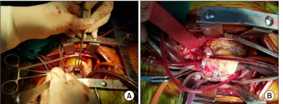
The operative findings showed vegetation on the P1 leaflet, which was then removed (Fig. 3A), and mitral valve replacement was performed. The fistula between the coronary artery and the MPA was obli- terated through external ligation (Fig. 3B). Follow-up TTE conducted 1 week after the operation showed a well-functioning mechanical mitral valve without a residual fistula. We planned to perform a post- operative coronary angio-CT a few months later, but the patient was lost to follow-up.
The patient provided written informed consent for the publication of clinical details and images.
Discussion
CAFs are abnormal connections between the coro- nary artery, heart chambers, and great vessels. CAFs were previously diagnosed using invasive coronary angiography. However, recent advances in coronary CT angiography have shown that the prevalence (0.9%) of this condition is somewhat higher than previously reported [1].
Most patients have no symptoms; however, among symptomatic patients, chest pain (39%) and dyspnea (25%) are common. Fistula most commonly arises in the left main artery/LAD (84%), followed by the RCA (38%), and it most commonly terminates in the MPA (89%) [2]. In our case, the fistula originated from the communicating artery between the proximal RCA and the proximal LAD, which constitutes VAR. VAR was first described by Raymond de Vieussens in 1706 and is thought to be an embryological remnant of an abnormal connection between the conus branch of the RCA and the LAD [3]. Aneurysms of VAR have been previously reported; however, reports of a fistula between VAR and the MPA are rare.
Although Lee and Cho [4] reported a single case sim- ilar to our case, the patient in that paper did not suffer from infective endocarditis.
The patient in our study had never been treated with dental therapy, and his hygiene was good. It was difficult to identify the factors causing endo- carditis in this patient, and given his circumstances, VAR was presumed to have caused this condition.
CAF with a right-sided origin can cause infective en- docarditis of left-sided valve through migration of in- fectious emboli via the pulmonary artery [5].
Imaging studies of CAFs include echocardiography, coronary angiography, and coronary angio-CT. Coronary angio-CT is far less invasive and more cost-effective than coronary angiography and is now widely used
Experience of Pathologic Vieussens’ Arterial Ring
− 427 −
Fig. 3. Vegetation was visible on the P1 area of the mitral valve (A).
Multiple pledgeted sutures were performed on the coronary artery fistula for external ligation (B).
as a diagnostic tool. In our case, because P1 vegeta- tion due to infective endocarditis was observed, we chose CT over coronary angiography.
It is well established that patients with sympto- matic CAF should undergo closure of medium to large fistulas. Latson [6] reported that it is better to close even small fistulas if: (1) the patient is consid- ered to be at high risk for later endocarditis, (2) lon- gitudinal follow-up is not feasible, or (3) the patient is undergoing an invasive procedure for another car- diac problem.
When considering the management of VAR, the treatment options are not markedly different from those for CAF. Both surgical correction or trans- catheter embolization are available. In our patient, surgical fistula closure was considered to be an ap- propriate next step because of the vegetation of the mitral valve due to infective endocarditis.
Conflict of interest
The authors declare that they have no competing interests.
ORCID
Sang Ho Lee: https://orcid.org/0000-0001-6093-9632 Taek Yong Ko: https://orcid.org/0000-0002-0096-0664 Seong Ho Cho: https://orcid.org/0000-0002-9833-6375
References
1. Lim JJ, Jung JI, Lee BY, Lee HG. Prevalence and types of coronary artery fistulas detected with coronary CT angiography. AJR Am J Roentgenol 2014;203:W237-43.
2. Verdini D, Vargas D, Kuo A, et al. Coronary-pulmonary ar- tery fistulas: a systematic review. J Thorac Imaging 2016;
31:380-90.
3. Loukas M, Clarke P, Tubbs RS, Kapos T. Raymond de Vieussens. Anat Sci Int 2007;82:233-6.
4. Lee HY, Cho SH. An unusual form of coronary artery fistu- la: a small aneurysm of Vieussens’ arterial ring communi- cating with the pulmonary artery. Korean J Thorac Cardiovasc Surg 2014;47:152-4.
5. Said SA. Characteristics of congenital coronary artery fis- tulas complicated with infective endocarditis: analysis of 25 reported cases. Congenit Heart Dis 2016;11:756-65.
6. Latson LA. Coronary artery fistulas: how to manage them.
Catheter Cardiovasc Interv 2007;70:110-6.