pISSN : 1229-5418
Implantology 2018; 22(4): 242-254
https://doi.org/10.32542/implantology.20180020
Received: November 9, 2018 Revised: December 20, 2018 Accepted: December 21, 2018
Copyright © 2018. The Korean Academy of Oral &
Maxillofacial Implantology
This is an Open Access article distributed under the terms of the Creative Commons Attribution Non-Commercial License (http://creativecommons.
org/licenses/by-nc/4.0/) which permits unrestricted non-commercial use, distribution, and reproduction in any medium, provided the original work is properly cited.
OPEN ACCESS
The aim of the current study was to evaluate the results of horizontal guided bone regeneration (GBR) with xenograft (deproteinized bovine bone mineral, DBBM), allograft (irradiated allogenic cancellous bone and marrow), titanium membrane, resorbable collagen membrane, and advanced platelet-rich fibrin (A-PRF) in the anterior maxilla. The titanium membrane was used in this study has a three-dimensional (3D) shape that can cover ridge defects. Case 1. A 32-year- old female patient presented with discomfort due to mobility and pus discharge on tooth #11.
Three months after extracting tooth #11, diagnostic software (R2 GATE diagnostic software, Megagen, Daegu, Korea) was used to establish the treatment plan for implant placement. At the first stage of implant surgery, GBR for horizontal augmentation was performed with DBBM (Bio-Oss
®, Geistlich, Wolhusen, Switzerland), irradiated allogenic cancellous bone and marrow (ICB cancellous
®, Rocky Mountain Tissue Bank, Denver, USA), 3D-titanium membrane (i-Gen
®, Megagen, Daegu, Korea), resorbable collagen membrane (Collagen membrane
®, Genoss, Suwon, Korea), and A-PRF because there was approximately 4 mm labial dehiscence
Abstract
상악 전치부 3D-티타늄 차폐막과
혈소판농축섬유소를 적용한 골유도재생술의 임상적 평가
이나연1†, 고미선1,2†, 정양훈1,2, 이정진4,5, 서재민4,5, 윤정호1,2,3*
1전북대학교 치과병원 치주과
2전북대학교 치과대학 치주과학교실, 구강생체과학연구소
3전북대학교 임상의학연구소, 전북대학교병원 의생명연구원
4전북대학교 치과병원 보철과
5전북대학교 치과대학 보철과학교실, 구강생체과학연구소
*Corresponding author: Jeong-Ho Yun, grayheron@hanmail.net
†
These authors contributed equally to this work.
Clinical Evaluation of Guided Bone Regeneration Using 3D-titanium Membrane and Advanced Platelet-Rich Fibrin on the Maxillary Anterior Area
Na-Yeon Lee1†, Mi-Seon Goh1,2†, Yang-Hun Jung1,2, Jung-Jin Lee4,5, Jae-Min Seo4,5, Jeong-Ho Yun1,2,3*
1Department of Periodontics, Chonbuk National University Dental Hospital, Korea
2Department of Periodontology, College of Dentistry and Institute of Oral Bioscience, Chonbuk National University, Korea
3Research Institute of Clinical Medicine of Chonbuk National University-Biomedical Research Institute of Chonbuk National University Hospital, Korea
4Department of Prosthodontics, Chonbuk National University Dental Hospital, Korea
5Department of Prosthodontics, College of Dentistry and Institute of Oral Bioscience, Chonbuk National University, Korea
after implant placement. Five months after placing the implant, the second stage of implant surgery was performed, and healing abutment was connected after removal of the 3D-titanium membrane. Five months after the second stage of implant surgery was done, the final prosthesis was then delivered. Case 2. A 35-year-old female patient presented with discomfort due to pain and mobility of implant #21. Removal of implant #21 fixture was planned simultaneously with placement of the new implant fixture. At the first stage of implant surgery, GBR for horizontal augmentation was performed with DBBM (Bio-Oss
®), irradiated allogenic cancellous bone and marrow (ICB cancellous
®), 3D-titanium membrane (i-Gen
®), resorbable collagen membrane (Ossix plus
®, Datum, Telrad, Israel), and A-PRF because there was approximately 7 mm labial dehiscence after implant placement. At the second stage of implant surgery six months after implant placement, healing abutment was connected after removing the 3D-titanium membrane. Nine months after the second stage of implant surgery was done, the final prosthesis was then delivered. In these two clinical cases, wound healing of the operation sites was uneventful. All implants were clinically stable without inflammation or additional bone loss, and there was no discomfort to the patient. With the non-resorbable titanium membrane, the ability of bone formation in the space was stably maintained in three dimensions, and A-PRF might influence soft tissue healing. This limited study suggests that aesthetic results can be achieved with GBR using 3D-titanium membrane and A-PRF in the anterior maxilla. However, long-term follow- up evaluation should be performed.
Keywords: Guided bone regeneration, Anterior teeth, Dental implant, Titanium membrane, Platelet rich fibrin
Ⅰ. 서론
상악 전치부의 경우 임플란트 식립은 환자의 심미적 요구와 기존의 해부학적 형태로 인해 임상가들 에게 도전적인 영역일 수 있다. 상악 전치부는 크게 해부학적, 병리학적 2가지의 원인으로 인한 조직 결 함을 가지게 된다. 해부학적 요인으로는 좁은 치조골 너비와 치조돌기의 순면 경사, 병리학적 요인으로 치아 외상, 급성 또는 만성 감염 및 결손치의 부적절한 시기의 수복으로 인한 치조골 결손부의 증가를 예로 들 수 있다1, 2. 그러나 위와 같은 어려움에도 불구하고, 상악 전치부에서 임플란트 식립은 구치부와 비교 시 성공률과 생존율 면에서 유사하다고 보고된다3, 4.
치아는 발치 후 잔존 치조골 폭경과 수직적 골 높이의 소실이 일어나고, 이로 인해 연조직의 변화 또 한 수반된다5. 따라서, 이상적인 위치에 임플란트를 식립하기 위해서는 결손 된 경조직과 연조직의 변화 를 고려해야 한다. 골 결손 부위를 재건하기 위한 골유도재생술(guided bone regeneration, GBR)은 예 지성 있는 치료로 여겨지며, 일차 치유 봉합(primary wound closure), 혈관 신생(angiogenesis), 공간 형성/
유지(space creation/maintenance), 그리고 초기 혈병과 임플란트의 안정성(stability of both the initial blood clot implant fixture)의 4가지 생물학적 원리에 기초하여 임상적인 성공을 이룰 수 있다6.
경조직 재건을 위한 차폐막의 사용은 필수적인데 현재 임상에서 환자와 술자의 편의를 위해 차폐막 의 제거를 위한 2차 수술이 필요 없는 흡수성 차폐막의 사용이 많이 이루어지고 있다. 그러나 비흡수성 차폐막의 경우 흡수성보다 공간 유지에 유리하고, 차폐막이 기능하는 시간을 조절할 수 있으며, 흡수되 면서 방출되는 물질에 의한 골 이식 부위의 영향이 없다는 장점이 있어 골 이식 부위에 따라 비흡수성 차폐막의 사용은 계속 이루어지고 있다7.
특히 이번 증례에서 사용한 티타늄 차폐막은 골 결손부 크기에 따라 선택할 수 있도록 3차원적(3 dimension, 3D)으로 디자인된 제품(i-Gen®, Megagen, Daegu, Korea)으로 이러한 3D-티타늄 차폐막 (i-Gen®)은 차폐막이 제거된 이후에 발생 가능한 골 소실의 양을 고려하여 임플란트 주위에 충분한 골 두께가 확보되도록 만들어졌다. 이렇게 3D-티타늄 차폐막을 사용하여 골유도재생술을 하면 만들어지 는 치조골의 형태를 예측 할 수 있다.
한편, 최근 의료계에서는 결손부 치료 시, 재생 및 상처 치유 과정을 효과적으로 증진시키기 위한 재 료 및 방법에 관해 활발히 연구 중이다. 그중 platelet-rich plasma (PRP)는 인간 혈액 샘플에서 유래한 1세대 지지체(scaffold)로 광범위한 연구가 이루어졌다. 그러나 PRP를 만들기 위해서는 항응고제와 bovine thrombin이 필요하고 제작 과정이 번거로운 단점이 있어 임상 적용에 제한이 있었다. 그래서 이 런 단점을 개선하고 간소화한 platelet-rich fibrin (PRF)가 개발되었다8.
PRF는 제작과정에 따라 여러 종류가 있는데 이번 증례에는 advanced platelet-rich fibrin (A-PRF)를 적용하였다. A-PRF는 기존의 PRF보다 느린 속도의 원심 분리 과정을 통해 제작되며, 더 많고 다양한
Fig. 1. Scanning electron microscope (SEM) analysis in three regions of the A-PRF. (A) Freeze dried A-PRF membrane was divided into three regions: region 1 was the distal portion from the red blood cell (RBC) thrombus, region 2 was the middle portion, and region 3 was adjacent to RBC fraction, (B) Fibrin clot revealed a dense and mature fibrin matrix, (C) Region 1 (SEM; original magnificationⅹ1,000), (D) Region 2 (SEM; original magnificationⅹ1,000), (E) Region 3 (SEM; original magnificationⅹ1,000), (F) Region 1 (SEM; original magnificationⅹ3,000), (G) Region 2 (SEM; original magnification
ⅹ3,000), (H) Region 3 (SEM; original magnificationⅹ3,000). (white arrows: leukocytes, white arrow heads: RBC).
Na-Yeon Lee et al. : Clinical Evaluation of Guided Bone Regeneration Using 3D-titanium Membrane and Advanced Platelet-Rich Fibrin on the Maxillary Anterior Area.
Implantology 2018
혈액 세포와 성장 인자를 포함하여 세포 활성과 치유를 증진시킨다는 연구보고가 있다9. 이번 연구에서 A-PRF의 구성을 확인 하기 위해 환자의 A-PRF membrane에 대해서 scanning electron microscope (SEM) 분석을 시행하였다. A-PRF membrane은 탈수 과정을 거친 후 영하 70도로 동결 건조되었다.
SEM 촬영을 위해 동결 건조시킨 검체를 platinum으로 coating (LEICA EM ACE200, sputter current 40 mA)후 membrane을 위쪽, 가운데, RBC 가까운 부분으로 나눈 뒤 전자현미경(FE-SEM, SUPRA 40VP, Carl Zeiss, Oberkochen, Germany)을 이용하여 1,000배와 3,000배로 확대하여 관찰했다(Fig. 1).
본 증례 보고에서는 충분한 협측골 두께를 얻기 위해 수평적 치조골 증대술을 동반하여 상악 전치부 에 임플란트를 식립한 2명의 환자에 대한 임상 결과를 평가하고자 한다. 골유도재생술에 사용한 재료로 는 동종골, 이종골 및 3D-티타늄 차폐막, 흡수성 콜라겐 차폐막을 사용하였고 연조직 및 창상의 초기 치유를 위해 A-PRF를 추가로 적용하였다.
Ⅱ. 증례 보고
1. 증례 1
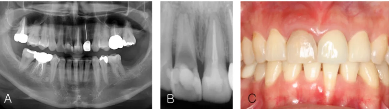
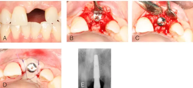
32세 여자 환자가 약 1년 전 신경치료 받은 #11 치아의 동요도와 염증을 주소로 내원하였다. 전신 질 환은 없었다. 임상 검사 및 방사선 사진 검사 시 #11 치아에 2도의 동요도, 치주 탐침 시 출혈 및 배농을 보였다. 치아 변색(Fig. 2C) 및 통증이 존재하고 예후가 불량할 것으로 평가되어 해당 치아의 발치 후 임 플란트 식립이 결정되었다.
발치 3개월 후, 임플란트 수술 전에 진단 프로그램 R2 GATE diagnostic software(Megagen, Daegu, Korea)을 이용하여 협설로 좁은 치조골에서 최적의 임플란트 식립 부위를 결정하였고(Fig. 3) 3D-티타 늄 차폐막 (i-Gen®, Megagen, Daegu, Korea)을 사용한 골유도재생술식을 계획하였다. 그 후, 임플란트 식립을 위한 R2 GATE Surgical Stent 제작을 의뢰하였다.
Fig. 2. At first visit. (A) Panoramic view x-ray, (B) Periapical x-ray, (C) Clinical photo, the color of tooth
#11 was changed.
Na-Yeon Lee et al. : Clinical Evaluation of Guided Bone Regeneration Using 3D-titanium Membrane and Advanced Platelet-Rich Fibrin on the
Maxillary Anterior Area. Implantology 2018
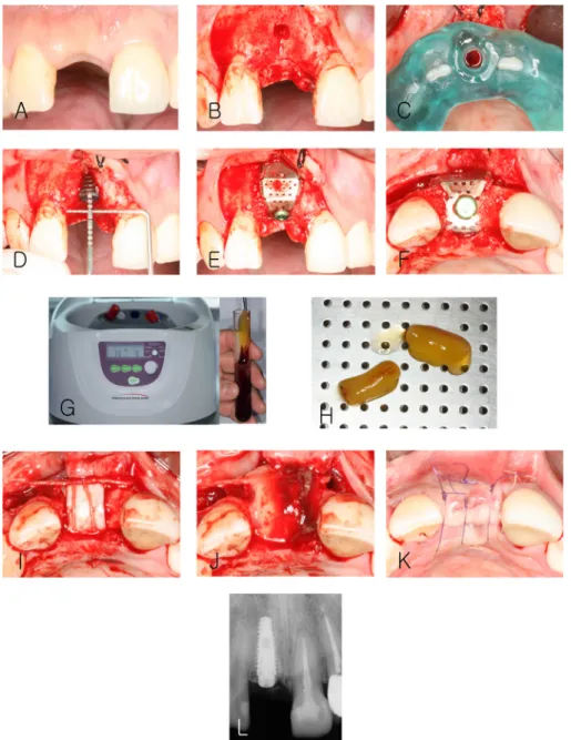
수술 전 환자의 정맥혈 20 mL를 채혈 튜브(BD Vacutainer® plus plastic serum tube, BD, Franklin Lakes, USA)에 담은 뒤 원심분리기(DuoQuattro Centrifuge, PRF process, Nice, France)를 사용하여 1,300 rpm으로 8분간 원심분리 후 A-PRF를 형성하였다(Fig. 4G). A-PRF는 fibrin clot만 분리한 뒤 틀 (A-PRF Box, PRF process, Nice, France)위에 올려(Fig. 4H) 트레이로 덮고 뚜껑을 닫아 membrane 형 태로 만들어 수술 시 사용할 수 있도록 준비하였다.
임플란트 식립을 위해 전층판막 거상 후 치조골을 확인하였다
(Fig. 4B).
R2GATE Surgical Stent를 위 치시킨 다음(Fig. 4C) R2GATE universal kit (Megagen, Daegu, Korea)를 이용하여 drilling 후 직경 4.0 mm, 길이 11.5 mm 임플란트 고정체(Anyone®, Megagen, Daegu, Korea)를 식립하였다. 임플란트 식립 후 순측 부위에 나사선 4개 정도(약 4 mm)가 노출되는 열개(dehiscence)가 관찰되어(Fig. 4D) 이종골 (Bio-Oss®, Geistlich, Wolhusen, Switzerland) 0.25 g과 irradiated allogenic cancellous bone and mar- row (ICB cancellous®, Rocky Mountain Tissue Bank, Denver, USA) 0.5 g을 혼합하여 적용한 후 3D-티 타늄 차폐막(i-Gen®)을 이용하여 공간 유지를 얻었다(Fig. 4E-F).
후에 상방에 흡수성 콜라겐 차폐막(Collagen membrane®, Genoss, Suwon, Korea)(Fig. 4I)으로 덮은 뒤 5-0 흡수성 합성 봉합사(Vicryl®, Ethicon, Somerville, USA)로 협측 판막 감장 절개한 부위 하방에 골막 봉합법을 사용하여 차폐막을 고정시켰다. 콜라겐 차폐막 위에 미리 준비해 둔 A-PRF membrane
(Fig. 4J)을 적용시킨 후 판막을 덮고 5-0 흡수성 단일 섬유 봉합사(Monosyn
®, B.Braun, Tuttlingen, Germany)로 봉합 하였다(Fig. 4K). 수술 직후 방사선 사진을 촬영하여 임플란트가 3차원적으로 적절한Fig. 3. Megagen R2GATE diagnostic software, planning for the optimal position of implant #11 and i-Gen
®.
Na-Yeon Lee et al. : Clinical Evaluation of Guided Bone Regeneration Using 3D-titanium Membrane and Advanced Platelet-Rich Fibrin on the
Maxillary Anterior Area. Implantology 2018
위치에 식립된 것을 확인하였다(Fig. 4L).
임플란트 식립 5개월 후 2차 수술을 시행하였다. 전층판막 거상하고(Fig. 5B), 3D-티타늄 차폐막 (i-Gen®)을 제거하였을 때, 임플란트 주위로 안정적인 신생골 형성이 관찰되었다(Fig. 5C-D). 임시 지 대주를 체결 후(Fig. 5F) 5-0 흡수성 단일 섬유 봉합사(Monosyn®)로 봉합하였다(Fig. 5G). 수술 직후 치 근단 방사선 사진 촬영 하여 임시 지대주 체결 상태를 확인하였다(Fig. 5H).
Fig. 4. Clinical photograph at the first stage of implant surgery. (A) Labial view before surgery, (B) After flap elevation, (C) Surgical stent positioned on #11, (D) Labial dehiscence after implant placement, (E) Labial view after application of bone graft material and 3D-titanium membrane, (F) Occlusal view after application of bone graft material and 3D-titanium membrane, (G) A-PRF fibrin matrix after centrifugation, (H) Prepared A-PRF, (I) Covering with collagen membrane, (J) Covering with A-PRF membrane, (K) Suture, (L) Periapical x-ray after the first stage of implant surgery.
Na-Yeon Lee et al. : Clinical Evaluation of Guided Bone Regeneration Using 3D-titanium Membrane and Advanced Platelet-Rich Fibrin on the Maxillary Anterior Area.
Implantology 2018
임시 보철물 장착한지 3개월 후 검진을 시행한 결과, 치은의 이상적인 emergence profile과 적절한 높 이가 잘 재현되고 전반적으로 보철물 주위의 치은 형태와 교합 상태가 양호했다. 2차 수술 5개월 후 최 종 지르코니아 크라운을 장착하였다(Fig. 6A). 치근단 방사선 사진 촬영 결과 임플란트 주위의 골 소실 양상은 관찰되지 않았다(Fig. 6B).
Fig. 5. Clinical photograph at the second stage of implant surgery. (A) Labial view before surgery, (B) After flap elevation, (C) After removal of the 3D-titanium membrane, (D) Removed 3D-titanium membrane, (E) A-PRF membrane, (F) Healing abutment connection, (G) Suture, (H) Periapical x-ray after the second stage of implant surgery.
Na-Yeon Lee et al. : Clinical Evaluation of Guided Bone Regeneration Using 3D-titanium Membrane and Advanced Platelet-Rich Fibrin on the Maxillary Anterior Area.
Implantology 2018
Fig. 6. (A) Clinical photograph on the labial view after delivery of the final prosthesis, (B) Periapical x-ray after delivery of the final prosthesis.
Na-Yeon Lee et al. : Clinical Evaluation of Guided Bone Regeneration Using 3D-titanium Membrane and Advanced Platelet-Rich Fibrin on the Maxillary Anterior Area.
Implantology 2018
2. 증례 2
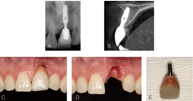
35세 여자 환자가 11년 전 식립 한 #21 임플란트의 동요도 및 통증을 주소로 내원하였다. 전신 질환은 없었다. 임상 검사 및 방사선 사진 검사 시 심한 치은 퇴축과 순측 골 소실이 관찰되었다(Fig. 7). 임상 검 사를 통해 실패한 임플란트로 평가되어, 임플란트를 제거하고 해당 부위에 임플란트를 재식립 하는 것 을 계획하였다.
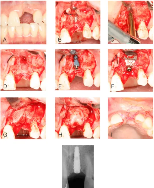
이전 케이스와 마찬가지로 수술 전 채혈을 한 뒤 A-PRF membrane을 준비하였다. 임플란트 제거 후, 식립을 위해 전층판막 거상 한 뒤 fixture removal kit (Neo fixture remover kit, Neobiotech, Seoul, Korea) 를 이용하여 기존 임플란트를 제거하였다(Fig. 8B-D).
환자의 요구를 고려하여 제거와 동시에 임플란트를 재식립하기로 하였으며 R2GATE universal kit를 이용하여 drilling 후 직경 4.0 mm, 길이 13 mm 임플란트 고정체(Anyone®)를 식립하였다(Fig. 9E). 임 플란트 식립 후 순측 부위 임플란트 고정체 길이의 2/3 정도(약 7 mm) 노출되는 열개가 관찰되어 이종 골(Bio-Oss®) 0.25 g과 irradiated allogenic cancellous bone and marrow (ICB cancellous®) 0.5 g을 혼 합하여 적용한 후 3D-티타늄 차폐막(i-Gen®)을 이용하여 공간 유지를 얻었다
(Fig. 8F).
이후에 상방에 흡수성 콜라겐 차폐막(Ossix plus®, Datum, Telrad, Israel)(Fig. 8G)으로 덮은 뒤 5-0 흡 수성 합성 봉합사(Vicryl®)로 협측 판막 감장 절개한 부위 하방에 골막 봉합법을 사용하여 차폐막을 고정 시켰다. 콜라겐 차폐막 위에 A-PRF membrane을(Fig. 8H) 덮은 뒤 5-0 흡수성 단일 섬유 봉합사(Mono- syn®)로 봉합하였다(Fig. 8I). 수술 직후 방사선 사진을 촬영하여 양호한 위치에 식립 된 임플란트를 확인 하였다(Fig. 8J).
Fig. 7. (A) Periapical x-ray, (B) CBCT sagittal view on #21 implant, (C) Clinical photograph on the labial view, (D) Clinical photograph on the labial view after removal of the prosthesis, (E) Removed prosthesis.
Na-Yeon Lee et al. : Clinical Evaluation of Guided Bone Regeneration Using 3D-titanium Membrane and Advanced Platelet-Rich Fibrin on the Maxillary Anterior Area.
Implantology 2018
임플란트 식립 6개월 후 2차 수술을 시행하였다. 전층판막 거상 후(Fig. 9B), 3D-티타늄 차폐막 (i-Gen®)을 제거하였고 임플란트 주위 신생골 형성을 확인하였다. 이후 임시 지대주 체결 뒤(Fig. 9C) 5-0 흡수성 단일 섬유 봉합사(Monosyn®)로 봉합하였다(Fig. 9D). 수술 직후 치근단 방사선 사진 촬영 하여 임시 지대주 체결 상태를 확인 하였다(Fig. 9E).
임시 보철물 장착한지 7개월 후에 검진을 시행한 결과, 처음 내원과 비교 시 #21 임플란트 치은 변연 이 안정적인 것을 확인하였고 전반적으로 보철물 주위의 치은 형태, 교합 상태가 양호한 것으로 판단되
Fig. 8. Clinical photograph at removal of failed implant and first stage of implant surgery. (A) Labial view before surgery, (B) After flap elevation, (C) Implant #21 removal, (D) After implant removal, (E) Labial dehiscence after implant placement, (F) Application of bone graft material and 3D-titanium membrane, (G) Covering with collagen membrane, (H) Covering with A-PRF membrane, (I) Suture, (J) Periapical x-ray after the first stage of implant surgery.
Na-Yeon Lee et al. : Clinical Evaluation of Guided Bone Regeneration Using 3D-titanium Membrane and Advanced Platelet-Rich Fibrin on the Maxillary Anterior Area.
Implantology 2018
어 2차 수술 9개월 후 최종 지르코니아 크라운을 장착하였다
(Fig. 10A). 치근단 방사선 사진 촬영 결과
골 소실 양상은 관찰되지 않았다(Fig. 10B). 하지만 치간유두의 부족, 임플란트 주위 치은연의 퇴축으로 인한 부조화 등 다소 비심미적인 결과를 보였다.Ⅲ. 총괄 및 고찰
상악 전치부 결손 부위의 임플란트 수복 시에는 만족스러운 결과를 얻기 위해 정확한 진단과 적절한 치료계획 및 치료가 이루어져야 한다2. 본 증례에서는 상악 전치부에 임플란트 식립 시, 3D-티타늄 차 폐막과 A-PRF를 적용하여 상악 전치부의 수평적 골유도재생술을 시행하였다.
골유도재생술은 골 이식재와 차폐막을 이용하여 이루어지며, 사용되는 골 이식재는 골 재생을 위한
Fig. 10. (A) Clinical photograph on the labial view after delivery of the final prosthesis, (B) Periapical x-ray after delivery of the final prosthesis.
Na-Yeon Lee et al. : Clinical Evaluation of Guided Bone Regeneration Using 3D-titanium Membrane and Advanced Platelet-Rich Fibrin on the Maxillary Anterior Area.
Implantology 2018
Fig. 9. Clinical photograph at the second stage of implant surgery. (A) Labial view before surgery, (B) After flap elevation, (C) After healing abutment connection, (D) Suture, (E) Periapical x-ray after the second stage of implant surgery.
Na-Yeon Lee et al. : Clinical Evaluation of Guided Bone Regeneration Using 3D-titanium Membrane and Advanced Platelet-Rich Fibrin on the Maxillary Anterior Area.
Implantology 2018
지지체(scaffold)로 작용한다. 골 이식재는 자가골, 동종골, 이종골, 합성골로 나누어지는데, 본 증례에서 는 골 전도성이 우수한 동종골(irradiated allogenic cancellous bone)과 이종골(deproteinized bovine bone mineral, DBBM)을 혼합하여 적용하였다10.
본 증례에서는 비흡수성 차폐막의 하나인 티타늄 차폐막과 상부에 흡수성 콜라겐 차폐막을 추가로 사 용하였다. 우수한 기계적 성질을 가진 티타늄 차폐막은 이식 부위의 공간 유지와 모양의 붕괴를 방지하 며, 이러한 공간 유지를 통해 창상의 안정화를 증진시켜 골 재생 효과를 증진시키고, 재생된 치조골의 형태학적 증진에 기여한다. 티타늄 차폐막 위에 콜라겐 차폐막을 추가로 사용함으로써 콜라겐 차폐막 의 추가적인 효과를 보고자 하였다. 콜라겐 차폐막은 우수한 생체 적합성과 감염에 저항성이 있으며 티 타늄 차폐막 내부로의 연조직 침투를 방지한다14. 뿐만 아니라 골 형성 세포의 이주 및 증식을 유도하고, 상피와 결합조직 세포를 배제시키는 차폐막의 기본적인 기능에 충실하다6. 따라서 결과적으로 향상된 골 형성을 기대할 수 있다.
또한, 티타늄 차폐막의 경우 골 결손부의 형태에 맞게 모양 형성이 용이하고, 외력에 저항할 수 있다 는 장점이 있지만, 자르고, 구부리는 과정에서 생긴 날카로운 모서리로 인해 차폐막이 노출될 수 있다는 단점이 있다. 이러한 티타늄 차폐막의 노출은 감염, 골 이식의 실패로 이어질 수 있다11. 현재 여러 회사 에서 개발되어 기성품으로 제작된 티타늄 차폐막이 임상에서 사용되고 있다. 미리 제작된 티타늄 차폐 막은 골 결손 부위에 맞는 제품을 선택하면 되기 때문에 수술 과정을 간편하게 해주고, 날카로운 모서리 로 인한 문제도 보완해준다.
협측골의 소실은 임플란트 실패와 높은 연관성을 보이는데, 장기적인 성공률을 고려하면 임플란트 식 립 시 가급적 2 mm의 협측 골 두께가 유지되어야 한다고 보고된 바 있다12. 이번 연구에서 사용한 3D-티 타늄 차폐막은 협측 골 두께가 적어도 2 mm 이상 확보될 수 있게 제작되어 결과적으로 임플란트 식립 시 안정적인 수준으로 치조정 부위에서 골폭이 확보될 수 있게 해준다.
그러나, 비흡수성 막의 주요한 합병증으로 여겨지는 차폐막의 노출은 기성품을 사용한다고 해도 일 어 날 수 있다. 예지성 있는 골유도재생술의 결과를 위해 일차 치유 봉합은 중요하며, 일차 치유 봉합의 실패는 차폐막 노출로 이어지고 이는 차폐막 하방의 골 형성에 유해한 영향을 준다6. 따라서 성공적인 일차 치유 봉합을 위해 연조직을 적절하게 다루는 것과 함께 적절한 봉합술이 필요하다.
임플란트 수술 후 초기 창상 및 연조직의 치유는 골유도재생술의 결과에 영향을 미칠 수 있는 중요한 부분으로 이러한 치유 과정을 보다 향상시키기 위해 본 증례에서는 A-PRF를 적용하였다. 2017년 발 표된 systematic review에 따르면 연조직 재생, 증대, 상처 치유를 위해 임상적으로 PRF가 많이 사용되 고 있으며, 그 결과 또한 긍정적인 것으로 보고 되었다13.
본 증례에 사용된 A-PRF를 SEM으로 분석한 결과 견고한 3차원 구조의 fibrin matrix가 관찰되었다.
Fibrin matrix는 지지체(scaffold) 역할을 함으로써 혈소판 및 다양한 혈액세포를 잡아줄 수 있고, 이러 한 세포들에서는 다양한 성장인자가 방출될 수 있다13. 가장 많은 세포 분포는 RBC 가까운 부분에서 관
찰되었다(Fig 1).
PRP, PRF를 비교한 연구 결과에 따르면, A-PRF에는 기존의 PRP, PRF보다 혈소판, B 및 T 림프구, 단핵 세포, 줄기세포 및 호중구, 과립구와 같은 다양한 혈액 세포와 성장 인자를 더 많이 포함하고 있으 며, 성장 인자의 시간에 따른 방출 정도를 비교한 결과에서도, 총 10일간의 기간 동안 A-PRF에서 가장 많은 양의 성장 인자의 방출이 오랫동안 지속적으로 관찰 되었다9. 성장 인자는 염증과 상처 치유에 중 요한 역할을 하며 세포 이동, 부착, 증식 및 분열을 자극하거나 억제할 수 있다13. 이 결과를 토대로 A-PRF를 적용한 경우, 상처 치유 촉진, 치유 기간 감소 등의 임상적 결과 향상을 기대할 수 있을 것으 로 사료된다.
두번째 증례에서 치간유두의 부족, 임플란트 주위 치은연의 퇴축으로 인한 부조화 등 심미적으로 아 쉬운 결과를 보였다. 이와 관련하여 Buser 등은 심미적인 임플란트 수복을 위해 implant shoulder의 깊 이는 인접치의 순측 백악법랑경계보다 1~2 mm 아래에 위치하는 것을 이상적인 위치라고 하였다. 본 증례에서는 임플란트 식립 시 치조골정의 위치가 이상적 위치에 비해 다소 근단측 및 순측에 위치하였 기 때문에 비심미적인 결과를 가져온 것으로 생각된다. 또한, 치간유두의 심미적인 외형 형성을 위해서 는 접촉면의 길이, 치조정으로부터 접촉점까지의 거리, 인접면 치은의 두께 등을 고려해야할 것이다1.
본 증례에서는 임상적, 방사선학적 평가만을 통해 골유도재생술의 성공여부를 평가한 한계점이 있으 며, 경과 관찰 기간이 짧아 향후 추가적인 장기간의 경과 관찰이 필요할 것으로 생각된다. 아울러, 골 이 식된 부위의 정확한 평가를 위해서는 조직학적 평가가 필요할 것으로 사료된다.
Ⅳ. 결론
본 증례에서는 상악 전치부의 단일치 결손부위에서 수복을 위해 골유도재생술(GBR)을 통한 임플란 트 식립을 시행하였다. 단기간의 관찰이지만, 임상적으로 환자의 불편감은 없었고, 염증 및 골 소실의 증 상 없이 심미적인 결과가 얻어졌다. 3D-티타늄 차폐막을 통해 골 재생이 이루어진 부위는 3차원적 모 양이 안정적으로 유지되었고, A-PRF는 초기 창상 및 연조직 치유를 향상시켜 골유도재생술의 예지성 을 높이는데 기여했을 것으로 사료된다.
Acknowledgements
본 연구는 전북대학교병원 의생명연구원의 학술연구비 지원에 의하여 수행되었음.
References
1. Buser D, Martin W, Belser UC. Optimizing esthetics for implant restorations in the anterior maxilla:
anatomic and surgical considerations. Int J Oral Maxillofac Implants. 2004; 19: 43-61.
2. Al-Sabbagh M. Implants in the esthetic zone. Dent Clin North Am. 2006; 50: 391-407.
3. Eckert SE, Wollan PC. Retrospective review of 1170 endosseous implants placed in partially eden- tulous jaws. J Prosthet Dent. 1998; 79: 415-421.
4. Belser UC, Schmid B, Higginbottom F, et al. Outcome analysis of implant restorations located in the anterior maxilla: a review of the recent literature. Int J Oral Maxillofac Implants. 2004; 19: 30-42.
5. Merheb J, Quirynen M, Teughels W. Critical buccal bone dimensions along implants. Periodontol 2000. 2014; 66: 97-105.
6. Wang HL, Boyapati L. “PASS” principles for predictable bone regeneration. Implant Dent. 2006; 15:
8-17.
7. Hämmerle CH, Jung RE. Bone augmentation by means of barrier membranes. Periodontol 2000.
2003; 33: 36-53.
8. Ghanaati S, Booms P, Orlowska A, et al. Advanced platelet-rich fibrin: a new concept for cell-based tissue engineering by means of inflammatory cells. J Oral Implantol. 2014; 40: 679-689.
9. Kobayashi E, Flückiger L, Fujioka-Kobayashi M, et al. Comparative release of growth factors from PRP, PRF, and advanced-PRF. Clin Oral Investig. 2016; 20: 2353-2360.
10. Fu JH, Wang HL. Horizontal bone augmentation: the decision tree. Int J Periodontics Restorative Dent. 2011; 31: 429-436.
11. Her S, Kang T, Fien MJ. Titanium membrane as an alternative to a membrane for ridge augmenta- tion. J Oral Maxillofac Surg. 2012; 70: 803-810.
12. Spray JR, Black CG, Morris HF, et al. The influence of bone thickness on facial marginal bone re- sponse: stage 1 placement through stage 2 uncovering. Ann Periodontol. 2000; 5: 119-128.
13. Miron RJ, Fujioka-Kobayashi M, Bishara M, et al. Platelet-rich fibrin and soft tissue wound healing:
a systematic review. Tissue Eng Part B Rev. 2017; 23: 83-99.
14. Funato A, Ishikawa T, Kitajima H, et al. A novel Combined Surgical Approach to Vertical Alveolar Ridge Augmentation with Titanium Mesh, Resorbable Membrane, and rhPDGF-BB: A Retrospec- tive Consecutive Case Series Int J Periodontics Restorative Dent. 2013; 33: 437-445.