• 검색 결과가 없습니다.

Department of Molecular Biology, College of Natural Sciences, Pusan National University, Busandaehak-ro 63beon-gil, Geumjeong-gu, Busan 609-735, Korea

N/A
N/A
Protected

Academic year: 2021

Share "Department of Molecular Biology, College of Natural Sciences, Pusan National University, Busandaehak-ro 63beon-gil, Geumjeong-gu, Busan 609-735, Korea"

Copied!
12
0
0

로드 중.... (전체 텍스트 보기)

전체 글

(1)

Vol. 24, No. 1, 21-32, April 2013 Original Article

Received on April 10, 2013. Revised on April 19, 2013. Accepted on April 21, 2013 Correspondence to: Jae-Hun Cheong

Department of Molecular Biology, College of Natural Sciences, Pusan National University, Busandaehak-ro 63beon-gil, Geumjeong-gu, Busan 609-735, Korea

Tel: 82-51-510-2277, Fax: 82-51-513-9258, E-mail: [email protected] Co-correspondence to: Hyung-Hoi Kim

Department of Laboratory Medicine, School of Medicine, Pusan National University, 179 Gudeok-ro, Seo-gu, Busan 602-739, Korea Tel: 82-51-240-7419, Fax: 82-51-247-6560, E-mail: [email protected]

단핵구에서 TNF-α Gene의 전사 활성을 증가시키는 HIF-1과 NF-κB의 기능적 상호작용

박민정

1

ㆍ이선민

2

ㆍ옥순정

2

ㆍ김혜림

2

ㆍ김형회

2

ㆍ정재훈

1

부산대학교 자연과학대학 분자생물학과1, 부산대학교 의과대학 진단검사의학교실2

Functional Interaction of HIF-1 and NF-κB Increasing the Transcriptional Activation of TNF-α Gene in Monocytes

Min-Jeong Park

1

, Sun-Min Lee

2

, Soon-Jung Ok

2

, Hye-Rim Kim

2

, Hyung-Hoi Kim

2

, Jae-Hun Cheong

1

Department of Molecular Biology, College of Natural Sciences1, Department of Laboratory Medicine, School of Medicine2, Pusan National University, Busan, Korea

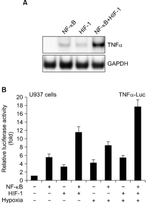
Background: Tumor necrosis factor alpha (TNF-α) is a pleiotropic cytokine fulfilling a broad variety of immunoregulatory functions. Monocytes and macrophages play a pivotal role in inflammation and immune regulation. NF-κB and HIF-1 are known to increase expression of the TNF-α gene in a separate way.

Methods: Human monocytic leukemia, U937 cells, were transfected using the standard electroporation method for intracellular expression of NF-κB and HIF-1. We performed analysis using the mammalian two-hybrid assay and co-immunoprecipitation assay for detection of protein interaction of both proteins. In addition, chromatin immuno- precipitation analysis was performed for examination of NF-κB and HIF-1 binding on the TNF-α gene promoter.

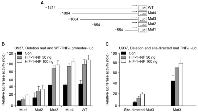
Results: Here we show that NF-κB and HIF-1 cooperatively induced an increase in expression of the TNF-α gene dependent on promoter activity by the direct protein interaction of these two transcription factors. Hypoxia signaling induced marked enhancement of the transactivation of TNF-α promoter by HIF-1 and NF-κB. A tandem NF-κB/HIF-1 binding site was identified within the TNF-α promoter, which acted as a strong enhancer element.

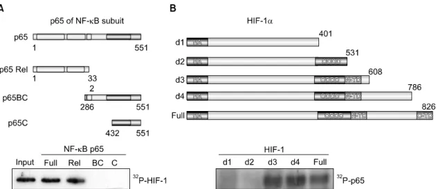
Physical association of the Rel domain of NF-κB and the N-TD domain of HIF-1 was required. Hypoxia treatment also resulted in a significant increase in the protein interaction of NF-κB and HIF-1 in vivo. Both transcription factors were recruited on the chromatin TNF-α promoter dependent on hypoxia stimuli.

Conclusion: The results of this study indicate that a variety of extracellular signals for activation of TNF-α gene expression might converge on the transcriptional regulation through the NF-κB/HIF-1 signaling pathway. (Korean J Blood Transfus 2013;24:21-32)

Key words: HIF-1, NF-κB, Hypoxia, TNF-α, Transactivation

(2)

Introduction

Tumor necrosis factor alpha (TNF-α) is a pleio- tropic cytokine fulfilling a broad variety of im- munoregulatory functions. Monocytes and macro- phages play a pivotal role in inflammation and im- mune regulation. Upon activation, monocytes pro- duce and release many inflammatory mediators such as IL-1, IL-6, IL-8, TNF-α, or arachidonic acid me- tabolites, as well as the anti-inflammatory mediators IL-10, soluble TNF receptor, and IL-1 receptor antagonist. Among the different products released, TNF-α is thought to be one of the most important mediators of inflammatory disease,

1)

and therefore the understanding of molecular mechanisms of TNF- α gene induction is of considerable medical interest.

The human TNF-α is a potent proinflammatory cy- tokine whose expression can be induced by cellular stimulation such as phorbol 12-myristate 13-acetate (PMA), lipopolysaccharide (LPS), virus infection, exposure to calcium ionophore or antigen and TNF- α itself.

2,3)

TNF-α promoter contains several binding sites, including AP-1, AP-2, NFAT, Sp1, Ets, and cyclic AMP response element (CRE).

4,5)

The CRE site is a PMA-responsive region and necessary to the regulation of TNF-α gene are NFAT, ATF-2/Jun, NF-κB, Ets/Elk, and Sp1 and coactivator CBP/p300 family.

6,7)

TNF-α has a key role in the regulation of the host defense responses against infection and tu- mor formation. But, abnormal expression of TNF-α contributes to the pathogenesis of numerous chronic inflammatory diseases including septic shock, dia- betes, rheumatoid arthritis, hepatitis, cachexia, auto- immune diseases.

8)

Therefore, it is important that TNF-α expression is controlled. But regulation of

human TNF-α expression in cells of monocytic line- age is quite complex, involving controls at both tran- scriptional and post- transcriptional levels.

Under hypoxic conditions, the diverse target genes are all transcriptionally up-regulated by the heterodimeric Hypoxia-inducible factors (HIF)-1.

The subunit of HIF-1 is the hypoxia-responsive component of the dimer, while HIF-1β is expressed constitutively. Under normoxic conditions, HIF-1α is rapidly degraded by the ubiquitin-proteasome pathway.

9)

Ubiquitination of HIF-1α is mediated by interaction with von Hippel-Lindau tumor suppressor protein (pVHL)

10,11)

and p53.

12,13)

HIF-1α is targeted for VHL E3 ligase complex-mediated destruction by proline hydroxylation of oxygen-dependent degrada- tion (ODD) region.

14)

However, p53 promotes Mdm- 2-mediated ubiquitination and proteasomal degrada- tion of the HIF-1α through direct interaction with HIF-1α in hypoxia.

12)

Under hypoxic conditions, HIF-1α is stabilized, which is determined by bal- ance between negative regulator such as p53 and positive unknown factor, and accumulated in the nucleus.

15)

Stabilized HIF-1α exerts its transcrip- tional activity by binding to the p300/CBP,

16)

steroid receptor coactivator-1 (SRC-1) family coactivators, nuclear redox regulator Ref-1,

17)

and molecular cha- perone heat shock protein 90 (HSP90).

18)

p300/CBP, SRC-1, and Ref-1 synergistically enhance HIF-1α- mediated transcriptional regulation under hypoxic conditions. The modulation of HIF-1α stability and activation requires interaction of these multiproteins with HIF-1α.

Here we aimed to analyze the mechanism by

which NF-κB activates transcription of TNF-α on

hypoxia signaling. In this study, we found that

(3)

NF-κB, although a relatively moderate activator of TNF transcription, is synergistic with HIF-1 in stim- ulating the TNF-α promoter transactivation. NF-κB and HIF-1 physically interact in vivo and in vitro.

NF-κB binds to a B binding site-related sequences, each in close proximity to functional HIF-1 binding sites in the TNF-α promoter. It was found that indis- pensable, positively acting elements of both tran- scription factors was in the TNF-α 5'-flanking region. Taken together, our data emphasize the im- portance of a cooperative interaction of the NF-κB and HIF-1 transcription factors for cellular hypoxia signaling.

Materials and Methods

1. Cell culture

Human monocytic leukemia, U937, cells were maintained in Dulbecco's modified Eagle's medium (DMEM) supplemented with 10% fetal bovine se- rum (Invitrogen, Carlsbad, CA) and 1% antibiotics.

For hypoxic condition, cells were incubated at 5%

CO

2

level with 1% O

2

balanced with N

2

in hypoxic chamber (Forma).

2. Transient transfection and lucifer- ase assay

U937 cells were transfected by the standard elec- troporation method. Cells were incubated with DNA precipitates for 16 h, washed, and maintained in complete medium 48 h prior to harvest. Relative lu- ciferase and β-galactosidase activities were de- termined as described.

19)

Basal promoter activity is reported as the activity observed after transfection of

the reporter plus an appropriate amount of empty ex- pression vector. In all cases, transfection data repre- sent the mean of three independent experiments.

3. Mammalian two-hybrid assay

U937 cells were seeded in 24-well plates with growth medium supplemented with 10% FBS and 1% antibiotics, and co-transfected with expression vectors encoding Gal4-DNA binding domain fusions (pCMX/Gal4N/pCMX/Gal4N-NF-κB series) and VP16-activation domain fusions (pCMX/VP16/pCMX/

VP16-HIF-1) as well as the previously described Gal4-tk-luc reporter plasmid. After 48 h, cells were harvested and the luciferase activity was normalized to the β-galactosidase expression. All the results rep- resent the average of at least three independent experiments.

4. Co-immunoprecipitation assay

Cell lysates (500 μg) were incubated with 1 μg of anti-NF-κB p65 antibody at 4

o

C for 2 h with gen- tle agitation. Immune complexes were collected on protein G-Sepharose beads (Gibco BRL). After washing three times with RIPA (−) buffer (1%

Triton X-100, 1% deoxycholate in PBS), the precip- itates were boiled with an equal volume of 2x LSB at 100

o

C for 3 min and analyzed by SDS-PAGE.

5. Western blot analysis

Cells were harvested on ice-cold lysis buffer (150

mM NaCl, 50 mM Tris-Cl, pH7.5, 1% NP-40, 1

mM EDTA, 10% glycerol) containing 1x protease

inhibitor at 4

o

C. The protein content of cell lysates

was determined with Bradford reagent (Bio-Rad) us-

ing bovine serum albumin (BSA) as standard. After

(4)

heating at 100

o

C for 5 min in 1x Laemmli sample buffer (LSB), the samples were separated by 10%

SDS-PAGE. The resulting gels were either stained with Commassie-blue or transferred to PVDF (Immobilon-P) membrane (Millipore). For Western blotting, the membrane was incubated with an- ti-HIF-1 (New England Biolab) and anti-TBP in TBS containing 1% non-fat dried milk for 1 h at RT.

After washing three times with cold TBS-T (TBS containing 0.04% Tween-20), the blotted membranes incubated with peroxidase-conjugated secondary an- tibody (Santa Cruz Biotechnology) for 30 min at RT. After washing three times with cold TBS-T, the protein bands were visualized by the enhanced chemiluminescence detection system according to the recommended procedure (Amersham Corp.).

6. Chromatin immunoprecipitation analy- sis

Cells were lysed for 5 min in L1 buffer (50 mM Tris pH 8.0, 2 mM EDTA, 0.1% NP-40 and 10%

glycerol) supplemented with protease inhibitors.

Nuclei were pelleted at 3,000 r.p.m. and resuspended in L2 buffer (50 mM Tris pH 8.0, 0.1% SDS and 5 mM EDTA). Chromatin was sheared by soni- cation, centrifuged and diluted 10 times in dilution buffer (50 mM Tris pH 8.0, 0.5% NP-40, 0.2 M NaCl and 0.5 mM EDTA). Extracts were pre- cleared for 3 h with 60 μL of a 50% suspension of salmon sperm-saturated protein A-agarose. Immuno- precipitations were carried out overnight at 4

o

C.

Immunocomplexes were collected with salmon sperm- saturated protein A for 30 min and washed three times (5 min each) with high-salt buffer (20 mM Tris pH 8.0, 0.1% SDS, 1% NP-40, 2 mM EDTA

and 0.5 M NaCl) followed by three washes in no salt buffer (1x TE). Immunocomplexes were ex- tracted in 1x TE containing 2% SDS, and protein DNA cross-links were reverted by heating at 65

o

C overnight. After proteinase K digestion, DNA was extracted with phenol-chloroform and precipitated in ethanol. About one-twentieth of the immunopre- cipitated DNA was used in each PCR. Quantitative duplex PCR assay was performed to analyze the amount of DNA precipitated by specified antibodies in proportion to input DNA. Two pairs of primers were used: Forward (5'-AAGTTTAGTCAATCAA- ACGTT-3') and Reverse (5'-TGCTTGGTAGCTA- GCCCTCCT-3') for the TNF-α promoter. The PCR conditions were as follows: 1.25 U of Taq DNA pol- ymerase (Amersham Biosciences), 100 ng of each primer, 200 μM dNTP, 2.5 μL of 10x Taq buffer and double-distilled water to a final volume of 25 μL: 94

o

C for 180 s; 34 cycles at 94

o

C for 45 s, 60°C for 60 s and 72

o

C for 60 s; final elongation at 72

o

C for 10 min.

Results

1. NF-κB and HIF-1 synergistically increase the TNF-α gene expre- ssion

To study the levels at which TNF-α production

is regulated by hypoxia, we first compared hypo-

xia-induced TNF-α mRNA in hypoxia-treated and

nontreated U937 cells. At first, we examined the

transactivation of TNF-α promoter by hypoxia stim-

uli and the effect of etopic expression of NF-κB and

HIF-1. As showin in Fig. 1A, hypoxia treatment in-

(5)

Fig. 1. NF-κB and HIF-1 synergistically increase the TNF-α gene expression. (A) Hypoxia and ectopic NF-κB/HIF-1 expression synergistically increased the mRNA production in U937 cells. U937 cells were transfected with the expressoin vector encoding NF-κB and HIF-1 in the presence or absence of hypoxia stimuli. The cell was harvested and applied to the Northern blot assay using TNF-α cDNA probe. (B) U937 cells were transfected with 20 ng of reporter plasmid, TNF-α gene promoter construct and indicated amount of expression vector encoding NF-κB and HIF-1. The cell was harvested, and cell lysates were assayed for luciferase activity. Trans- activation potentials were calculated by taking the luciferase/

β

-galactosidase activity ratio for each con- struct and were plotted relative to the activity of the reporter vector alone. Data are representative of four independent experiments.

creased the transactivation of the TNF-α promoter without the overexpression of both transcription factors. Furthermore, the overexpression of both NF-κB and HIF-1 transcription factors synergisti- cally enhanced an increase of the hypoxia-induced transactivation of TNF-α promoter (Fig. 1A). To verify the effect of hypoxia on the TNF-α gene ex- pression directly, U937 cells were stimulated with hypoxia (2% O

2

), and TNF-α gene induction was evaluated by Northern blot analysis (Fig. 1A). In U937 cells, hypoxia only weakly induces TNF-α transcripts, whereas after transfection of NF-κB or HIF-1, much higher levels of TNF-α mRNA are seen in response to hypoxia (Fig. 1B). Co-trans- fection of NF-κB and HIF-1 in cells synergistically increased the hypoxia-induced transactivation of TNF-α promoter. This finding, together with the transient response, characterizes the TNF-α gene as an immediate-early gene in response to hypoxia stimulation and two different activation pathways of NF-κB and HIF-1 give a significant reaction for TNF-α gene expression together.

2. Identification of the cooperative HIF-1/NF-κB responsive region in TNF-α promoter

To identify the cooperative HIF-1/NF-κB re-

sponsive region in the TNF-α promoter, a series of

transactivation assay was performed using system-

atic deleted TNF-α promoters dependent on the ten-

tative intact HIF-1/NF-κB binding motif. The de-

leted DNA constructs of TNF-α promoter were de-

scribed in Fig. 2A. Each construct was transfected

into U937 cells. Except p -854/+68 and -554/+68,

other deletion constructs of TNF-α promoter still

(6)

Fig. 2. Identification of the cooperative HIF-1/NF-κB responsive region in TNF-α promoter. (A) A schematic representation of the deletion mutant of TNF-α reporter constructs used in transient transfection assays. (B) Luciferase activity of the deletion mutant promoter in U937 cells. U937 cells were transient transfected with the indicated promoter-deletion constructs, and 48 h post-transfection, cell lysates were assayed for luciferase activity. (C) Luciferase activity of the site-directed mutant promoter in U937 cells. HIF-1/NF-κB binding site of p -1004/-854 mut3 construct were mutated with site-directed mutation method. U937 cells were transient transfected with mut3 reporter plasmids containing either wild type or mutated type, and 48 h post-transfection, cell lysates were assayed for luciferase activity.

had activity in a dose-dependent manner of HIF-1/

NF-κB expression vector in transient transfection.

The promoter activity based on a luciferase ex- pression was dramatically decreased between p -854/+68 and p -1004/+68, suggesting that there is a positive regulation site for the cooperative HIF-1/NF-κB transcriptional activation between -1004 and -854. This result indicates that -1004 and -854 region of TNF-α promoter may be a crucial tandem NF-κB/HIF-1 binding site in regulating tran-

scription of TNF-α (Fig. 2B). We found a cognate

binding sequence for HIF-1 and NF-κB in the pro-

moter region between -1004 and -854 (one site, -998

to -992 (AGCCTCCA) for NF-κB and one site, -964

to -960 (ACGTT) for HIF-1). To determine whether

TNF-α promoter region between -1004 and -854 has

any specific regulatory binding site for NF-κB and

HIF-1, site-directed mutagenesis in the luciferase

construct of mut3 was examined in the transient

transactivation assay, resulting in large decrease of

(7)

Fig. 3. Identification of each interaction region of HIF-1 and NF-κB p65. (A) A schematic diagram of the truncated NF-κB mutant constructs was shown in upper panel. HIF-1 were labeled with [

35

S]-methionine by in vitro translation and incubated with glutathione beads containing GST-p65(1-551), GST-p65(1-332), GST-p65(286-551), and GST-p65(432-551) as indicated. The bound proteins were resolved by SDS-PAGE and autoradiography. Beads were washed and specifically bound material was eluted with reduced glutathione and resolved by SDS-PAGE. Approximately 10% of the labeled proteins used in binding reactions were loaded as input. (B) A schematic diagram of the truncated HIF-1 mutant constructs was shown in upper panel. NF-κB p65 protein was labeled with [

35

S]-methionine by in vitro translation and incubated with glutathione beads containing GST-HIF-1(1-401), GST-HIF-1(1-531), GST-HIF-1(1-608), GST-HIF-1(1-786), and GST-HIF-1(full) as indicated. The bound proteins were resolved by SDS-PAGE and autoradiography.

NF-κB/HIF-1 response. Thus, -1004/-854 region of a potential NF-κB/HIF-1-binding site the reporter plasmid was changed into a mutated one (AGCCT- CCA to AGTTTCCA and ACGTT to ATTTT). As shown in Fig. 2C, a site-directed mutation resulted in complete loss of luciferase activity compared to the wild type of TNF-α promoter (mut3) construct in U937 cells. The result suggests that both tran- scription factors together bind to their cognate se- quence in the near promoter region of TNF-α gene.

3. NF-κB interacts with HIF-1

To further characterize the interacting region of

NF-κB against HIF-1 in vitro, GST pull-down assay

was performed. GST fusion proteins encoding the

full-length, 1-332aa, 286-551aa, and 432-551aa of

NF-κB p65 were expressed in Escherichia coli, im-

mobilized on Glutathione Sepharose 4B beads, and

incubated with

35

S-labeled full-length HIF-1 pro-

duced by in vitro translation system. The N-terminal

containing Rel domain of NF-κB interacts with

HIF-1 protein (Fig. 3A). The reciprocal strategy was

(8)

Fig. 4. Hypoxia increases the protein interaction of HIF-1 and NF-κB. (A) U937 cells were transiently transfected with Gal4N-NF-κB p65 and VP16N-HIF-1 for mammalian two-hybrid assay. 48 h after trans- fection in the three different oxygen concentration (2, 10, and 20%), cells were harvested for luciferase activities. (B) U937 cells were treated in the absence or presence of hypoxia stimuli. The cell extracts were loaded to co-immunoprecipitation with pre-immune IgG and specific antibodies against MyoD and NF- kB p65. (C) ChIP analysis of factor occupancy on TNF-α promoters. Following formaldehyde cross-link- ing, soluble chromatin was prepared. After IP with antibodies against the indicated proteins (Myo-D, HIF-1, and NF-κB), precipitated DNAs were used in PCR analysis. Input shows the starting chromatin extracts.

used to delineate the region of HIF-1 required for interaction with NF-κB using the same GST pull- down assay. For this study, the C-terminal serial truncated proteins of HIF-1 were produced as GST-fusion ones as shown in Fig. 3B. The two trun- cated mutants (d1 and d2) did not bind to radio- labeled NF-κB protein, but the d3 protein, which cover from N-termini to 608aa of HIF-1, interacted with NF-κB (Fig. 3B). This result showed that the region between 531aa and 608aa of HIF-1 is in- volved in protein-protein interaction with NF-κB p65.

4. HIF-1 and NF-κB are associated in the chromatin TNF-α promoter by hypoxia

Since the results of Fig. 1 showed that hypoxia synergistically increased the transcriptional activa- tion of HIF-1 and NF-κB, we addressed whether hy- poxia affects the protein interaction of two tran- scription factor in cells by mammalian two-hybrid assay. For this, the expression plasmids of Gal4N- HIF-1 and VP16N-NF-κB p65 were transfected into U937 cells and the luciferase activity was evaluated.

As shown in Fig. 4A, the protein interaction of two

proteins gradually increased in hypoxia-dependent

manner. The results of co-immunoprecipitation also

showed consistently that hypoxia significantly in-

creased the direct interaction of HIF-1 and NF-κB

(Fig. 4B). To further confirm that hypoxia induces

a functional transcriptional protein complex with

HIF-1 and NF-κB at the TNF-α gene promoter site,

it was addressed whether these factors interact and

are assembled on promoters in cells by using Chip

assays. After providing hypoxia stimuli, cells were

(9)

lysed and solubilized chromatin was immunopre- cipitated, initially with antibodies against Myo-D, NF-κB, or HIF-1, and recovered DNAs were ampli- fied by PCR using TNF-α promoter-specific pri- mers. It is clear from the data in Fig. 4C that we could detect binding of NF-κB and HIF-1 to the TNF-α promoter, whereas Myo-D antibody im- munoprecipitations did not produce any signal.

Collectively, these findings support the notion that the hypoxia-induced transactivation may coopera- tively control the recruitment of essential compo- nents of the transcriptional activation machinery and consequently the efficiency of specific transcrip- tional activation of TNF-α.

Discussion

In the present study, the activity of TNF-α pro- moter was up-regulated by NF-κB and HIF-1 via di- rect protein interaction. The TNF-α promoter con- tains a variety of cis-acting elements recognized by a number of transcription factors involved in in- duction of the proinflammatory response. The TNF-α promoter itself contains NF-κB and HIF-1 bindng sites and is subject to positive regulation, a property that is important for the amplification of the in- flammatory response.

There is evidence supporting the involvement of the inflammatory cascade in the pathogenesis of is- chemic brain injury. Inflammation triggered by cen- tral nervous system (CNS) ischemia is characterized by polymorphonuclear recruitment, followed by monocytes and by microglial activation, requiring the expression of specific adhesion molecules and chemotactic factors. The inflammatory response is

mediated by cytokines that are produced and se- creted by microglia and astrocytes.

Monocytes/macrophages in ischemic tissues are involved in inflammation and suppression of adap- tive immunity via secretion of proinflammatory cy- tokines and reduced ability to trigger T cells, respectively. We subjected human mononuclear cells and mouse macrophages to hypoxia and reoxygena- tion, the main constituents of ischemia and re- perfusion, and added LPS to simulate bacterial trans- location, which frequently accompanies ischemia.

Hypoxia selectively reduced the surface expression of CD80 (P<0.01), and synergistically with LPS, it enhanced TNF-α secretion (P<0.003). Cumulative- ly, these results suggest that hypoxia simultaneously affects monocytes/macrophages to enhance inflam- mation and reduce their ability to initiate adap- tive-immunity responses associated with ischemic injury.

In addition, regions of reduced oxygen have been reported in cancers, including breast,

20,21)

prostate,

22)

melanoma,

23,24)

and cervical cancers,

25,26)

although the oxygen levels are very heterogeneous within in- dividual tumors. Thus examination of the effects of hypoxia on monocyte inflammatory mediator pro- duction has relevance to many pathological sit- uations in which monocytes are present. Whatever may be the pathological consequences, it is clear that hypoxia is an important but often neglected de- terminant of inflammatory mediator production and one that potentially may influence a broad range of events that occur in monocyte-containing lesions.

Therefore, the effect of hypoxia on the activities of

other cells at sites of inflammation or ischemia also

warrants investigation.

(10)

Summary

배경: 종양 괴사 인자 알파(TNF-α)는 광범위하 고 다양한 면역 조절 기능을 수행하는 다면발현 (pleiotropic) 사이토카인이다. 단핵구와 대식세포 는 염증 및 면역 조절에 중추적인 역할을 한다.

NF-κB와 HIF-1은 다른 방식으로 TNF-α 유전자 발현을 증가하는 것으로 알려져 있다.

방법: NF-κB와 HIF-1의 세포 내 발현을 위해 인간 단핵구 세포주인 U937 세포를 표준전기천 공방법으로 형질 전환시켰고, 두 단백질의 상호 작용을 확인하기 위해 mammalian two-hybrid as- say와 Co-immunoprecipitation assay로 분석하였다.

또한, NF-κB와 HIF-1이 binding 하는 TNF-α 유전 자의 프로모터를 확인하기 위해 chromatin im- munoprecipitation 분석을 시행하였다.

결과: 두 전사인자 NF-κB와 HIF-1의 직접 단백 질 상호 작용에 의해 프로모터 활성도에 따른 TNF-α 유전자 발현이 협동적으로 증가하는 것으 로 나타났다. 저산소 상태에서 HIF-1과 NF-κB에 의한 TNF-α 프로모터의 활성도가 상당히 증가되 었고, tandem NF-κB/HIF-1 binding site가 강력한 인핸서로 작용하는 것이 TNF-α 프로모터 내에서 확인되었다. NF-κB의 Rel도메인과 HIF-1의 N-TD 도메인은 물리적으로 서로 연결되어야 했다. Hy- poxia 치료에서도 생체 내 NF-κB와 HIF-1 단백질 상호 작용이 상당히 증가되었고, 두 전사 인자는 hypoxia 자극에 의해 염색질 TNF-α 프로모토에 결합되었다.

결론: 이러한 결과는 TNF-α gene활성화를 위 한 다양한 세포외 신호 전달이 NF-κB/HIF-1 신호 경로를 통해 전사 조절이 될 수 있음을 나타낸다.

References

1. Beutler B, Krochin N, Milsark IW, Luedke C, Cerami A. Control of cachectin (tumor necro- sis factor) synthesis: mechanisms of endotoxin resistance. Science 1986;232:977-80

2. Tracey KJ, Wei H, Manogue KR, Fong Y, Hesse DG, Nguyen HT, et al. Cachectin/

tumor necrosis factor induces cachexia, ane- mia, and inflammation. J Exp Med 1988;167:

1211-27

3. Goldfeld AE, McCaffrey PG, Strominger JL, Rao A. Identification of a novel cyclosporin- sensitive element in the human tumor necrosis factor alpha gene promoter. J Exp Med 1993;

178:1365-79

4. Rhoades KL, Golub SH, Economou JS. The regulation of the human tumor necrosis factor alpha promoter region in macrophage, T cell, and B cell lines. J Biol Chem 1992;267:22102-7 5. Tsai EY, Jain J, Pesavento PA, Rao A, Goldfeld

AE. Tumor necrosis factor alpha gene regula- tion in activated T cells involves ATF-2/Jun and NFATp. Mol Cell Biol 1996;16:459-67 6. Falvo JV, Brinkman BM, Tsytsykova AV, Tsai

EY, Yao TP, Kung AL, et al. A stimulus- specific role for CREB-binding protein (CBP) in T cell receptor-activated tumor necrosis factor alpha gene expression. Proc Natl Acad Sci U S A 2000;97:3925-9

7. Tsai EY, Falvo JV, Tsytsykova AV, Barczak AK, Reimold AM, Glimcher LH, et al. A lipopolysaccharide-specific enhancer complex involving Ets, Elk-1, Sp1, and CREB binding protein and p300 is recruited to the tumor necrosis factor alpha promoter in vivo. Mol Cell Biol 2000;20:6084-94

8. Arias J, Alberts AS, Brindle P, Claret FX,

Smeal T, Karin M, et al. Activation of cAMP

(11)

and mitogen responsive genes relies on a common nuclear factor. Nature 1994;370:226-9 9. Kallio PJ, Wilson WJ, O'Brien S, Makino Y,

Poellinger L. Regulation of the hypoxia-indu- cible transcription factor 1alpha by the ubi- quitin-proteasome pathway. J Biol Chem 1999;

274:6519-25

10. Ohh M, Park CW, Ivan M, Hoffman MA, Kim TY, Huang LE, et al. Ubiquitination of hy- poxia-inducible factor requires direct binding to the beta-domain of the von Hippel-Lindau protein. Nat Cell Biol 2000;2:423-7

11. Maxwell PH, Wiesener MS, Chang GW, Clifford SC, Vaux EC, Cockman ME, et al. The tumour suppressor protein VHL targets hy- poxia-inducible factors for oxygen-dependent proteolysis. Nature 1999;399:271-5

12. Ravi R, Mookerjee B, Bhujwalla ZM, Sutter CH, Artemov D, Zeng Q, et al. Regulation of tumor angiogenesis by p53-induced degrada- tion of hypoxia-inducible factor 1alpha. Genes Dev 2000;14:34-44

13. An WG, Kanekal M, Simon MC, Maltepe E, Blagosklonny MV, Neckers LM. Stabilization of wild-type p53 by hypoxia-inducible factor 1alpha. Nature 1998;392:405-8

14. Ivan M, Kondo K, Yang H, Kim W, Valiando J, Ohh M, et al. HIFalpha targeted for VHL- mediated destruction by proline hydroxyla- tion: implications for O2 sensing. Science 2001;

292:464-8

15. Ema M, Hirota K, Mimura J, Abe H, Yodoi J, Sogawa K, et al. Molecular mechanisms of transcription activation by HLF and HIF1- alpha in response to hypoxia: their stabiliz- ation and redox signal-induced interaction with CBP/p300. EMBO J 1999;18:1905-14 16. Arany Z, Huang LE, Eckner R, Bhattacharya

S, Jiang C, Goldberg MA, et al. An essential role for p300/CBP in the cellular response to

hypoxia. Proc Natl Acad Sci U S A 1996;93:

12969-73

17. Carrero P, Okamoto K, Coumailleau P, O'Brien S, Tanaka H, Poellinger L. Redox-regulated recruitment of the transcriptional coactivators CREB-binding protein and SRC-1 to hypoxia- inducible factor 1alpha. Mol Cell Biol 2000;20:

402-15

18. Minet E, Mottet D, Michel G, Roland I, Raes M, Remacle J, et al. Hypoxia-induced activ- ation of HIF-1: role of HIF-1alpha-Hsp90 in- teraction. FEBS Lett 1999;460:251-6

19. Cheong J, Coligan JE, Shuman JD. Activating transcription factor-2 regulates phosphoeno- lpyruvate carboxykinase transcription through a stress-inducible mitogen-activated protein kinase pathway. J Biol Chem 1998;273:22714-8 20. Runkel S, Wischnik A, Teubner J, Kaven E, Gaa J, Melchert F. Oxygenation of mammary tumors as evaluated by ultrasound-guided computerized-pO2-histography. Adv Exp Med Biol 1994;345:451-8

21. Vaupel P, Schlenger K, Knoop C, Höckel M.

Oxygenation of human tumors: evaluation of tissue oxygen distribution in breast cancers by computerized O2 tension measurements. Can- cer Res 1991;51:3316-22

22. Movsas B, Chapman JD, Horwitz EM, Pinover WH, Greenberg RE, Hanlon AL, et al. Hypoxic regions exist in human prostate carcinoma.

Urology 1999;53:11-8

23. Lartigau E, Randrianarivelo H, Avril MF, Margulis A, Spatz A, Eschwège F, et al. Intra- tumoral oxygen tension in metastatic melano- ma. Melanoma Res 1997;7:400-6

24. Rofstad EK, Måseide K. Radiobiological and

immunohistochemical assessment of hypoxia

in human melanoma xenografts: acute and

chronic hypoxia in individual tumours. Int J

Radiat Biol 1999;75:1377-93

(12)

25. Knocke TH, Weitmann HD, Feldmann HJ, Selzer E, Pötter R. Intratumoral pO2-measure- ments as predictive assay in the treatment of carcinoma of the uterine cervix. Radiother Oncol 1999;53:99-104

26. Hockel M, Schlenger K, Aral B, Mitze M,

Schaffer U, Vaupel P. Association between

tumor hypoxia and malignant progression in

advanced cancer of the uterine cervix. Cancer

Res 1996;56:4509-15

수치

Fig. 1.  NF-κB  and  HIF-1  synergistically  increase  the  TNF-α  gene  expression.  (A)  Hypoxia  and  ectopic  NF-κB/HIF-1  expression  synergistically  increased  the  mRNA  production  in  U937  cells
Fig. 2.  Identification  of  the  cooperative  HIF-1/NF-κB  responsive  region  in  TNF-α  promoter
Fig.  3.  Identification  of  each  interaction  region  of  HIF-1  and  NF-κB  p65.  (A)  A  schematic  diagram  of  the  truncated  NF-κB  mutant  constructs  was  shown  in  upper  panel
Fig.  4.  Hypoxia  increases  the  protein  interaction  of  HIF-1  and  NF-κB.  (A)  U937  cells  were  transiently  transfected  with  Gal4N-NF-κB  p65  and  VP16N-HIF-1  for  mammalian  two-hybrid  assay

참조

관련 문서