ISSN: 2233-601X (Print) ISSN: 2093-6516 (Online)
Received: January 4, 2016, Revised: March 21, 2016, Accepted: March 23, 2016, Published online: December 5, 2016
Corresponding author: Kyung-Hwan Kim, Department of Thoracic and Cardiovascular Surgery, Seoul National University Hospital, Seoul National University College of Medicine, 101 Daehak-ro, Seoul 03080, Korea
(Tel) 82-2-2072-3971 (Fax) 82-2-765-7117 (E-mail) [email protected]
© The Korean Society for Thoracic and Cardiovascular Surgery. 2016. All right reserved.
This is an open access article distributed under the terms of the Creative Commons Attribution Non-Commercial License (http://creativecommons.org/
licenses/by-nc/4.0) which permits unrestricted non-commercial use, distribution, and reproduction in any medium, provided the original work is properly cited.
Clinical Implication of Aortic Wall Biopsy in Aortic Valve Disease with Bicuspid Valve Pathology
Yong Han Kim, M.D. 1 , Ji Seong Kim, M.D. 1 , Jae-Woong Choi, M.D. 1 , Hyoung Woo Chang, M.D. 2 , Kwon Joong Na, M.D. 1 , Jun Sung Kim, M.D., Ph.D. 3 ,
Kyung-Hwan Kim, M.D., Ph.D. 1
1
Department of Thoracic and Cardiovascular Surgery, Seoul National University Hospital, Seoul National University College of Medicine,
2Department of Thoracic and Cardiovascular Surgery, Samsung Medical Center, Sungkyunkwan University School of Medicine,
3Department of Thoracic and Cardiovascular Surgery, Seoul National University Bundang Hospital,
Seoul National University College of Medicine
Background: Although unique aortic pathology related to bicuspid aortic valve (BAV) has been previously re- ported, clinical implications of BAV to aortopathy risk have yet to be investigated. We looked for potential differences in matrix protein expressions in the aortic wall in BAV patients. Methods: Aorta specimens were obtained from 31 patients: BAV group (n=27), tricuspid aortic valve (TAV) group (n=4). The BAV group was categorized into three subgroups: left coronary sinus-right coronary sinus (R+L group; n=13, 42%), right coronary sinus-non-coronary sinus (R+N group; n=8, 26%), and anteroposterior (AP group; n=6, 19%). We analyzed the expression of endothelial nitric oxide synthase (eNOS), matrix metalloproteinase (MMP)-9, and tissue inhibitor of matrix metalloproteinase (TIMP)-2. Results: Based on the mean value of the control group, BAV group showed decreased expression of eNOS in 72.7% of patients, increased MMP-9 in 82.3%, and de- creased TIMP in 79.2%. There was a higher tendency for aortopathy in the BAV group: eNOS (BAV:TAV)=
53%±7%:57%±11%, MMP-9 (BAV:TAV)=48%±10%:38%±1%. The AP group showed lower expression of eNOS than the fusion (R+L, R+N) group did; 48%±5% vs. 55%±7% (p=0.081). Conclusion: Not all patients with BAV had expression of aortopathy; however, for patients who had a suspicious form of bicuspid valve, aortic wall biopsy could be valuable to signify the presence of aortopathy.
Key words: 1. Aortic valve
2. Bicuspid aortic valve 3. Matrix metalloproteinase 9
4. Tissue inhibitor of metalloproteinase-2 5. Endothelial nitric oxide synthase
Introduction
Bicuspid aortic valve (BAV) disease is the most common congenital heart anomaly, with an estimated prevalence of 0.5% to 2% [1-3]. Male predominance
with a ratio of approximately 3:1 has been reported, and recent clinical studies show a high genetic herit- ability, with a 9% prevalence of the disease in first-degree relatives of patients with BAV disease [4]. Though patients with BAV disease can live with-
https://doi.org/10.5090/kjtcs.2016.49.6.443
Yong Han Kim, et al
Table 1. Baseline characteristics of all study patients Characteristic Tricuspid
aortic valve
Bicuspid aortic valve p-value
Total no. 4 (12.9) 27 (87.1)
Sex (male:female) 2:2 19:8 0.433
Age (yr) 64±10 63±10 0.941
Hypertension 4 (75.0) 13 (48.1) 0.332
Diabetes mellitus 0 4 (14.8) 0.426
Marfan 0 0 -
Ex-smoker 0 3 (11.1) 0.499
Current smoker 0 4 (14.8) 0.426
Predominant AS 2 (50.0) 21 (77.8) 0.439
More than moderate AS 2 21
Predominant AR 2 (50.0) 6 (22.2) 0.685
More than moderate AR 2 4
Ascending aorta diameter (mm) 48±6 43±5 0.153 Values are presented as number (%), mean±standard deviation, or number.
AS, aortic stenosis; AR, aortic regurgitation.
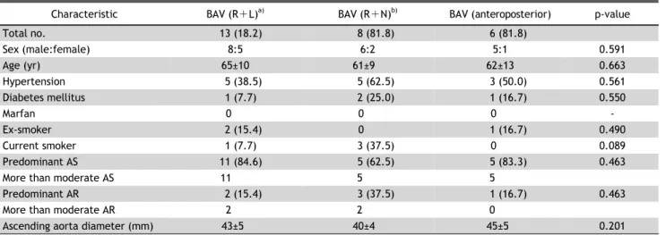
Table 2. Baseline characteristics of each BAV subgroup
Characteristic BAV (R +L)
a)BAV (R +N)
b)BAV (anteroposterior) p-value
Total no. 13 (18.2) 8 (81.8) 6 (81.8)
Sex (male:female) 8:5 6:2 5:1 0.591
Age (yr) 65±10 61±9 62±13 0.663
Hypertension 5 (38.5) 5 (62.5) 3 (50.0) 0.561
Diabetes mellitus 1 (7.7) 2 (25.0) 1 (16.7) 0.550
Marfan 0 0 0 -
Ex-smoker 2 (15.4) 0 1 (16.7) 0.490
Current smoker 1 (7.7) 3 (37.5) 0 0.089
Predominant AS 11 (84.6) 5 (62.5) 5 (83.3) 0.463
More than moderate AS 11 5 5
Predominant AR 2 (15.4) 3 (37.5) 1 (16.7) 0.463
More than moderate AR 2 2 0
Ascending aorta diameter (mm) 43±5 40±4 45±5 0.201
Values are presented as number (%), mean±standard deviation, or number.
BAV, bicuspid aortic valve; AS, aortic stenosis; AR, aortic regurgitation.
a)
Left coronary sinus-right coronary sinus.
b)Right coronary sinus- non-coronary sinus.
out clinical significance, complications by BAV disease are relatively common in adulthood. Therefore, BAV disease accounts for higher morbidity and mortality than other congenital heart diseases.
BAV disease is associated with significant valvular disease such as aortic stenosis or regurgitation.
Patients with BAV disease are also at increased risk of aortopathy such as aortic dilatation, aneurysmal change, or dissection. Previous studies have demon-
strated that aortopathy could occur without valve dysfunction [5,6]. In BAV, the connective tissue of the aortic media has abnormal properties and the colla- gen metabolism is disturbed. Extracellular matrix proteins or enzymes associated with aortopathy have been identified: an increased level of matrix metal- loproteinases (MMPs), decreased level of endothelial nitric oxide synthase (eNOS), and decreased level of tissue inhibitor of matrix metalloproteinase (TIMP) [7-9].
The objective of this study was to evaluate poten- tial differences in matrix protein expression in the aortic wall according to various types of BAV disease.
Methods
1) Patient characteristics
From January 2009 to December 2012, samples of the aortic wall were obtained from 31 patients who underwent open heart surgery for aortic stenosis or regurgitation and associated ascending aorta dilatation.
Intraoperative assessment of the aortic valve was
performed to identify whether the patients had BAV
disease. Four patients (12.9%) were identified with
tricuspid aortic valve (TAV) disease, and 27 patients
(87.1%) were identified with BAV disease. Patients in
the BAV group were categorized into three sub-
groups according to the classification proposed by
Sievers and Schmidtke [10]: left coronary sinus-right
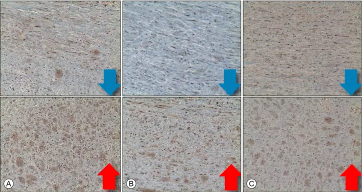
Fig. 1. Typical findings of immunohistochemistry staining for target molecule expression. Direction of arrow means ‘increase’ or
‘decrease’. (A) Endothelial nitric oxide synthase. (B) Matrix metalloproteinase-9. (C) Tissue inhibitor of matrix metalloproteinase-2.
coronary sinus (R+L group; n=13, 42%), right coro- nary sinus-non-coronary sinus (R+N group; n=8, 26%), and anteroposterior (AP group; n=6, 19%). We considered the R+L and R+N groups to be non-AP groups. The R+L and R+N groups had a BAV with one raphe and two fused leaflets, and the AP group included true bicuspid valves in the AP orientation with no raphe.
Patients’ preoperative characteristics are described in Table 1. The characteristics of each subgroup of BAV are additionally described in Table 2. All pa- tients with predominant aortic stenosis and asso- ciated ascending aorta dilatation had a degree of aortic stenosis being more than moderate.
Aortic tissue samples were approximately 3.0×3.0 mm
2. Samples were obtained from the proximal end of the resected ascending aorta or from the same site during aortotomy repair when the ascending aorta was not replaced.
2) Immunohistochemistry
Fresh samples were fixed with formalin and em- bedded in paraffin. The paraffin block containing the tissue sample was cut into thin sections of 4 to 5 μm.
These sections were stained with primary and secon- dary antibodies. The sections were mounted and ex- amined with a optical microscope that could take digital images. The whole area of a single slice was scanned using a ×100 objective, and magnified (×400) as digital images. The digital images were quanti- tatively analyzed using ImageJ software (National Institutes of Health, Bethesda, MD, USA). The area of secondary antibody expression was calculated with color picking tools. Only the media layer of the as- cending aorta was analyzed. The person who ob- tained and analyzed the images could not access pa- tient characteristics.
3) Statistical analysis
All statistical analyses were performed using the
PASW SPSS statistics software package ver. 18.0
(SPSS Inc., Chicago, IL, USA). Continuous data were
presented as mean±standard deviation. Categorical
data were presented as an absolute number or per-
centage. A p-value<0. 05 was considered to be stat-
istically significant. The comparisons of preoperative
characteristics and percentage of expression of the
target molecule between groups were performed us-
Yong Han Kim, et al
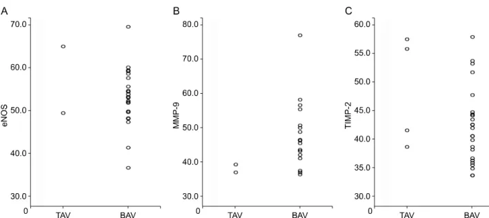
Fig. 2. (A–C) The scatter plot showing comparison of percentage of target molecule expression between TAV and BAV group. TAV, tricus- pid aortic valve; BAV, bicuspid aortic valve; eNOS, endothelial nitric oxide synthase; MMP, matrix metalloproteinase; TIMP, tissue in- hibitor of matrix metalloproteinase.
Fig. 3. Increased or decreased expression of eNOS, MMP-9, and TIMP-2. Reference: mean value of control group. eNOS, endothe- lial nitric oxide synthase; MMP, matrix metalloproteinase; TIMP, tissue inhibitor of matrix metalloproteinase.
ing univariate analysis. All parameters for this analy- sis were analyzed with non-parametric tests (Kruskal-Wallis test or Mann-Whitney U-test) as ap- plicable.
4) Research ethics
The study protocol was reviewed by the institu- tional review board of Seoul National University Hospital (approval no. H-0907-052-287) and the Helsinki Declaration was adhered to throughout the study. Patients provided their informed consent to participate after having received a complete descrip- tion of the study.
Results
Fig. 1 shows typical findings of immunohistoche- mistry staining for target protein expression. Fig. 2 shows the scatter plot that compares the percentage of target molecule expression between TAV and BAV groups. There were no significant statistical differ- ences between the two groups; eNOS (p=0.705), MMP-9 (p=0.111), and TIMP-2 (p=0.726). We set the mean percentage of the target molecule expression of the TAV group as the reference, and analyzed wheth- er the target molecule expression in the BAV group was increased or not in comparison.
We checked the increase or decrease of the target molecule level from the reference value (Fig. 3). The reference indicates the mean value of the control group. The expression of eNOS was decreased in the BAV group (16/22, 72.7%), MMP-9 was increased in the BAV group (14/17, 82.3%), and TIMP-2 was de- creased in the BAV group (19/24, 79.2%).
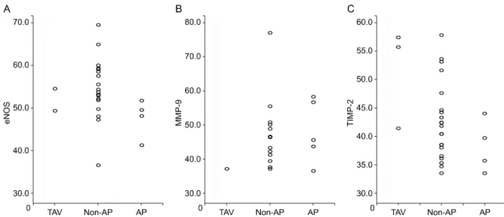
Fig. 4 shows the scatter plot that compares the
percentage of target molecule expression between
TAV, non-AP, and AP groups. There were no sig-
nificant statistical differences identified between the
three groups; eNOS (p=0.094), MMP-9 (p=0.377), and
TIMP-2 (p=0.411). Four patients (66.7%) from the
AP group were available to undergo evaluation of
eNOS expression, and all 4 patients (100%) showed
Table 3. Comparison of matrix protein expression in the media layer of AP and non-AP BAV Endothelial nitric
oxide synthase (%) Matrix metalloproteinase-9 (%) Tissue inhibitor of matrix metalloproteinase-2 (%)
Non-AP BAV (n=21) 55±7 47±10 43±7
AP BAV (n=6) 48±5 48±9 38±5
p-value 0.081 0.889 0.234
AP, anteroposterior; BAV, bicuspid aortic valve.
Fig. 4. (A–C) The scatter plot showing comparison of percentage of target molecule expression between TAV, non-AP, and AP group. TAV, tricuspid aortic valve; AP, anteroposterior; eNOS, endothelial nitric oxide synthase; MMP, matrix metalloproteinase; TIMP, tissue in- hibitor of matrix metalloproteinase.
a decreased level of eNOS compared to the TAV group. Five patients (83.3%) from the AP group were available to undergo evaluation of MMP-9 expression;
2 patients (40%) showed an increased level of MMP-9 and 3 patients (60%) showed a decreased level of MMP-9 compared to the TAV group.
Comparison of target molecule expressions in AP and non-AP groups in comparison to the BAV group showed decreased expression of eNOS in the AP group (p=0.081) (Table 3).
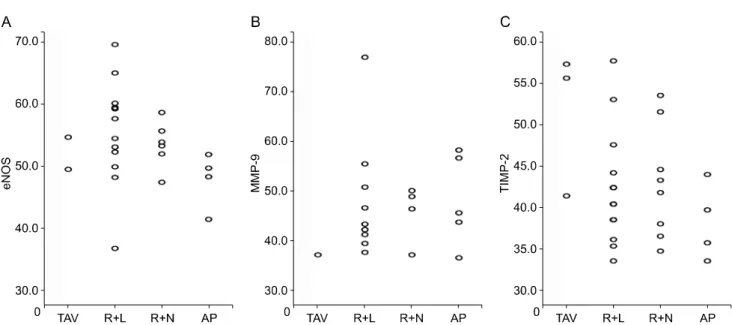
Fig. 5 shows the scatter plot that compares the percentage of target molecule expression among the TAV, R+L, R+N, and AP groups. There were no sig- nificant statistical differences among the four groups:
eNOS (p=0.115), MMP-9 (p=0.582), and TIMP-2 (p=
0.212) (Table 4).
Fig. 6 shows the scatter plot that demonstrates the correlation between the diameter of the ascending aorta and target molecule expression. There was no significant statistical correlation identified in any of
the molecules, meaning aorta dilatation is not corre- lated with target molecule expression.
Discussion
Our study demonstrates a decrease in eNOS/TIMP-2, and increase in MMP-9 in around 75% of patients in the BAV group (Fig. 2). Although we could not corre- late BAV with aortopathy (Fig. 1), our data indicate the importance of aortic wall biopsy in aortic valve diseases. The prevalence of aortic dilatation in pa- tients with BAV disease ranges from 33% to 80%, and is most frequently observed in the ascending aorta [11].
There was no significant difference in the percent-
age of target molecule expression of eNOS, MMP-9,
and TIMP-2 between TAV, non-AP, and AP groups
(Fig. 3). These findings indicate that the subtype of
BAV disease does not influence the degree of target
molecule expression. In contrast, Ikonomidis et al.
Yong Han Kim, et al
Table 4. Overall comparison of matrix protein expression in the media layer Endothelial nitric
oxide synthase (%)
Matrix metalloproteinase-9 (%)
Tissue inhibitor of matrix metalloproteinase-2 (%)
Bicuspid (R +L, N)
a)(n=13) 56±9 48±12 42±7
Bicuspid (R +N, L)
b)(n=8) 54±4 46±6 43±7
Bicuspid (anteroposterior) (n=6) 48±5 48±9 38±5
Control (n=4) 57±11 38±1 51±9
p-value 0.115 0.582 0.212
a)