• 검색 결과가 없습니다.

Surgical Repair of Ventricular Septal Defect after Myocardial Infarction: A Single Center Experience during 22 Years

N/A
N/A
Protected

Academic year: 2021

Share "Surgical Repair of Ventricular Septal Defect after Myocardial Infarction: A Single Center Experience during 22 Years"

Copied!
6
0
0

로드 중.... (전체 텍스트 보기)

전체 글

(1)

http://dx.doi.org/10.5090/kjtcs.2013.46.6.433 ISSN: 2233-601X (Print) ISSN: 2093-6516 (Online)

Department of Thoracic and Cardiovascular Surgery, Asan Medical Center, University of Ulsan College of Medicine Received: April 12, 2013, Revised: July 16, 2013, Accepted: July 22, 2013

Corresponding author: Cheol Hyun Chung, Department of Thoracic and Cardiovascular Surgery, Asan Medical Center, University of Ulsan College of Medicine, 88 Olympic-ro 43-gil, Songpa-gu, Seoul 138-736, Korea

(Tel) 82-2-3010-3946 (Fax) 82-2-3010-6966 (E-mail) [email protected]

C

The Korean Society for Thoracic and Cardiovascular Surgery. 2013. All right reserved.

CC

This is an open access article distributed under the terms of the Creative Commons Attribution Non-Commercial License (http://creative- commons.org/licenses/by-nc/3.0) which permits unrestricted non-commercial use, distribution, and reproduction in any medium, provided the original work is properly cited.

Surgical Repair of Ventricular Septal Defect after Myocardial Infarction: A Single Center Experience during 22 Years

Sung Jun Park, M.D., Joon Bum Kim, M.D., Sung-Ho Jung, M.D., Suk Jung Choo, M.D., Cheol Hyun Chung, M.D., Jae Won Lee, M.D.

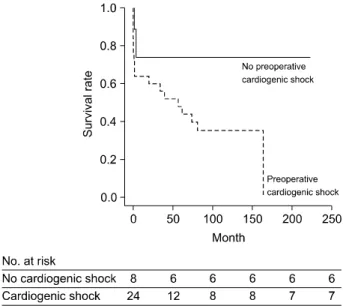
Background: Surgical repair of post-infarct ventricular septal defect (VSD) is considered one of the most challeng- ing procedures having high surgical mortality. This study aimed to evaluate the outcomes of the surgical repair of post-infarct VSD. Methods: From May 1991 to July 2012, 34 patients (mean age, 67.1±7.9 years) underwent sur- gical repair of post-infarct VSD. A retrospective review of clinical and surgical data was performed. Results: VSD repair involved the infarct exclusion technique using a patch in all patients. For coronary revascularization, 12 pa- tients (35.3%) underwent concomitant coronary artery bypass graft, 3 patients (8.8%) underwent preoperative percu- taneous coronary intervention, and 9 patients (26.5%) underwent both of these procedures. The early mortality rate was 20.6%. Six patients (17.6%) required reoperation due to residual shunt or newly developed VSD. During fol- low-up (median, 4.8 years; range, 0 to 18.4 years), late death occurred in nine patients. Overall, the 5-year and 10-year survival rates were 54.4%±8.8% and 44.3%±8.9%, respectively. According to a Cox regression analysis, preoperative cardiogenic shock (p=0.069) and prolonged cardiopulmonary bypass time (p=0.008) were independent predictors of mortality. Conclusion: The early surgical outcome of post-infarct VSD was acceptable considering the high-risk nature of the disease. The long-term outcome, however, was still dismal, necessitating comprehensive opti- mal management through close follow-up.

Key words: 1. Myocardial infarction

2. Ventricular heart septal defects 3. Surgery

4. Mortality 5. Risk factors

INTRODUCTION

The development of ventricular septal defect (VSD) is an uncommon complication following acute myocardial infarction (MI) occurring in 0.2% of patients with MI [1], but it is one of the most serious and life-threatening complications.

Although surgical repair of post-infarct VSD is a challenging

procedure having high surgical mortality of 19% to 60% in previous reports [1-16], there is still no alternative therapeutic option. For instance, the outcomes of medically treated pa- tients are reported as extremely poor, having a mortality rate of 90% or more [1].

Recently, data regarding the outcomes of post-infarct VSD

from two nationwide large-scale registries have been reported

(2)

in Europe and the United States [9,10]; however, there has been no report regarding this issue from a reasonably sized population consisting of other ethnic groups such as the Korean population. Therefore, the present study aimed to evaluate the early and late outcomes of the surgical repair of post-infarct VSD performed in Korea and to determine the predictive factors of mortality.

METHODS 1) Patients

Between May 1991 and July 2012, 34 adult patients under- went the surgical repair of post-infarct VSD at the Asan Medical Center, Seoul, Korea. From transthoracic echo- cardiography and coronary angiography (CAG) profiles, pre- operative, operative, and postoperative variables were col- lected retrospectively. These included demographic character- istics, co-morbidities, preoperative hemodynamic status (presence of shock, requirements of intra-aortic balloon pump, extracorporeal membrane oxygenation [ECMO]), aortic cross clamp time, and cardiopulmonary bypass (CPB) time. The study was approved by the institutional ethics committee/re- view board of the Asan Medical Center, and the requirement for informed patient consent was waived in view of the retro- spective nature of the study.

2) Surgical technique

The operations were performed by six cardiac surgeons.

All of the VSD repairs were performed by using the in- farct-exclusion technique first described by David and colleagues. The ventriculotomy incisions were made in the in- fracted area of the left ventricular or right ventricular free wall. Then, the interventricular septal defect was excluded us- ing a Dacron (DuPont, Wilmington, DE, USA), Teflon (Impra Inc., subsidiary of LR Bard, Tempe, AZ, USA), or bovine pericardium patch. The patch was sutured to un-infarcted tis- sue to avoid dehiscence or recurrence of VSD. The ven- triculotomy was closed and reinforced by Teflon strips beside the suture line. Concomitant coronary arterial bypass graft surgery (CABG) was performed in 21 patients.

3) Statistical analysis

Categorical variables were presented as frequencies and percentages, and were compared using the chi-squared test or Fisher’s exact test. Continuous variables were expressed as mean±standard deviation and compared using the Student un- paired t-test. To determine the predictors of mortality, a Cox regression model was used for multivariable analyses.

Variables with a p-value of ≤0.20 in the univariable analy- ses were candidates for the multivariable models. The multi- variable analyses involved a backward elimination technique, and only variables with a p-value of ≤0.10 were used in the final model. Results were expressed as a hazard ratio (HR) with 95% confidence intervals (CI). All reported p-values were two-sided, and p-values of <0.05 were considered to indicate statistical significance. Statistical analyses were per- formed with PASW SPSS ver. 18.0 (SPSS Inc., Chicago, IL, USA).

RESULTS 1) Baseline characteristics

The baseline characteristics of the patients are summarized in Table 1. Preoperative cardiogenic shock was present in 25 patients (73.5%). The cardiogenic shock was defined as a systolic blood pressure of <90 mmHg for at least 30 mi- nutes or the need for inotropic drugs to maintain a systolic blood pressure of ≥90 mmHg. The median time interval from the MI to the operation was 8.5 days (range, 0 to 187 days), and the median time interval from the VSD diagnosis to the operation was 3 days (range, 0 to 187 days). Nine pa- tients (26.5%) underwent emergency surgical repair within 24 hours of the VSD diagnosis, 16 patients (47.1%) between 2 days and a week, and 9 patients (26.5%) after a week.

A preoperative CAG was performed in all of the patients

with post-infarct VSD. Twenty-three patients (67.6%) were

revascularized of culprit vessels. Twelve patients (35.3%) un-

derwent concomitant CABG during surgical repair of VSD,

three patients (8.8%) underwent preoperative percutaneous

coronary intervention (PCI), and nine patients (26.5%) under-

went both preoperative PCI and concomitant CABG.

(3)

Table 1. Baseline characteristics of the patients who underwent surgical repair of post-infarct VSD (n=34)

Variable Value

Age (yr) Male gender

Body surface area (m

2

) Hypertension

Diabetes mellitus

Previous myocardial infarction Preoperative cardiogenic shock Preoperative intraaortic balloon pump

Preoperative extracorporeal membrane oxygenation Echocardiographic data

LV end-systolic dimension (mm) LV end-diastolic dimension (mm) LV ejection fraction

LV mass (g)

Myocardial infarction to operation (day) VSD diagnosis to operation (day) Aorta cross clamp time (min) Cardiopulmonary bypass time (min) No. of diseased vessels

One-vessel disease Two-vessel disease Three-vessel disease

Left main coronary artery disease Anatomy of culprit lesion

Left anterior descending artery Right coronary artery

Location of VSD Anterior Posterior VSD size (mm)

67.1±8.0 13 (38.2) 1.62±0.14 12 (35.3) 10 (29.4) 1 (2.9) 25 (73.5) 23 (67.6) 1 (2.9)

36.8±7.28 52.9±11.6 43.7±8.6 210.3±57.1

8.5 (0–187) 3 (0–187) 85.2±45.2 165.0±88.3

16 (47.1) 11 (32.4) 7 (20.6) 2 (5.9)

28 (82.4) 6 (17.6)

28 (82.4) 6 (17.6) 13.9±7.3 Values are presented as mean±standard deviation, number (%) or median (range).

VSD, ventricular septal defect; LV, left ventricle.

Table 2. Early operative outcomes (n=34)

Variable Value

30-day mortality In-hospital mortality

No. of patients with major complications

Residual shunt or recurrent ventricular septal defect Left ventricular free wall rupture

Re-exploration for bleeding New dialysis

Low cardiac output syndrome

a)

7 (20.6) 10 (29.4)

9 (26.5) 1 (2.9) 1 (2.9) 3 (8.8) 7 (20.6) Values are presented as number (%).

a)

Postoperative requirement of mechanical support such as intra- aortic balloon pump or extracorporeal membrane oxygenation.

2) Early outcomes

The early operative outcomes are summarized in Table 2.

Postoperative ECMO was required in three patients; one of them also required renal replacement therapy. Further, all three of them died of low cardiac output syndrome (LCOS).

Residual shunt or recurrent VSD was observed in nine pa- tients (26.5%) in the postoperative echocardiography fol- low-up. Among them, five patients required reoperation be- cause of a large hemodynamically significant defect, and two patients underwent concomitant tricuspid valve (TV) replace- ment during reoperation because of TV papillary muscle in-

farct necrosis in one patient and infective endocarditis of TV in the other. Among the nine patients with residual VSD, three patients died in hospital and another three patients died after discharge during the follow-up period.

Another patient underwent reoperation on the day after the first operation due to left ventricular free wall rupture. The left ventricular free wall rupture was newly developed, not a consequence of ventriculotomy dehiscence.

The 30-day mortality rate was 20.6% (n=7), and the cause of death was profound cardiac failure in all these cases. The patients in these cases were in cardiogenic shock at the time of operation. The other three patients died in hospital more than 30 days after the operation, and the causes of death were sepsis in two patients and subdural hemorrhage in the third patient. This resulted in the in-hospital mortality rate of 31.2% (n=10).

3) Overall outcomes and predictors of mortality

During the follow-up period (median, 4.8 years; range, 0 to 18.4 years), nine patients died after discharge. Overall, the 5-year and 10-year survival rates were 54.4%±8.8% and 44.3%±8.9%, respectively (Fig. 1).

Multivariable analyses revealed that the prolonged CPB time (HR, 1.07; 95% CI, 1.02 to 1.13; p=0.008) is an in- dependent factor associated with the overall mortality.

Preoperative cardiogenic shock (HR, 3.91; 95% CI, 0.90 to

17.01; p=0.069) also showed an association with the overall

mortality; however, it was not statistically significant (Table

3). Fig. 2 shows higher survival rates of patients who were

(4)

Fig. 1. Kaplan-Meier survival curve. Five-year survival rate, 54.4%±

8.8%; ten-year survival rate, 44.3%±8.9%.

Table 3. Multivariable risk factors analyses for overall mortality

Hazard ratio

95%

confidence interval

p-value

Preoperative cardiogenic shock Prolonged cardiopulmonary

bypass time

3.91 1.07

0.90–17.01 1.02–1.13

0.069 0.008

Fig. 2. Kaplan-Meier survival curve according to the presence of cardiogenic shock at the time of the operation.

not in preoperative cardiogenic shock at the time of operation than of the patients who were in cardiogenic shock.

DISCUSSION

After the introduction of reperfusion therapy such as early PCI or thrombolysis, the incidence of VSD development fol- lowing MI significantly decreased by 5- to 10-fold [1,17];

however, the outcomes of medically treated patients are re-

ported as extremely poor, having a mortality rate of 90% or more [1]. Thus, surgical repair is the only option to treat post-infarct VSD. Surgical repair of post-infarct VSD is still one of the most challenging cardiac procedures, despite recent advancements in surgical techniques, cardiac anesthesia, and myocardial protection during cardiac surgery.

The recurrence rates of VSD after the initial surgical repair of post-infarct VSD have been reported to range from 10% to 44% [3,9,14,18]. In the present study, nine patients (26.5%) had complications of residual shunt or recurrent VSD after surgery at postoperative echocardiogram, and five of them re- quired reoperation because of a large hemodynamically sig- nificant defect. The myocardial fragility and vague margin of the infracted tissue may be attributable to the relatively high incidence of residual shunt. A study suggests that residual VSD occurrence would be reduced with a double patch tech- nique as compared to a single patch technique [14]. However, the operative periods were distinct between the two operative techniques in the study: the single patch technique in the first half and double patch technique in the second half period.

Moreover, the study could not show a statistically significant difference between the two techniques. A comparison of the recurrence rate according to surgical methods was not con- ducted in most of the studies reported thus far because of the small number of subjects considered and the long study peri- od owing to the rare incidence of post-infarct VSD. In this present study, a comparison of surgical techniques for the re- currence of VSD was also not conducted because all of the VSD repairs were performed using a uniform surgical technique. Nevertheless, we believe that achieving complete exclusion of the fragile-infarcted myocardial tissue by fixing the patch on the healthy, viable myocardium may be the best way to reduce the risk of residual VSD.

Although a recent report demonstrated a significant im-

(5)

provement of surgical outcomes in the second half of the 25-year study period [6], there were no remarkable changes in the surgical outcomes of post-infarct VSD in the other pre- vious studies [1-5,7-18]. The dismal outcomes may be attrib- utable to the disease nature of post-infarct VSD: the surgical outcomes of post-infarct VSD are predominantly affected by the preoperative patients’ hemodynamic stability. Most of the studies on post-infarct VSD showed a strong association of shock at the time of operation and early mortality [3,13-16].

It has been commonly believed that if patients are hemody- namically stable, surgical repair should be delayed until the formation of the myocardial scar tissue and the development of the margins of the infracted muscle because the myocar- dium is considerably fragile; this may facilitate technical as- pects of the surgical procedure. However, a majority of the studies demonstrated no association of the surgical timing of VSD and the surgical outcomes [3,6,11-13], and the current guideline of American College of Cardiology/American Heart Association recommends immediate repair of post-infarct VSD after diagnosis without delay, irrespective of the hemo- dynamic status because rapid deterioration can occur in many patients [19]. The association of the earlier surgical timing and operative mortality in some reports may be mostly attrib- utable to the hemodynamic status requiring earlier surgery, not to the technical difficulty. Our findings also suggest asso- ciations between preoperative cardiogenic shock and early and overall outcomes, whereas there were no associations between the surgical timing and the outcomes.

The posterior location of VSD has been found to be asso- ciated with poor prognosis in most of the previous studies [3,9,18,20]. Surgical difficulties in the exposure of the septal rupture site and the right heart failure are mainly attributed to poor prognosis. However, David et al. [7] and a recent report [8] showed a different result; they revealed that posterior VSD is significantly associated with improved surgical outcomes. The authors suggest that the infarct exclusion tech- nique may address the difficulties of posterior VSD repair and prevent right heart failure by the physiologically sound character of the technique. In the previous two studies, how- ever, the sample size was small, involving only 44 patients and 32 patients, respectively; thus, the sample size may have been too small to evaluate the casual relationship between the

VSD location and operative risk. Considering the relatively small sample size of 34 patients in the present study, it can be understood in the same context that the location of the VSD was not associated with the surgical outcomes in the present study.

The present study showed an early mortality rate of 20.6%

and in-hospital mortality of 31.2%. The results are com- parable to the previous reports of post-infarct VSD that re- ported early mortality ranging from 19% to 60% [1-16].

Consistent with other studies [3,13-16], our current findings suggest a strong association between preoperative cardiogenic shock and early and overall mortality. All of the patients who died within the first 30 days after the operation were in car- diogenic shock at the time of operation. However, because of a small sample size, the statistical significance was marginal (p=0.069).

Prolonged CPB time was found to be a risk factor of over- all mortality in several previous studies [5,6]. However, pro- longed CPB time was not a predictor of cumulative mortality in 30-day survivors in the cited study [6], and the authors suggested that the prolonged CPB might have been con- founded by underlying factors. In our study, prolonged CPB time was an independent factor associated with overall mor- tality in the multivariable analysis. We also believe that the result cannot be attributed to the effect of CPB itself, but the underlying clinical situations of prolonged CPB such as diffi- culty in weaning from CPB might have contributed to the mortality. For instance, two patients required more than 400 minutes of CPB time; these patients were given ECMO as a result of CPB weaning failure and died of LCOS.

This study is subject to the limitations inherent in the ret- rospective studies of observational data from a single center.

The study results might have been affected by unmeasured confounders, procedure bias, or detection bias. Although the number of subject patients was not small compared with pre- vious studies on post-infarct VSD, the absolute number of pa- tients enrolled was small to draw robust conclusions.

In conclusion, using the infarct exclusion technique, we

found that the early surgical outcome of post-infarct VSD

was acceptable considering the high-risk nature of the

disease. However, the long-term outcome was still dismal, ne-

cessitating comprehensive optimal management through close

(6)

follow-up. Preoperative cardiogenic shock and prolonged CPB time were significant and independent predictors of the over- all mortality.

CONFLICT OF INTEREST

No potential conflict of interest relevant to this article was reported.

REFERENCES

1. Crenshaw BS, Granger CB, Birnbaum Y, et al. Risk factors, angiographic patterns, and outcomes in patients with ven- tricular septal defect complicating acute myocardial infarction. GUSTO-I (Global Utilization of Streptokinase and TPA for Occluded Coronary Arteries) Trial Investigators.

Circulation 2000;101:27-32.

2. Barker TA, Ramnarine IR, Woo EB, et al. Repair of post-in- farct ventricular septal defect with or without coronary ar- tery bypass grafting in the northwest of England: a 5-year multi-institutional experience. Eur J Cardiothorac Surg 2003;

24:940-6.

3. Deja MA, Szostek J, Widenka K, et al. Post infarction ven- tricular septal defect - can we do better? Eur J Cardiothorac Surg 2000;18:194-201.

4. David TE, Armstrong S. Surgical repair of postinfarction ventricular septal defect by infarct exclusion. Semin Thorac Cardiovasc Surg 1998;10:105-10.

5. Dalrymple-Hay MJ, Monro JL, Livesey SA, Lamb RK.

Postinfarction ventricular septal rupture: the Wessex experience. Semin Thorac Cardiovasc Surg 1998;10:111-6.

6. Lundblad R, Abdelnoor M, Geiran OR, Svennevig JL.

Surgical repair of postinfarction ventricular septal rupture:

risk factors of early and late death. J Thorac Cardiovasc Surg 2009;137:862-8.

7. David TE, Dale L, Sun Z. Postinfarction ventricular septal rupture: repair by endocardial patch with infarct exclusion.

J Thorac Cardiovasc Surg 1995;110:1315-22.

8. Papadopoulos N, Moritz A, Dzemali O, et al. Long-term re- sults after surgical repair of postinfarction ventricular septal rupture by infarct exclusion technique. Ann Thorac Surg 2009;87:1421-5.

9. Jeppsson A, Liden H, Johnsson P, Hartford M, Radegran K.

Surgical repair of post infarction ventricular septal defects:

a national experience. Eur J Cardiothorac Surg 2005;27:

216-21.

10. Arnaoutakis GJ, Zhao Y, George TJ, Sciortino CM, McCarthy PM, Conte JV. Surgical repair of ventricular sep- tal defect after myocardial infarction: outcomes from the Society of Thoracic Surgeons National Database. Ann Thorac Surg 2012;94:436-43.

11. Killen DA, Piehler JM, Borkon AM, Gorton ME, Reed WA.

Early repair of postinfarction ventricular septal rupture. Ann Thorac Surg 1997;63:138-42.

12. Deville C, Fontan F, Chevalier JM, Madonna F, Ebner A, Besse P. Surgery of post-infarction ventricular septal defect:

risk factors for hospital death and long-term results. Eur J Cardiothorac Surg 1991;5:167-74.

13. Lemery R, Smith HC, Giuliani ER, Gersh BJ. Prognosis in rupture of the ventricular septum after acute myocardial in- farction and role of early surgical intervention. Am J Cardiol 1992;70:147-51.

14. Labrousse L, Choukroun E, Chevalier JM, et al. Surgery for post infarction ventricular septal defect (VSD): risk factors for hospital death and long term results. Eur J Cardiothorac Surg 2002;21:725-31.

15. Cerin G, Di Donato M, Dimulescu D, et al. Surgical treat- ment of ventricular septal defect complicating acute my- ocardial infarction: experience of a north Italian referral hospital. Cardiovasc Surg 2003;11:149-54.

16. Cox FF, Morshuis WJ, Plokker HW, et al. Early mortality after surgical repair of postinfarction ventricular septal rup- ture: importance of rupture location. Ann Thorac Surg 1996;

61:1752-7.

17. Yip HK, Fang CY, Tsai KT, et al. The potential impact of primary percutaneous coronary intervention on ventricular septal rupture complicating acute myocardial infarction.

Chest 2004;125:1622-8.

18. Skillington PD, Davies RH, Luff AJ, et al. Surgical treat- ment for infarct-related ventricular septal defects. Improved early results combined with analysis of late functional status.

J Thorac Cardiovasc Surg 1990;99:798-808.

19. Cannon CP, Brindis RG, Chaitman BR, et al. 2013 ACCF/

AHA key data elements and definitions for measuring the clinical management and outcomes of patients with acute coronary syndromes and coronary artery disease: a report of the American College of Cardiology Foundation/American Heart Association Task Force on clinical data standards (writing committee to develop acute coronary syndromes and coronary artery disease clinical data standards). J Am Coll Cardiol 2013;61:992-1025.

20. Anderson DR, Adams S, Bhat A, Pepper JR. Post-infarction

ventricular septal defect: the importance of site of infarction

and cardiogenic shock on outcome. Eur J Cardiothorac Surg

1989;3:554-7.

수치

Table 1. Baseline characteristics of the patients who underwent  surgical repair of post-infarct VSD (n=34)
Fig. 2. Kaplan-Meier survival curve according to the presence of  cardiogenic shock at the time of the operation.

참조

관련 문서