http://dx.doi.org/10.5090/kjtcs.2014.47.2.124 ISSN: 2233-601X (Print) ISSN: 2093-6516 (Online)
1
Department of Thoracic and Cardiovascular Surgery, Korea University Medical Center;
2Thoracic Surgery & Thoracic Surgical Oncology Clinic, Fortis-Hospital;
3Korea Artificial Organ Center, Korea University
Received: August 8, 2013, Revised: October 14, 2013, Accepted: October 15, 2013
Corresponding author: Sung Ho Lee, Department of Thoracic and Cardiovascular Surgery, Korea University Medical Center, 73 Inchon-ro, Seongbuk-gu, Seoul 136-705, Korea
(Tel) 82-2-920-5837 (Fax) 82-2-928-8793 (E-mail) [email protected]
C
The Korean Society for Thoracic and Cardiovascular Surgery. 2014. All right reserved.
CC
This is an open access article distributed under the terms of the Creative Commons Attribution Non-Commercial License (http://creative- commons.org/licenses/by-nc/3.0) which permits unrestricted non-commercial use, distribution, and reproduction in any medium, provided the original work is properly cited.
Is There a Role for a Needle Thoracoscopic Pleural Biopsy under Local Anesthesia for Pleural Effusions?
Ho Sung Son, M.D.
1, Sung Ho Lee, M.D.
1, Laleng Mawia Darlong, M.S.
2, Jae Seong Jung, M.D.
1, Kyung Sun, M.D.
1, Kwang Taik Kim, M.D.
1, Hee Jung Kim, M.D.
1,
Kanghoon Lee, M.D.
1, Seung Hun Lee, M.D.
1, Jong Tae Lee
3Background: A closed pleural biopsy is commonly performed for diagnosing patients exhibiting pleural effusion if prior thoracentesis is not diagnostic. However, the diagnostic yield of such biopsies is unsatisfactory. Instead, a thoracoscopic pleural biopsy is more useful and less painful. Methods: We compared the diagnostic yield of nee- dle thoracoscopic pleural biopsy performed under local anesthesia with that of closed pleural biopsy. Sixty-seven patients with pleural effusion were randomized into groups A and B. Group A patients were subjected to closed pleural biopsies, and group B patients were subjected to pleural biopsies performed using needle thoracoscopy un- der local anesthesia. Results: The diagnostic yields and complication rates of the two groups were compared. The diagnostic yield was 55.6% in group A and 93.5% in group B (p<0.05). Procedure-related complications developed in seven group A patients but not in any group B patients. Of the seven complications, five were pneumothorax and two were vasovagal syncope. Conclusion: Needle thoracoscopic pleural biopsy under local anesthesia is a simple and safe procedure that has a high diagnostic yield. This procedure is recommended as a useful diagnostic modality if prior thoracentesis is non-diagnostic.
Key words: 1. Biopsy
2. Pleural disease 3. Pleural effusion 4. Tumor, malignant
5. Video-assisted thoracic surgery (VATS)
INTRODUCTION
If thoracentesis is not diagnostic for a patient with pleural effusion, it is important to perform tissue diagnosis for ensur- ing adequate treatment. Currently, blind closed pleural biopsy is usually conducted for this purpose because the procedure is simple and can be performed readily under local anesthesia.
However, the diagnostic yield of this procedure is low. A ret-
rospective study of 414 patients with malignant pleural effu-
sion revealed that closed pleural biopsies were of diagnostic
utility in only 7.1% of the patients [1]. It is known that per-
forming video-assisted thoracoscopic surgery (VATS) on
pleural effusion increases the diagnostic yield, even when
both thoracentesis and closed pleural biopsy (both conducted
prior) are non-diagnostic [2,3]. In recent times, the diagnostic
yield afforded by VATS in this context has approached 100%
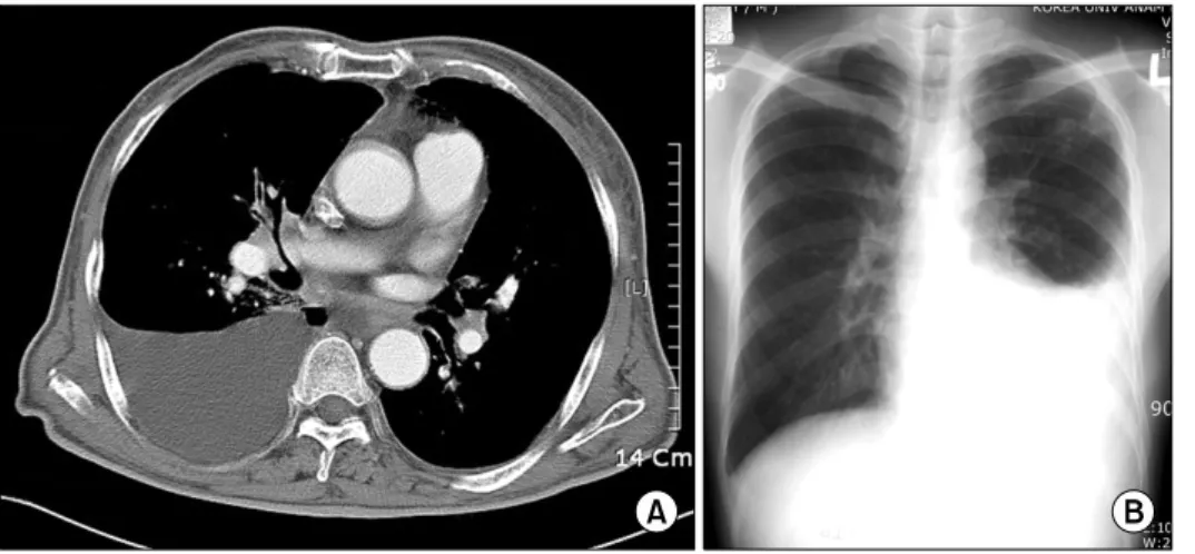
Fig. 1. Radiological findings upon preprocedural evaluation. (A) Chest computed tomography of a 78-year- old male patient. A biopsy revealed the presence of metastatic squamous cell carcinoma. (B) A chest posterior–
anterior view of a 27-year-old male patient. Tubercular pleurisy was diag- nosed by biopsy.
[4]. Thus, even as early as 1997, the American Thoracic Society recommended that thoracoscopy should be used for evaluating lung cancer prior to treatment if two prior cyto- logical examinations of pleural effusion in patients with sus- pected malignancies have been negative [5]. Although con- ventional thoracoscopy using a tube having a diameter of 5 to 10 mm affords high diagnostic accuracy, patients are vul- nerable to the risks associated with general anesthesia.
Conventional thoracoscopy performed under local anesthesia is poorly accepted by patients because of the pain and dis- comfort associated with the passage of relatively large instru- ments through narrow intercostal spaces. However, the use of a 2-mm-diameter instrument in a technique called needle thoracoscopy reduces the procedure-related discomfort, trau- ma, and the torque exerted on narrow intercostal spaces upon the introduction of the instrument into the pleural cavity. To evaluate the utility of performing this procedure under local anesthesia for diagnosing pleural effusion, we prospectively compared its diagnostic yield with that of closed pleural biopsy.
METHODS
Sixty-seven patients underwent pleural biopsies over an 18-month period. The criterion for patient inclusion was that their pleural effusion remained undiagnosed after thoracentesis. Patients were randomized into two groups.
Group A contained 36 patients with hydrothorax who under- went closed pleural biopsies. Group B contained 31 patients
with hydrothorax who underwent pleural biopsies via needle thoracoscopy under local anesthesia. A pre-procedural workup included chest radiography, chest computed tomography (CT), thoracentesis for pleural fluid analysis, a bacteriological study, and cytological evaluation (Fig. 1). In group A patients, closed pleural biopsies were performed aseptically using Abrams biopsy needles after administering local anesthesia (via infiltration of 5 to 10 mL of 1% [w/v] lidocaine) at the bedside. Oxygen was provided continuously via a facial mask, and pulse oximetry, electrocardiogram (ECG), and blood pressure monitoring were conducted throughout the procedure. A pleural biopsy was considered adequate if at least three specimens containing pleural tissue were obtained.
Chest radiography was performed immediately after each pro- cedure for identifying any iatrogenic pneumothorax. Group B patients were subjected to video-assisted thoracoscopic pleural biopsy in a fully equipped operating room, under local anes- thesia, and by using a thoracoscope and biopsy forceps, each having a diameter of 2 mm (Auto Suture, Norwalk, CT, USA), introduced via a trocar of the same diameter (Miniport; Auto Suture). Oxygen was provided continuously via a facial mask, and pulse oximetry, ECG, and blood pres- sure monitoring were conducted throughout the procedure.
The procedure was usually performed with the patient in the
lateral decubitus position, but the supine or semi-lateral decu-
bitus positions were occasionally employed. Local anesthesia
was administered via the infiltration of 1% (w/v) lidocaine
(range, 10 to 15 mL) at the site of the thoracoscopic port’s
placement. The entry point was selected according to the lo-
Fig. 2. Gross finding of pleural nodules yielded by 2-mm-diameter needle thoracoscopy. The multiple yellowish lesions were diag- nosed as metastatic adenocarcinoma.
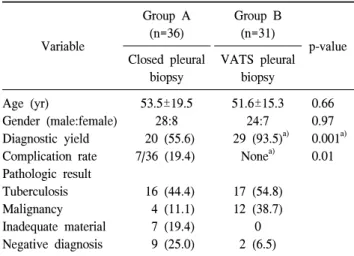
Table 1. Patient profiles and summary of the results
Variable
Group A (n=36)
Group B (n=31)
p-value Closed pleural
biopsy
VATS pleural biopsy Age (yr)
Gender (male:female) Diagnostic yield Complication rate Pathologic result Tuberculosis Malignancy Inadequate material Negative diagnosis
53.5±19.5 28:8 20 (55.6) 7/36 (19.4)
16 (44.4) 4 (11.1) 7 (19.4) 9 (25.0)
51.6±15.3 24:7 29 (93.5)
a)None
a)17 (54.8) 12 (38.7)
0 2 (6.5)
0.66 0.97 0.001
a)0.01
Values are presented as mean±standard deviation or number (%).
VATS, video-assisted thoracoscopic surgery.
a)