ISSN 2234-3806 • eISSN 2234-3814
Ann Lab Med 2014;34:247-251
http://dx.doi.org/10.3343/alm.2014.34.3.247
A Case of Isolated Lymphoblastic Relapse of the Central Nervous System in a Patient with Chronic Myelogenous Leukemia Treated with Imatinib
Mi-Jung Park, M.D.1, Pil-Whan Park, M.D.1, Yiel-Hea Seo, M.D.1, Kyung-Hee Kim, M.D.1, Ja-Young Seo, M.D.1, Ji-Hun Jeong, M.D.1, Moon Jin Kim, M.D.1, Jin-Woo Jeong, M.D.1, Jeong-Yeal Ahn, M.D.1, and Jinny Park, M.D.2
Departments of Laboratory Medicine1 and Internal Medicine2, Gachon University Gil Medical Center, Incheon, Korea
Isolated central nervous system (CNS) relapse is a rare, unpre- dictable event in patients with CML. Some studies have reported cases of isolated CNS relapse in CML [1-13]. However, very few studies have reported on isolated CNS relapse in the blast phase (BP) of CML in Korea [14, 15]. We report a case of BP CML with extramedullary lymphoblast proliferation in the CNS without evidence of disease in the peripheral blood (PB) and bone marrow (BM).
A 54-yr-old man was diagnosed as having BCR/ABL1 (b2a2 type)-positive CML in July 2012. He was considered to be in the chronic phase on the basis of his blast count on PB smear and BM aspiration, and subsequently, he was administered imatinib (400 mg/day). Three months after diagnosis, he demonstrated a complete hematologic response, major cytogenetic response, and no major molecular response according to the National Comprehensive Cancer Network Guidelines in Oncology for CML (karyotype 46,XY,t(9;22)(q34;q11.2)[7]/46,XY[23], BCR- ABL1 fusion transcript of 0.933% based on the International Scale [IS]) [16]. Seven months after initial diagnosis, he was re- admitted with a complaint of headache since two months. Diffu- sion brain magnetic resonance imaging with magnetic reso- nance angiography revealed abnormal leptomeningeal en-
hancement of both paramedian gyri, suggesting involvement of leukemic cells. In cerebrospinal fluid (CSF) study, his CSF was turbid and had increased number of white blood cells (WBCs) (3.5×10⁹/L), and almost all WBCs were lymphoid cells. Wright stain of cytospin-smeared CSF showed lymphoid cells with high nuclear-to-cytoplasmic ratio and coarse chromatin pattern with prominent nucleoli. Lymphoid cells were negative on periodic acid-Schiff staining. In flow cytometric analysis, the lymphoid cells had an early pre-B phenotype with an aberrant CD33 ex- pression (positive for CD45, CD34, CD33, terminal deoxynucle- otidyl transferase (TDT), HLA-DR, CD19, and CD10). The result of reverse transcriptase-PCR for detecting BCR-ABL1 fusion transcript was positive (b2a2 type) in CSF. On complete blood count analysis, Hb level was 13.3 g/dL, WBC count was 11.41×
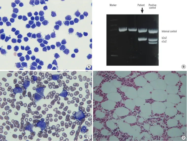
109/L, and platelet count was 167 ×109/L. There was no mor- phologic evidence of lymphoblasts in PB and BM samples (Fig.
1). The patient was treated with dasatinib, intrathecal metho- trexate, and cranial irradiation therapy. One week after initiation of treatment, CSF showed decreased WBCs (0.002×10⁹/L) with no evident malignant cells. Two months after relapse, patient demonstrated a complete hematologic response, complete cy- togenetic response, and major molecular response (karyotype
Received: June 28, 2013
Revision received: November 4, 2013 Accepted: February 13, 2014 Corresponding author: Jeong-Yeal Ahn
Department of Laboratory Medicine, Gachon University Gil Medical Center, 21 Namdong-daero 774beon-gil, Namdong-gu, Incheon 405-760, Korea Tel: +82-32-460-3831, Fax: +82-32-460-3415
E-mail: jyahn@gilhospital.com
© The Korean Society for Laboratory Medicine.
This is an Open Access article distributed under the terms of the Creative Commons Attribution Non-Commercial License (http://creativecommons.org/licenses/by-nc/3.0) which permits unrestricted non-commercial use, distribution, and reproduction in any medium, provided the original work is properly cited.
46,XY[30], BCR-ABL1 fusion transcript of 0.011% by IS). His laboratory findings are summarized in Table 1.
This patient showed optimal response to imatinib therapy that was administered according to the European Leukemia Net rec- ommendation [17]. However, spontaneous, isolated CNS re- lapse occurred after 7 months of imatinib therapy. In previous reports, isolated CNS relapse was observed in patients with imatinib-treated CML or Philadelphia chromosome-positive acute lymphoblastic leukemia [1-15]. This phenomena raised the possibility that imatinib may poorly penetrate the blood-brain barrier. Some studies have demonstrated that imatinib does not reach therapeutic levels in CSF, and therefore, imatinib therapy alone may lead to a potential risk of CNS involvement [1, 10].
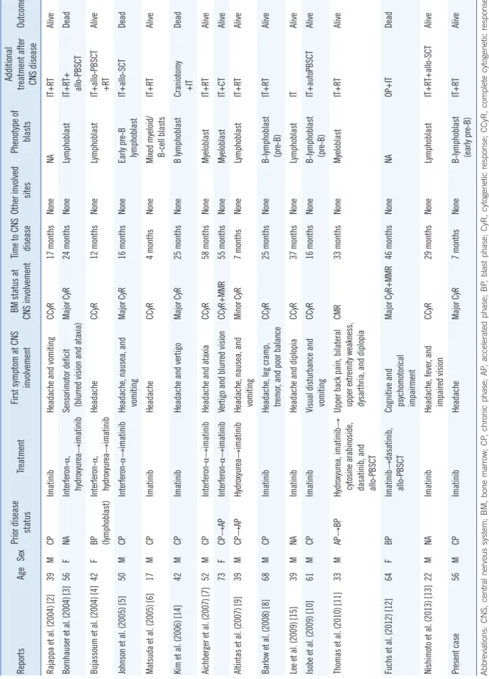
We present a summary of 15 cases of isolated CNS relapse in CML patients in Table 2. Isolated CNS relapse was predomi- nantly observed in men (M:F=11:4) with a median age of 42 yr (range, 17-73 yr). The major presenting symptom of isolated CNS relapses was headache with or without other neurologic symptoms. Nine of 15 cases (60%) had complete cytogenetic response state at CNS relapse. The median time from treatment initiation to CNS relapse was 25 months (range, 4-58 months) in 15 cases. Findings from these cases show that isolated CNS relapse has no correlation with a response to imatinib therapy.
No other sites were involved, except for CNS in all of the 15 cases, and eight cases had B-cell lymphoid phenotype of blasts [2-15]. In our case, the patient was a middle-aged man experi-
A B
C D
Marker Patient
Internal control b2a2 e1a2 Positive
control
Fig. 1. Cerebrospinal fluid (CSF) findings at seven months after initial diagnosis of CML. (A) Cytospin-smeared CSF showing numerous lym- phoid cells with high nuclear-to-cytoplasmic ratio and coarse chromatin pattern with prominent nucleoli (Wright stain, ×1,000). (B) The ma- jor BCR-ABL1 fusion transcript (b2a2 type) was detected in the CSF sample using reverse transcriptase-PCR. (C) Peripheral blood smear showing slightly increased white blood cell count including neutrophils with left-shiftness. No malignant lymphoid cells are seen (Wright stain,
×1,000). (D) Bone marrow aspirate smear showing myeloid hyperplasia without definite lymphoblasts (Wright stain, ×1,000).
encing headaches as the initial manifestation of CNS relapse.
Excluding the case in a 17-yr-old patient [6], the time interval from the diagnosis to CNS relapse in our case was shorter (7 months) than the previously reported cases. Most of the previ- ous cases had received additional treatment with intrathecal chemotherapy and cranial irradiation, and had showed good re- sponse [2-15]. Nishimoto et al. [13] reported a case with suc- cessful treatment with dasatinib and allogeneic hematopoietic stem cell transplantation (HSCT). Our patient was also treated with dasatinib after CNS relapse in BP of CML, and is currently being prepared to receive HSCT. Brain imaging, CSF study, or CNS prophylaxis are not routinely undertaken in patients with chronic phase CML at diagnosis. To our knowledge, there are no recommendations on the evaluation of CNS disease in CML patients. There are prognostic classifications using four factors, such as age, size of spleen, platelet count, and blast count at diagnosis and they remain valid for imatinib treatment [18-20].
Our patient was at high risk according to risk stratifications by Hasford et al. [19] and Sokal et al. [20] (Hasford score 1,509 and Sokal score 1.35). However, risk factors of CNS relapse have not been investigated. Further studies are needed to iden- tify risk factors of CNS relapse.
To our knowledge, this is the third report of isolated CNS lym- phoblast proliferation in a Korean CML patient. It is important to consider CNS relapse in chronic phase CML patients optimally treated with imatinib, especially in patients presenting with neu- rological symptoms, including headache. This emphasizes the
need for brain imaging study and CSF monitoring in imatinib- treated CML patients even without evidence of disease progres- sion on PB smear and BM studies.
REFERENCES
1. Leis JF, Stepan DE, Curtin PT, Ford JM, Peng B, Schubach S, et al.
Central nervous system failure in patients with chronic myelogenous leukemia lymphoid blast crisis and Philadelphia chromosome positive acute lymphoblastic leukemia treated with imatinib (STI-571). Leuk Lymphoma 2004;45:695-8.
2. Rajappa S, Uppin SG, Raghunadharao D, Rao IS, Surath A. Isolated central nervous system blast crisis in chronic myeloid leukemia. Hema- tol Oncol 2004;22:179-81.
3. Bornhauser M, Jenke A, Freiberg-Richter J, Radke J, Schuler US, Mohr B, et al. CNS blast crisis of chronic myelogenous leukemia in a patient with a major cytogenetic response in bone marrow associated with low levels of imatinib mesylate and its N-desmethylated metabolite in cere- bral spinal fluid. Ann Hematol 2004;83:401-2.
4. Bujassoum S, Rifkind J, Lipton JH. Isolated central nervous system re- lapse in lymphoid blast crisis chronic myeloid leukemia and acute lym- phoblastic leukemia in patients on imatinib therapy. Leuk Lymphoma 2004;45:401-3.
5. Johnson NA, Fetni R, Caplan SN. Isolated central nervous system re- lapse in patients with chronic myeloid leukemia on imatinib mesylate.
Leuk Lymphoma 2005;46:629-30.
6. Matsuda M, Morita Y, Shimada T, Miyatake J, Hirase C, Tanaka M, et al.
Extramedullary blast crisis derived from 2 different clones in the central nervous system and neck during complete cytogenetic remission of chronic myelogenous leukemia treated with imatinib mesylate. Int J He- matol 2005;81:307-9.
7. Aichberger KJ, Herndlhofer S, Agis H, Sperr WR, Esterbauer H, Rabitsch W, et al. Liposomal cytarabine for treatment of myeloid central Table 1. Brief laboratory data of the case
At diagnosis:
CML onset After
3 months After
6 months After 7 months:
CNS relapse After
9 months After 12 months Peripheral blood samples
WBC (×109/L) 98.75 3.46 5.69 11.41 2.75 2.5
PLT (×109/L) 351 157 206 167 171 57
Blast (%) 0 0 0 0 0 0
Bone marrow samples
Blast (%) 1.8 0.2 1.0 0.4 0.4 2.4
Karyotype 46,XY,t(9;22)
(q34;q11.2)[20]
46,XY,t(9;22)(q34;q11.2) [7]/46,XY[23]
46,XY,t(9;22)(q34;q11.2) [1]/46,XY[11]
46,XY,t(9;22)(q34;q11.2) [3]/46,XY[27]
46,XY[30] 46,XY[20]
BCR-ABL1 fusion gene (RT-PCR) Positive (b2a2) Positive (b2a2) Positive (b2a2) Positive (b2a2) Positive (b2a2) Negative
BCR-ABL1 fusion gene (qPCR) IS% 26.280 0.933 0.317 NA 0.011 0
CSF samples
WBCs (×10/L) NA NA NA 3.527 4 NA
BCR-ABL1 fusion gene (RT-PCR) NA NA NA Positive (b2a2) Negative NA
Abbreviations: CNS, central nervous system; WBC, white blood cell; PLT, platelet; RT-PCR, reverse transcriptase PCR; qPCR, quantitative PCR; CSF, cere- brospinal fluid; IS, international scale; NA, not available.
Table 2. Cases of isolated CNS blast crisis in CML ReportsAgeSexPrior disease statusTreatmentFirst symptom at CNS involvementBM status at CNS involvementTime to CNS diseaseOther involved sitesPhenotype of blasts
Additional treatment after CNS diseaseOutcome Rajappa et al. (2004) [2]39MCPImatinibHeadache and vomitingCCyR17 monthsNoneNAIT+RTAlive Bornhauser et al. (2004) [3]56FNAInterferon-α, hydroxyurea→imatinibSensorimotor deficit (blurred vision and ataxia)Major CyR24 monthsNoneLymphoblastIT+RT+ allo-PBSCTDead Bujassoum et al. (2004) [4]42FBP (lymphoblast)Interferon-α, hydroxyurea→imatinibHeadacheCCyR12 monthsNoneLymphoblastIT+allo-PBSCT +RTAlive Johnson et al. (2005) [5]50MCPInterferon-α→imatinibHeadache, nausea, and vomitingMajor CyR16 monthsNoneEarly pre-B lymphoblastIT+allo-SCTDead Matsuda et al. (2005) [6]17MCPImatinibHeadacheCCyR4 monthsNoneMixed myeloid/ B-cell blastsIT+RTAlive Kim et al. (2006) [14]42MCPImatinibHeadache and vertigoMajor CyR25 monthsNoneB lymphoblastCraniotomy +ITDead Aichberger et al. (2007) [7]52MCPInterferon-α→imatinibHeadache and ataxiaCCyR58 monthsNoneMyeloblastIT+RTAlive 73FCP→APInterferon-α→imatinibVertigo and blurred visionCCyR+MMR55 monthsNoneMyeloblastIT+CTAlive Altintas et al. (2007) [9]39MCP→APHydroxyurea→imatinibHeadache, nausea, and vomitingMinor CyR7 monthsNoneLymphoblastIT+RTAlive Barlow et al. (2008) [8]68MCPImatinibHeadache, leg cramp, tremor, and poor balanceCCyR25 monthsNoneB-lymphoblast (pre-B)IT+RTAlive Lee et al. (2009) [15]39MNAImatinibHeadache and diplopiaCCyR37 monthsNoneLymphoblastITAlive Isobe et al. (2009) [10]61MCPImatinibVisual disturbance and vomitingCCyR16 monthsNoneB-lymphoblast (pre-B)IT+autoPBSCTAlive Thomas et al. (2010) [11]33MAP→BPHydroxyurea, imatinib→ cytosine arabinoside, dasatinib, and allo-PBSCT
Upper back pain, bilateral upper extremity weakness, dysarthria, and diplopia
CMR33 monthsNoneMyeloblastIT+RTAlive Fuchs et al. (2012) [12]64FBPImatinib→dasatinib, allo-PBSCTCognitive and psychomotorical impairment
Major CyR+MMR46 monthsNoneNAOP+ITDead Nishimoto et al. (2013) [13]22MNAImatinibHeadache, fever, and impaired visionCCyR29 monthsNoneLymphoblastIT+RT+allo-SCTAlive Present case56MCPImatinibHeadacheMajor CyR7 monthsNoneB-lymphoblast (early pre-B)IT+RTAlive Abbreviations: CNS, central nervous system; BM, bone marrow; CP, chronic phase; AP, accelerated phase; BP, blast phase; CyR, cytogenetic response; CCyR, complete cytogenetic response; MMR, major molecular response; IT, intrathecal chemotherapy; RT, cranial irradiation; PB, peripheral blood; SCT, stem cell transplantation; NA, not available.
nervous system relapse in chronic myeloid leukaemia occurring during imatinib therapy. Eur J Clin Invest 2007;37:808-13.
8. Barlow A, Robertson M, Doig A, Stewart W, Drummond MW. Isolated central nervous system lymphoid blast crisis in chronic myeloid leukae- mia in major molecular remission. Br J Haematol 2008;142:327.
9. Altintas A, Cil T, Kilinc I, Kaplan MA, Ayyildiz O. Central nervous system blastic crisis in chronic myeloid leukemia on imatinib mesylate therapy:
a case report. J Neurooncol 2007;84:103-5.
10. Isobe Y, Sugimoto K, Masuda A, Hamano Y, Oshimi K. Central nervous system is a sanctuary site for chronic myelogenous leukaemia treated with imatinib mesylate. Intern Med J 2009;39:408-11.
11. Thomas A, Stein CK, Gentile TC, Shah CM. Isolated CNS relapse of CML after bone marrow transplantation. Leuk Res 2010;34:e113-4.
12. Fuchs M, Reinhöfer M, Ragoschke-Schumm A, Sayer HG, Böer K, Witte OW, et al. Isolated central nervous system relapse of chronic my- eloid leukemia after allogenic hematopoietic stem cell transplantation.
BMC Blood Disord 2012;12:9.
13. Nishimoto M, Nakamae H, Koh KR, Kosaka S, Matsumoto K, Morita K, et al. Dasatinib maintenance therapy after allogeneic hematopoietic stem cell transplantation for an isolated central nervous system blast crisis in chronic myelogenous leukemia. Acta Haematologica 2013;130:
111-4.
14. Kim HJ, Jung CW, Kim K, Ahn JS, Kim WS, Park K, et al. Isolated blast crisis in CNS in a patient with chronic myelogenous leukemia maintain- ing major cytogenetic response after imatinib. J Clin Oncol 2006;24:
4028-9.
15. Lee KW, Song MK, Seol YM, Choi YJ, Shin HJ, Chung JS, et al. Isolated central nervous system blast crisis in chronic myeloid leukemia. Korean J Med 2009;77(S2):S441-4.
16. Cortes J, Goldman JM, Hughes T. Current issues in chronic myeloid leukemia: monitoring, resistance, and functional care. J Natl Compr Canc Netw 2012;10(S3):S1-13.
17. Baccarani M, Cortes J, Pane F, Niederwieser D, Saglio G, Apperley J, et al. Chronic myeloid leukemia: an update of concepts and management recommendations of European LeukemiaNet. J Clin Oncol 2009;27:
6041-51.
18. Forrest DL, Trainor S, Brinkman RR, Barnett MJ, Hogge DE, Nevill TJ, et al. Cytogenetic and molecular responses to standard-dose imatinib in chronic myeloid leukemia are correlated with Sokal risk scores and du- ration of therapy but not trough imatinib plasma levels. Leuk Res 2009;
33:271-5.
19. Hasford J, Pfirrmann M, Hehlmann R, Allan NC, Baccarani M, Kluin- Nelemans JC, et al. A new prognostic score for survival of patients with chronic myeloid leukemia treated with interferon alfa. Writing Commit- tee for the Collaborative CML Prognostic Factors Project Group. J Natl Cancer Inst 1998;90:850-9.
20. Sokal JE, Cox EB, Baccarani M, Tura S, Gomez GA, Robertson JE, et al.
Prognostic discrimination in “good-risk” chronic granulocytic leukemia.
Blood 1984;63:789-99.