Received February 9, 2012, Revised July 5, 2012, Accepted for publication August 26, 2012
Corresponding author: You Chan Kim, Department of Dermatology, Ajou University School of Medicine, 206 WorldCup-ro, Yeongtong-gu, Suwon 443-749, Korea. Tel: 82-31-219-5190, Fax: 82-31-219-5189, E-mail: maychan@ajou.ac.kr
This is an Open Access article distributed under the terms of the Creative Commons Attribution Non-Commercial License (http://
creativecommons.org/licenses/by-nc/3.0) which permits unrestricted non-commercial use, distribution, and reproduction in any medium, provided the original work is properly cited.
ORIGINAL ARTICLE
Photodynamic Therapy with Ablative Carbon Dioxide Fractional Laser for Treating Bowen Disease
Sue Kyung Kim, Ji-Youn Park, Hyo Sang Song, You-Sun Kim1, You Chan Kim
Department of Dermatology, 1Institute for Medical Sciences, Ajou University School of Medicine, Suwon, Korea
Background: Topical photodynamic therapy (PDT) has been increasingly used to treat malignant skin tumors including the Bowen disease. However, patients could be displeased with the long incubation time required for conventional PDT. Objective: We evaluated the efficacy and safety of PDT with a short incubation time of ablative CO2 fractional laser pretreatment for treating Bowen disease. Methods: Ten patients were included. Just before applying the topical photosensitizer, all lesions were treated with ablative CO2
fractional laser, following the application of methyl aminolevulinate and irradiation with red light (Aktilite CL 128). Histological confirmation, rebiopsy, and clinical assessments were performed. Adverse events were also recorded. Results: Five of the ten (50%) lesions showed a complete response (CR) within three PDT sessions. After four treatment sessions, all lesions except one penile shaft lesion (90%) achieved clinical and histological CR or clinical CR only. The average number of treatments to CR was 3.70±1.70. The treatments showed favorable cosmetic outcomes and no serious adverse events. Conclusion: The results suggest that pretreatment with an ablative fractional CO2 laser before PDT has similar treatment efficacy and requires a shorter photosensitizer incubation time compared with the conventional PDT method. (Ann Dermatol 25(3) 335∼
339, 2013)
-Keywords-
Ablative carbon dioxide fractional laser, Bowen disease, Photodynamic therapy, Pretreatment
INTRODUCTION
Topical photodynamic therapy (PDT) has been increa- singly used to treat malignant skin tumors, including Bo- wen disease (BD)1. Clinicians prefer PDT because of its minimal invasiveness and favorable cosmetic outcomes.
PDT produces superior cosmetic results compared with those of cryotherapy and 5-fluorouracil2.
Transcutaneous penetration of photosensitizer is important to improve clinical outcomes of PDT. Therefore, a long incubation time (usually 3∼4 hours) is needed after app- lying the photosensitizer; however, patients are often dis- pleased with the long incubation time. Pretreatments of hyperkeratotic skin lesions by curettage, debulking, tape stripping, micro-dermabrasion or laser ablation, are useful procedures to increase photosensetizer absorption3,4. Ablative fractional lasers have been used to treat photo- damaged facial skin, rhytides, and various types of scars.
Ablative fractional laser creates microscopic vertical holes of ablated tissue, each surrounded by a thin layer of coagulated tissue, which constitutes microscopic treat- ment zones5. Therefore, the penetration of topically appl- ied photosensitizer can be enhanced through these cha- nnels formed by the ablative fractional laser.
Based on this concept, we evaluated the efficacy and safety of PDT with a short photosensitizer incubation time after ablative CO2 fractional laser pretreatment when deal- ing with BD.
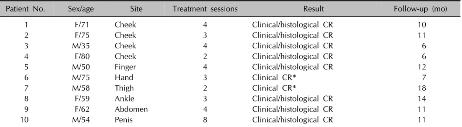
Table 1. Summary of the baseline characteristics and the results of photodynamic therapy following pretreatment with an ablative CO2 fractional laser
Patient No. Sex/age Site Treatment sessions Result Follow-up (mo)
1 F/71 Cheek 4 Clinical/histological CR 10
2 F/75 Cheek 3 Clinical/histological CR 11
3 M/35 Cheek 4 Clinical/histological CR 6
4 F/80 Cheek 2 Clinical/histological CR 6
5 M/50 Finger 4 Clinical/histological CR 12
6 M/75 Hand 3 Clinical CR* 7
7 M/58 Thigh 2 Clinical CR* 18
8 F/59 Ankle 3 Clinical/histological CR 14
9 F/62 Abdomen 4 Clinical/histological CR 11
10 M/54 Penis 8 Clinical/histological CR 11
F: female, M: male, CR: complete response. *Additional skin biopsy was refused by the patient.
MATERIALS AND METHODS
Patients
Ten Korean patients with BDs were enrolled. All BD lesions were confirmed by skin biopsy. Appropriate informed consents were obtained from all subjects before obtaining biopsy specimens. This study was approved by the institutional review board (IRB number: AJIRB-MED- MDB-11-173).
Treatment protocol
Just before applying the topical photosensitizer, all lesions were treated with a single pass of an ablative CO2 frac- tional laser (eCO2; Lutronic Inc., Seoul, Korea). Treatment parameters were as follows: pulse energy, 50 mJ; spot density, 100 spots/cm2, and power, 30 W. Immediately after each fractional laser treatment, the methyl amino- levulinate (MAL) (Metvix; Galderma, Sophia Antipolis, France) was applied to the lesion, and covered with an occlusive film (Tegaderm; 3M, Minneapolis, MN, USA) within a short incubation period (70 minutes). Before irradiation, the fluorescence of the lesion treated with photosensitizer was detected with a ultraviolet light emi- tting device (Yanus; PSI Inc., Suwon, Korea). Then, the lesion was irradiated with red light from the Aktilite CL 128 (Galderma) at a light dose of 37 J/cm2. Each session was repeated every two weeks.
Efficacy evaluation
Clinical improvement in the lesions was evaluated before each session. Two investigators evaluated clinical impro- vements either as a complete response (CR, complete removal of lesion), a partial response (PR, 25∼99%
reduction in lesion) or no response (NR, 0∼24% redu- ction in lesion). If a lesion showed a clinical CR, histo- logical improvement was also evaluated after 8 weeks
from the last session. Additional PDT was performed when the lesion showed a clinical PR or NR. Adverse events such as pain, phototoxic reactions (e.g., erythema, crusting, and ulceration), and pigment changes were also recorded.
RESULTS
Among the 10 patients with BD whom have completed the evaluation, five were male and five were female. Their ages ranged from 35 to 80 years (mean, 61.9 years;
median, 60.5 years). The ten lesions were located on the face (n=4), extremities (n=4), trunk (n=1), and penis (n=1). The baseline clinical characteristics of the ten patients including lesion locations are shown in Table 1.
Five of the ten (50%) lesions showed a CR within three PDT sessions (two lesions showed a clinical CR and three lesions showed both clinical and histological CR) (Fig. 1, 2).
All lesions except the penile shaft lesion (90%) achieved a clinical and histological CR or clinical CR after four treatment sessions. However, the one patient with the penile shaft lesion showed a clinical and histological CR after eight treatment sessions. The average number of treatments to CR was 3.70±1.70. Except for the patient with the penile shaft lesion, with eight treatment sessions, the average number of treatments to CR decreased to 3.20±0.80 (Table 1).
Treatments were generally well-tolerated, except for some patients who experienced a mild burning sensation during the procedure. No additional complications, such as pigment changes or scarring, were observed. No recurren- ces of the BD lesions were observed during the 6∼18 months of follow-up.
Fig. 1. Clinical clearance of Bowen disease on right cheek with ablative CO2 fractional laser pretreatment following methyl aminolevulinate-photodynamic therapy (MAL-PDT). (A) Before treatment, (B) after treatment.
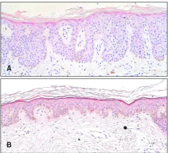
Fig. 2. Histological clearance of Bowen disease following pre- treatment with a ablative CO2 fractional laser following methyl aminolevulinate-photodynamic therapy (MAL-PDT) (H&E, ×200).
(A) Before treatment, (B) after treatment.
DISCUSSION
Among the various therapeutic modalities for treating BD, PDT is a promising therapeutic alternative, because of its good cosmetic outcomes6. However, a relatively long incubation time is one of the disadvantages of PDT;
therefore, trials to increase photosensitizer penetration to shorten the incubation time are needed. Up until today, clinicians have tried to develop novel photosensitizer delivery methods such as biomodulation with drugs and using the stratum corneum (e.g., drug-vehicle optimi- zation, liposomes, iontophoresis and electroporation), as well as physical modulation techniques such as micro-
needling or topical keratolytic agents, that mediate intra- dermal delivery of photosensitizer4,7-9. Ablative lasers, including the CO2 laser and the erbium YAG laser, have been used to increase photosensitizer penetrations10-12. Biomodulation with drugs and using the stratum corneum shows limited deep penetrations into the skin. Various physical modulation techniques including curettage can induce bleeding and oozing of tissue fluids within a lesion, following elimination of the photosensitizer from the lesion. Compared to these pretreatment techniques, which have several limitations, ablative fractional laser treatment has certain advantages. Because of greater penetration depth of the ablative fractional laser, photosensitizer may reach into deeper skin layers. Addi- tionally, photosensitizers are distributed evenly through- out the entire lesion with uniformly-distributed vertical holes made by the ablative fractional laser. Moreover, an ablative fractional laser rarely causes bleeding and oozing with their coagulation character. Based on this mecha- nism, the ablative fractional CO2 laser could be used to shorten the conventional PDT incubation time. Recently, ablative fractional laser treatment greatly facilitates the delivery of topical MAL deep into the skin of an animal model through the cone-shaped channels created by the laser5,13. Based on these results, we used ablative frac- tional CO2 laser treatment before MAL-PDT for Bowen disease to shorten the incubation period.
Previous reports demonstrated that 73∼93% of patients with BD achieve clinical CR after two MAL- PDT se- ssions14-17, which suggests a higher clearance rate than that of the present study. However, one of the limitations from previous studies is that only the clinical assessment was being evaluated without histological confirmation.
Moreover, the Asian skin type (skin type IV/V) is more
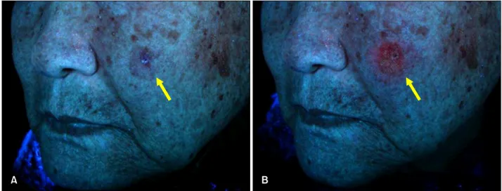
Fig. 3. Fluorescence assessment of the lesion (arrows). (A) Before application of photosensitizer, (B) after 70 minutes as incubation time.
resistant to light penetrations than the Caucasian skin type (skin type II/III)18. According to a multi-center study in Korea, about 60% of patients with BD achieved CR19. Additionally, 3∼4 treatment sessions are usually needed to achieve both clinical and histological CR of lesions using topical PDT and conventional methods during the 6 years at our center20. Therefore, the combined treatment of pretreating with an ablative fractional CO2 laser and topical PDT with a shorter incubation time (70 minutes) showed similar treatment efficacy compared with the conventional PDT method, which requires a 3-hour incubation time.
A fluorescence photograph was taken after applying MAL to evaluate MAL penetration. The fluorescence of the pen- etrated photosensitizer applied with a shorter incubation time was similar to that of the conventional method (Fig. 3).
Several limitations should be noted. Because of the small sample sizes, further larger studies will be needed to con- firm the results. It is obvious that adding the ablative fractional CO2 laser pretreatment contributed to the reduc- tion of incubation time; however, a comparative study with various incubation times will be necessary to dete- rmine the most suitable time. In this study, we have fixed the incubation time as 70 minutes (38.9% of conventional 3-hour incubation time). In the preliminary study, we have performed the 5-aminolevulinic acid (ALA)-PDT with abla- tive carbon dioxide fractional laser of 90 minutes as 37.5% of 4 hours for incubation time in actinic keratosis (AK), and the CR was up to 90% (unpublished data). The CR was similar to the previously published data of AK, ranging from 71 to 100%1. It is well known that the efficacy of MAL-PDT is prior to that of ALA-PDT in the case of BD2. Therefore, we have chosen MAL instead of
ALA with similar percentages of incubation time as 33.3%
(70 minutes) for 3 hour (the conventional incubation time of MAL is 3 hour). The parameter that we have chosen was based on the published data21. We expect that further study should be performed for accurate incubations and laser parameters with maximal efficacy and minimal adverse event.
The risk of ablative fractional laser exists such as thermal injury, coagulation, or tissue necrosis. Our patients, how- ever, received fractional laser only in lesional areas, and no serious effects mentioned above were noted either at the normal area or the lesional area. Furthermore, in our case, each lesion was not huge enough for patients to receive pretreatment like anesthetic cream or injection. Also, we assumed that the anesthetic cream could contribute the efficacy of PDT as pH changes22, and thus, we did not use any local anesthesia. The laser treatment time was very short and patients only complained about minimal pain, and there was no other adverse event. If the lesions were huge, multiple times of anesthesia might be necessary.
PDT itself is not a first-line therapy for all BD patients.
Also, there had been no head-to-head study to compare the CR of various treatment modalities. Considering the sizes and numbers of the lesions or the medical conditions of patients, the surgical management or alternative mana- gement, like PDT, can be individualized. In our cases, some patients were old aged with generally poor con- ditions to undergo such surgical management, and they demanded for more conservative treatment instead of sur- gical management. Other patients did not want to receive surgery because expected cosmetic results.
In summary, the present data suggests that ablative frac- tional CO2 laser pretreatment before PDT has similar
treatment efficacy and requires a shorter photosensitizer incubation time compared with the conventional PDT method. Long treatment times, as being one of the major disadvantages of topical PDT, can be reduced with this combined treatment.
ACKNOWLEDGMENT
This research was supported by the Basic Science Re- search Program through the National Research Foundation of Korea (NRK), funded by grant 2010-0022412 from the Ministry of Education, Science, and Technology.
REFERENCES
1. Morton CA, McKenna KE, Rhodes LE; British Association of Dermatologists Therapy Guidelines and Audit Subcommi- ttee and the British Photodermatology Group. Guidelines for topical photodynamic therapy: update. Br J Dermatol 2008;159:1245-1266.
2. Morton C, Horn M, Leman J, Tack B, Bedane C, Tjioe M, et al. Comparison of topical methyl aminolevulinate photody- namic therapy with cryotherapy or Fluorouracil for treat- ment of squamous cell carcinoma in situ: results of a multicenter randomized trial. Arch Dermatol 2006;142:
729-735.
3. Gerritsen MJ, Smits T, Kleinpenning MM, van de Kerkhof PC, van Erp PE. Pretreatment to enhance protoporphyrin IX accumulation in photodynamic therapy. Dermatology 2009;218:193-202.
4. Fang JY, Lee WR, Shen SC, Fang YP, Hu CH. Enhancement of topical 5-aminolaevulinic acid delivery by erbium:YAG laser and microdermabrasion: a comparison with iontopho- resis and electroporation. Br J Dermatol 2004;151:132- 140.
5. Haedersdal M, Sakamoto FH, Farinelli WA, Doukas AG, Tam J, Anderson RR. Fractional CO(2) laser-assisted drug delivery. Lasers Surg Med 2010;42:113-122.
6. Lehmann P. Methyl aminolaevulinate-photodynamic thera- py: a review of clinical trials in the treatment of actinic keratoses and nonmelanoma skin cancer. Br J Dermatol 2007;156:793-801.
7. Clementoni MT, B-Roscher M, Munavalli GS. Photo- dynamic photorejuvenation of the face with a combination of microneedling, red light, and broadband pulsed light.
Lasers Surg Med 2010;42:150-159.
8. de Leeuw J, van der Beek N, Bjerring P, Neumann HA.
Photodynamic therapy of acne vulgaris using 5-amino- levulinic acid 0.5% liposomal spray and intense pulsed light in combination with topical keratolytic agents. J Eur Acad Dermatol Venereol 2010;24:460-469.
9. Fang YP, Wu PC, Tsai YH, Huang YB. Physicochemical and safety evaluation of 5-aminolevulinic acid in novel
liposomes as carrier for skin delivery. J Liposome Res 2008;18:31-45.
10. Smucler R, Vlk M. Combination of Er:YAG laser and photodynamic therapy in the treatment of nodular basal cell carcinoma. Lasers Surg Med 2008;40:153-158.
11. Forster B, Klein A, Szeimies RM, Maisch T. Penetration enhancement of two topical 5-aminolaevulinic acid for- mulations for photodynamic therapy by erbium:YAG laser ablation of the stratum corneum: continuous versus frac- tional ablation. Exp Dermatol 2010;19:806-812.
12. Fukui T, Watanabe D, Tamada Y, Matsumoto Y. Photody- namic therapy following carbon dioxide laser enhances efficacy in the treatment of extramammary Paget's disease.
Acta Derm Venereol 2009;89:150-154.
13. Haedersdal M, Katsnelson J, Sakamoto FH, Farinelli WA, Doukas AG, Tam J, et al. Enhanced uptake and photo- activation of topical methyl aminolevulinate after fractional CO2 laser pretreatment. Lasers Surg Med 2011;43:804- 813.
14. Calzavara-Pinton PG, Venturini M, Sala R, Capezzera R, Parrinello G, Specchia C, et al. Methylaminolaevulinate- based photodynamic therapy of Bowen's disease and squamous cell carcinoma. Br J Dermatol 2008;159:137- 144.
15. Cox NH, Eedy DJ, Morton CA. Guidelines for management of Bowen's disease. British Association of Dermatologists.
Br J Dermatol 1999;141:633-641.
16. Cox NH, Eedy DJ, Morton CA; Therapy Guidelines and Audit Subcommittee, British Association of Dermatologists.
Guidelines for management of Bowen's disease: 2006 update. Br J Dermatol 2007;156:11-21.
17. Varma S, Wilson H, Kurwa HA, Gambles B, Charman C, Pearse AD, et al. Bowen's disease, solar keratoses and superficial basal cell carcinomas treated by photodynamic therapy using a large-field incoherent light source. Br J Dermatol 2001;144:567-574.
18. Ho SG, Chan HH. The Asian dermatologic patient: review of common pigmentary disorders and cutaneous diseases.
Am J Clin Dermatol 2009;10:153-168.
19. Kim YC, Kim IH, Lee MG, Cho KH. Clinical efficacy and tolerance of Metvix®-photodynamic therapy in the treat- ment of actinic keratosis, basal cell carcinoma, and Bo- wen’s disease in South Korea. Korean J Dermatol 2008;60 (Suppl. 2):108
20. Kim YJ, Kang HY, Lee ES, Kim YC. Photodynamic therapy for treatment of Bowen's disease. Korean J Dermatol 2007;
45:237-241.
21. España A, Solano T, Quintanilla E. Bleomycin in the treatment of keloids and hypertrophic scars by multiple needle punctures. Dermatol Surg 2001;27:23-27.
22. Borelli C, Herzinger T, Merk K, Berking C, Kunte C, Plewig G, et al. Effect of subcutaneous infiltration anesthesia on pain in photodynamic therapy: a controlled open pilot trial.
Dermatol Surg 2007;33:314-318.