Human Leptin Protein Induces Proliferation of A549 Cells via Inhibition of PKR-Like ER Kinase and Activating Transcription
Factor-6 Mediated Apoptosis
Qun Lai and Yan Sun
Department of Thoracic Surgery, General Hospital of Zaozhuang Mining Group, Shandong, Zaozhuang, China.
Received: May 7, 2013 Revised: June 2, 2013 Accepted: June 3, 2013
Corresponding author: Dr. Qun Lai, Department of Thoracic Surgery,
General Hospital of Zaozhuang Mining Group, Qi lian shan Road 12, Shandong,
Zaozhuang 277000, China.
Tel: 86-0632-4060240, Fax: 86-0632-4060004 E-mail: [email protected]
∙ The authors have no financial conflicts of interest.
© Copyright:
Yonsei University College of Medicine 2013 This is an Open Access article distributed under the terms of the Creative Commons Attribution Non- Commercial License (http://creativecommons.org/
licenses/by-nc/3.0) which permits unrestricted non- commercial use, distribution, and reproduction in any medium, provided the original work is properly cited.
Purpose: To investigate the anti-apoptotic mechanism of leptin in non-small cell lung cancer. Materials and Methods: The influences of leptin on apoptosis were in- vestigated, analyzing the mechanism that triggers growth of A549 cells. The effects of leptin on cell proliferation were examined by XTT analysis. Leptin, C/EBP ho- mologous protein (CHOP), phosphorylated-PKR-like ER kinase (p-Perk), inositol requiring proteins-1, spliced X-box transcription factor-1 (XBP1), cleaved activating transcription factor-6 (ATF6), eukaryotic translation initiation factor-2α, caspase-12 and CHOP protein were detected in four groups by western blot, and endoplasmic reticulum (ER) stress related mRNA were detected by reverse transcription PCR.
Results: The expression of leptin in A549 and leptin transfected cells inhibited cispl- atin activated ER stress-associated mRNA transcription and protein activation. Two ER stress unfolded protein response pathways, PERK and ATF6, were involved, and XBP1 and tumor necrosis factor receptor-associated factor 2 (TRAF2) were in- creased significantly when treated with cisplatin in A549-siRNA against leptin cells.
Furthermore, CHOP expression was inhibited upon leptin expression in A549, LPT- PeP and LPT-EX cells. Conclusion: Leptin serves as an important factor that pro- motes the growth of A549 cells through blocking ER stress-mediated pathways. This blocking is triggered by p-Perk and ATF6 via inhibition of CHOP expression.
Key Words: Apoptosis, ER stress, cell growth, leptin, TRAF2, XPB1
INTRODUCTION
Lung cancer is a widespread disease, with a high incidence rate, and is a leading cause of mortality worldwide. In particular, non-small cell lung cancer (NSCLC) accounts for more than 80% of all lung cancers.1 In clinical, NSCLC is divided into 3 types, including squamous cell carcinoma, adenocarcinoma and large cell lung cancer.2 NSCLC commonly develops resistance to radiation and chemotherapy, and often presents at stages too late for surgical therapy. Current therapy methods are very limited, so the effective methods are urgent to be involved to decrease the inci- dence of pulmonary neoplasms.3 Therefore, exploring targets for NSCLC therapy has led to the development of promising methods for potential clinical use.
conditions were as follows: 94°C for 1.5 min, followed by 94°C for 30 s, 63°C for 30 s and 72°C for 30 s for a total of 30 cycles, and a final extension at 72°C for 10 min.
Cell culture and transfection
Lung adenocarcinoma cell line A549 and non-tumorigenic human bronchial epithelial cell line BEAS2B were pur- chased from American Type Culture Collection (ATCC, Rockville, MD, USA). Cells were cultured in complete cul- ture medium (RPMI 1640 containing 10% FCS and 200 IU/mL penicillin/100 μg/mL streptomycin). All cells were cultured at 37°C with 5% CO2. A549 and BEAS2B cells were plated into 6-well or 96-well plates (Falcon, Tokyo, Ja- pan) 24 h before transfection (for the BEAS2B cells). Dif- ferent amounts of plasmids (2 μg DNA per well in a 6-well plate and 0.2 μg DNA per well in a 96-well plate) were transfected into the BEAS2B monolayer cells with Lipo- fectamineTM 2000 transfection reagent (Invitrogen, Carls- bad, CA, USA). A549 and BEAS2B cells were harvested by trypsin/EDTA in ethylenediaminetetraacetic acid 24 h or 48 h after transfection, and subsequently pelleted by short centrifugation and suspended in the lysis buffer as de- scribed by Wang, et al.13 Cells were supplemented with complete proteasomal inhibitor mixture (Merck, Darm- stadt, Germany).
Small interfering RNA (siRNA) transfection
siRNA against leptin (siLPT) were purchased from Invitro- gen Inc. Transient transfection was carried out with Lipo- fectamineTM 2000 transfection reagent according to the manufacturer’s protocol. A549 cells were seeded on 6-well plates for RNA or protein preparation and 96-well plates for DNA fragmentation or cell growth assays (named A549- siLPT). After 24 hours of incubation, media were replaced with serum-free RPMI 1640 containing siRNA (100 nmol/
L) and transfection reagent. Cells were harvested for assays daily for three consecutive days after transfection with the siRNA duplexes. The experiments were repeated on the at least three separate occasions.
Western blots
The cell lysates were separated by 15% SDS-PAGE and electro-transferred onto nitrocellulose membranes. After blocking with 5% defatted milk in phosphate buffered saline overnight at 4°C, the membranes were incubated with 1 : 2000 leptin specific monoclonal antibody (mAb) (Santa Cruz, Dallas, TX, USA), 1 : 1000 goat pAb anti-human Endoplasmic reticulum (ER) is a central organelle in
cells, which plays an important role in protein folding and maturation, and lipid synthesis. The ER can be affected by a variety of toxic insults.4-6 There is an increasing evidence that ER stress acts a significant function in the apoptosis regulation. Two specific signaling pathways were involving the ER stress process, such as, unfolded protein responses (UPR) and ER-associated protein degradation7,8 UPR path- way participates in the activation of some specific proteins, including activating transcription factor 6 (ATF6), PKR- like ER kinase (PERK), and inositol requiring proteins 1 (IRE1).9 The above three pathways of UPR activate several transcription factors, such as eukaryotic translation initiation factor-2α (eIF-2α) and X-box transcription factor-1 (XBP1).
The pro-apoptotic transcription factor C/EBP homologous protein (CHOP)/GADD153, which suppresses the tran- scription of Bcl-2, can also be induced by a combination of the PERK/ATF4 and ATF6 pathways.
Leptin, originally described as an adipocyte-derived hor- mone regulating food intake and energy expenditure, is a pleiotropic hormone that plays both a proliferative and an anti-apoptotic role in several conditions, such lung cancer,10 breast cancer,11 and gastric cancer.12 Previously, the long iso- form leptin receptor was identified in normal human lung tissue, suggesting that lung is a peripheral site of action for leptin. The circulating levels of leptin and/or overexpres- sion of leptin mRNA are increased in adipose tissue. How- ever, the anti-apoptosis effect and mechanism of leptin in lung cancer remain unknown. Accordingly, the present study attempted to establish an understanding of the anti-apoptot- ic mechanisms involving leptin in NSCLC.
MATERIALS AND METHODS
Plasmid construction
Leptin gene was amplified by the PCR technique, using cDNA from human adipocyte cells isolated from the subcu- taneous fat of patients; written informed consent and ap- proval from an Institutional Review Board were obtained prior to conducting this study. PCR was conducted with the forward primer (5’-GCGAATTCATGGTTCCAATC CAAAAAG TCCAAGAGG-3’, at BamH I site) and re- verse primer (5’-TATGGATCCTCA GCACCCAGGGCT GA GG-3’, at Not I site). The PCR product was ligated to vector pMD18-T and sub-cloned into vector pcDNA3.1(+), yielding recombinant plasmid pcDNA3.1-LPT. The PCR
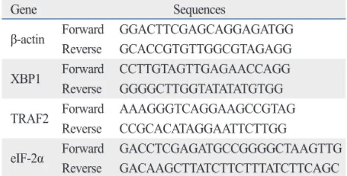
ed factors in the tested A549, A549-siLPT and BEAS2B cells, a series of hemi-quantitative RT-PCR assays were performed, including XBP1, eIF-2α and tumor necrosis factor receptor-associated factor 2 (TRAF2). The specific primers for XBP1, eIF-2α and TRAF2 were synthesized according to previous studies (Table 1).14-16 In parallel, indi- vidual β-actin was selected as an internal control. With an RNAsimple Total RNA Kit (TIANGEN, Beijing, China), total cellular RNA (including A549, A549-siLPT and leptin transfected BEAS2B cells) was prepared. Reverse tran- scription was performed using SuperScriptTM III First- Strand Synthesis System (Invitrogen, Carlsbad, CA, USA) according to the manufacturer’s protocol. Two μL of RT re- action products were amplified by PCR in a volume of 50 μL under the following conditions: 94°C for 40 s, 60°C for 30 s and 72°C for 30 s. After electrophoresis on 1.5% aga- rose gel, the gel images of each PCR product were digitally captured with a CCD camera and analyzed with NIH Imag- er beta version 2. Relative transcriptional values of each factor in hemi-quantitative RT-PCR were presented as a ra- tio of the signal value of the specific PCR product and that of the individual β-actin.
Leptin peptide treatments
Human leptin peptide (100 nm) (sequence: ASN-VAL-ILE- GLN-ILE-SER-ASN-ASP-LEU-GLU-ASN-LEU-ARG, LPT-PeP) (Bio Vision, Milpitas, CA, USA) was employed into BEAS2B cells 4 hours before treated with cisplatin or without. Later, the ER stress-associated proteins were de- tected by western blot or XTT analysis.
Statistic analysis
Quantitative analysis of immunoblotting bands was per- formed using the computer-assisted software Image Total CHOP, 1 : 2000 mice mAb anti-human p-Perk, 1 : 3000 mice
mAb anti-human IRE1, 1 : 600 mAb anti-human β-actin (Santa Cruz, Dallas, TX, USA), 1 : 1000 polyclonal Ab anti- human caspase 3 (Santa Cruz, Dallas, TX, USA), 1 : 1000 mAb anti-full length and spliced XBP1 (Stressgen, Pennsyl- vania, USA), 1 : 1000 mAb anti-full length and cleaved ATF6 (Santa Cruz, Dallas, TX, USA), and 1 : 1000 anti-eIF- 2α (Santa Cruz, Dallas, TX, USA) for 2 h at room tempera- ture, after which there were incubated with 1 : 4000 horse- radish peroxidase -conjugated anti-mouse, 1 : 1000 anti- rabbit or anti-goat IgG (Santa Cruz, Dallas, TX, USA). The reactive signals were visualized by ECL kit (PE Applied Bio- systems, Foster City, CA, USA).
Proliferation analysis
To measure the effect of leptin on cell proliferation, the A549 cells were cultured and transiently transfected with individual leptin-expressing plasmids. XTT assay was employed to measure the cell proliferation using cytotoxicity detection kit (Cayman, San Diego, CA, USA). Briefly, 24 h or 48 h after transfection, the XTT activity from each well was ter- minated by adding 1N HCl to dissolve the formazan prod- uct. The multi-well plates were read at 490 nm on an ELISA plate reader (Thermo, Waltham, MA, USA). Every analysis was performed for at least six wells in duplication.
Detection of apoptosis
Cell apoptosis was detected by flow cytometry analysis that monitored Annexin V-FITC binding and propidium iodide uptake simultaneously, according to the manufacturer’s in- structions (Sigma, Shanghai, China). The samples were an- alyzed by fluorescence on a FACScan flow cytometer (Beckman, Miami, FL, USA). Potential DNA fragmenta- tion was examined by the TUNEL apoptosis detection kit (Chemicon, Temecula, CA, USA) following the manufac- turer’s instructions. Apoptosis and death bodies were stained as brilliant blue.
Cisplatin treatment
1×10⁵ BEAS2B cells or A549 cells were grown in 6-well plates and treated with cisplatin for 24 hours or 48 hours at a final concentration of 1 μmol/L. Cell viability was quanti- fied using the XTT analysis as described above. Data were collected from three independent experiments in triplicate.
RNA extraction and hemi-quantitative RT-PCR To evaluate the transcriptional status of the ER stress-relat-
Table 1. The Sequences of the Primers for the ER-Stress Related Genes
Gene Sequences
β-actin Forward GGACTTCGAGCAGGAGATGG Reverse GCACCGTGTTGGCGTAGAGG XBP1 Forward CCTTGTAGTTGAGAACCAGG Reverse GGGGCTTGGTATATATGTGG TRAF2 Forward AAAGGGTCAGGAAGCCGTAG
Reverse CCGCACATAGGAATTCTTGG
eIF-2α Forward GACCTCGAGATGCCGGGGCTAAGTTG Reverse GACAAGCTTATCTTCTTTATCTTCAGC ER, endoplasmic reticulum; XBP1, X-box transcription factor-1; eIF-2α, eukaryotic translation initiation factor-2α.
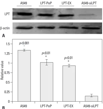
pressing BEAS2B cells (LPT-EX), the expression levels of leptin were also high. For the siRNA interfered control group (A549-siLPT), vector pcDNA3.1 carrying siRNA was transfected into the cells, and no leptin bands were de- tected (Fig. 1). Furthermore, intrinsic expressed leptin in the A549 cells was higher than that expressed in the LPT- PeP and LPT-EX groups (p<0.05).
Leptin expression promotes the proliferation of human lung cell lines
To observe the influence of leptin expression on the prolif- eration ability in abnormal and normal lung cell lines, the proliferation activities were measured with XTT analysis 24 hours post transfection (or incubation). XTT analysis results revealed no difference in the proliferation viabilities among A549, LPT-PeP and LPT-EX groups, both treated with and without cisplatin. However, in the inhibited leptin expression preparations of the A549-siLPT group, when treated with cisplatin, proliferation viabilities decreased significantly (p<0.001) 24 hours after transfection (Fig.
Tech (Pharmacia, St. Paul, MN, USA). The immunoblotting bands were scanned with Typhoon (Pharmacia, St. Paul, MN, USA), digitalized, and saved in TIF format. The gray values of each bands were evaluated detailed. All data were calculated as a mean±SD. Statistical analysis was acted uti- lizing the t-test. All p-values that less than 0.05 were con- sidered as significant differences.
RESULTS
Leptin expression in cells and interfered leptin in A549- siLPT cells
The levels of leptin in A549 tumor cells, siRNA interfered A549-siLPT cells and leptin-expressing BEAS2B cells were detected to explore the pathogenicity of leptin in NSCLC. In the preparations of A549 cells, high levels of leptin were intrinsically expressed 24 h after culturing. The peptide LPT-PeP also activated high levels of leptin in BEAS2B cells 24 hours post incubation. In the leptin-ex-
0 0.25 0.5 0.75 1 1.25 1.5
Relative value
A549 LPT-PeP LPT-EX A549-siLPT
p<0.001
†
p<0.01
* p<0.01
* β-actin
LPT
A549 LPT-PeP LPT-EX A549-siLPT
Fig. 1. Detection of intrinsic leptin in A549, LPT-PeP, LPT-EX, and A549-siLPT cells with western blot assay. (A) Intrinsic or expressed leptin detected with leptin-specific monoclonal antibody. Various leptin proteins are indi- cated above the western blot bands. Protein molecular weights are shown on the right. (B) Statistical analysis. In the figure, LPT-PeP represents the leptin in peptide incubated BEAS2B cells, LPT-EX represents the expressed leptin in BEAS2B cells, and A549-siLPT represents the leptin in the inter- fered A549 cells. The relative value of each preparation is calculated by each gray numerical value of the specific product vs. that of β-actin. The average data of each preparation are evaluated based on three indepen- dent reactions and represented as mean±SD. Statistical differences of the data of cisplatin treatment compared with that of non-treatment are illus- trated as *p<0.01, †p<0.001, respectively. siLPT, siRNA against leptin.
Fig. 2. Cell proliferation effects of leptin in the four groups. (A) A549, LPT- PeP, LPT-EX and A549-siLPT cells were incubated with cisplatin, respec- tively. The cell proliferations were measured by the XTT method. (B) TUNEL analysis of leptin expressing cells treated with or without cisplatin. The ar- rows show TUNEL positive cells. The average data of each preparation are evaluated based on three independent reactions and represented as mean±SD. Statistical differences of the data of cisplatin treatment com- pared with that of non-treatment are illustrated as *p<0.001. siLPT, siRNA against leptin.
A
B
A549 LPT-PeP LPT-EX A549-siLPT
Without cisplatin
Withcisplatin 0 20 40 60 80 100 120
Cell proliferation (%)
A549 LPT-PeP LPT-EX A549-siLPT
A
B
p<0.001 * Without cisplatin With cisplatin
cells was significantly increased compared with cells not treated with cisplatin (p<0.001) (Fig. 3). Accordingly, we discerned cell apoptosis might be blocked by the expression of leptin.
Two ER stress UPR pathways are involved in the inhibition of apoptosis by leptin
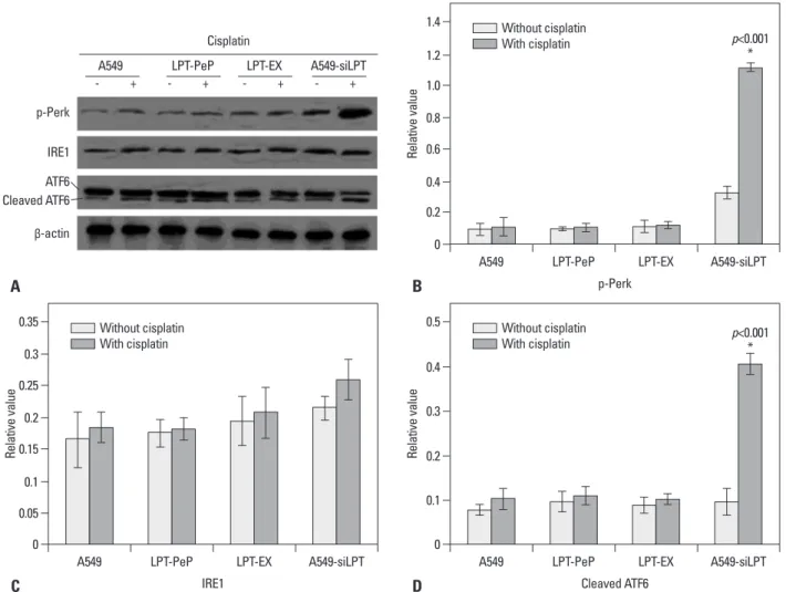
In order to evaluate changes in UPR pathway factors, three main UPR factors, p-Perk, IRE1 and ATF6, were detected by western blot assay. As shown in Fig. 3, the expression of leptin blocked the phosphorylation of Perk and cleaving of ATF6 protein. Therefore, the amounts of p-Perk and cleaved ATF6 were not triggered by the treatment of cisplatin in the A549, PLT-PeP and PLT-EX groups (Fig. 4). However, in the A549-siPLT group, for the blocking of leptin expres- sion, the phosphorylation of Perk and cleaving of ATF6 were significantly increased upon treatment with cisplatin (p<0.05) (Fig. 4).
2A). Cell death and apoptosis were also detected by the TUNEL method, and we found that cisplatin did not affect cells death and apoptosis of A549, LPT-PeP and LPT-EX cells, which expressed leptin protein (Fig. 2B). The results also indicated that cisplatin could induce cell death and apoptosis in A549-siLPT cells, lacking leptin expression.
Accordingly, we determined that leptin expression in BEAS2B cells saved dead cells and promoted cell prolifer- ation.
Leptin inhibits the early and late apoptosis of lung cells In order to investigate the mechanism of leptin-induced cell proliferation, we employed flow cytometry analysis to de- tect early and late apoptosis in all cell groups. For the A549, LPT-PeP, LPT-EX groups, there were no significant differ- ences in early or late apoptosis between cells treated with cisplatin and those that were not (p>0.05). However, when treated with cisplatin, early or late apoptosis in A549-siLPT
A
B
Without cisplatin
With cisplatin
PI
Annexin-V
Fig. 3. Early and late apoptosis phenomena were observed in cisplatin treatment and non-treatment groups. (A) Annexin V/PI double staining assays of cells incubated with or without cisplatin. (B) Statistical analysis. Y axis indicates the numbers of PI stained cells. X axis indicates the numbers of Annexin V-FITC strained cells. Results for three independent experiments are shown. The mean data of each preparation were the results of three independent western blots scan and calculation, and indicated as mean±SD. Statistical differences of the data of cisplatin treatment compared with that of non-treatment are il- lustrated as *p<0.01, †p<0.001, respectively. PI, propidium iodide; siLPT, siRNA against leptin.
p<0.001
†
p<0.01
*
0 0 0 0
8 8 8
20
2 2 2 5
10 10 10
25
4 4 4 10
12
12 12
4540 3530
6 6 6
15 14
14 16
14
16 50
Apoptosis (%) Apoptosis (%) Apoptosis (%) Apoptosis (%)
Early
apoptosis Early
apoptosis Early
apoptosis Early
apoptosis
apoptosisLate Late
apoptosis Late
apoptosis Late
apoptosis Without cisplatin With cisplatin
A549 LPT-PeP LPT-EX A549-siLPT
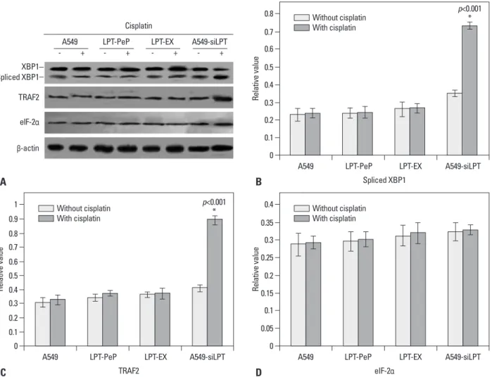
tected. The results were similar to those above for mRNA transcription. In the A549-siLPT group, the levels of spliced XBP1 and TRAF2 proteins increased significantly when treated with cisplatin (p<0.05) (Fig. 6). However, cisplatin treatment did not affect the protein levels of XBP1 and TRAF2 in the A549, LPT-PeP and LPT-EX groups.
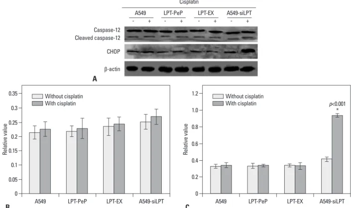
Leptin expression blocked CHOP triggered apoptosis To detect potential changes in the ER stress induced apop- tosis related markers after expression or incubation with leptin, the cellular levels of cleaved caspase 12 and CHOP protein were evaluated by individual western blot assay (Fig.
7A). Absent cisplatin treatment, there were no changes in caspase 12 and CHOP for all groups (Fig. 7B and C). Addi- tionally, we observed no significant differences in cleaved caspase 12 between the cisplatin treated cells compared with non-treated cells for all three groups (Fig. 7B). When UPR downstream mRNA and proteins were highly
inhibited in leptin expressed cells
In order to further investigate the pathways of apoptosis, the mRNA and proteins associated with ER stress (UPR path- way), including spliced XBP1, TRAF2, and eIF-2α, were an- alyzed with hemi-quantitative RT-PCR 12 h after transfec- tion. As shown in Fig. 5, when treated with cisplatin, the mRNA levels of XBP1 and TRAF2 were increased signifi- cantly in A549-siLPT cells, compared with cells not treated with cisplatin (p<0.05). Additionally, the levels of eIF-2α remained comparable among the preparations for all three groups (Fig. 5). Interestingly, there were no significant dif- ferences in spliced XBP1 and TRAF2 between cells treated with cisplatin and those that were not, among the A549, LPT-PeP and LPT-EX groups (Fig. 5).
Meanwhile, downstream ER stress associated proteins (spliced XBP1, TRAF2, and eIF-2α proteins) were also de-
0
0 0
0.2
0.05 0.1
0.4 0.6
0.2 0.1
0.15
0.8
0.2
1.0
0.3 0.25
1.2
0.3 0.4
1.4
0.5 0.35
Relative valueRelative value
Relative value
A549
A549 A549
p-Perk
Cleaved ATF6 IRE1
LPT-PeP
LPT-PeP LPT-PeP
LPT-EX
LPT-EX LPT-EX
A549-siLPT
A549-siLPT A549-siLPT
B A
D C
p<0.001
*
p<0.001
* Without cisplatin
With cisplatin
Without cisplatin With cisplatin Without cisplatin
With cisplatin
Fig. 4. Detection of three UPR pathway proteins. (A) Detection of IRE1, p-Perk and cleaved ATF6. Statistical analysis of p-Perk (B), IRE1 (C) and Cleaved ATF6 (D). The average gray value of each preparation was calculated by the gray numerical value of each blot vs. that of β-actin. The mean data of each prepara- tion were the results of three independent western blots scan and calculation, and indicated as mean±SD. Statistical differences of the data of cisplatin treatment compared with that of non-treatment are illustrated as *p<0.001. p-Perk, phosphorylated-PKR-like ER kinase; ER, endoplasmic reticulum; ATF6, ac- tivating transcription factor-6; UPR, unfolded protein response; IRE1, inositol requiring proteins-1; siLPT, siRNA against leptin.
β-actin p-Perk
IRE1 ATF6 Cleaved ATF6
Cisplatin
- + - + - + - +
A549 LPT-PeP LPT-EX A549-siLPT
cisplatin (Fig. 7C). However, no changes in CHOP were dis- covered between the cisplatin treated cells compared with treated with cisplatin, CHOP levels in A549-siLPT cells
were significantly higher than that of those untreated with
Fig. 5. Hemi-quantification PCR detects the mRNA levels of ER stress (UPR) associated genes. (A) Detection of spliced XBP1 mRNA. (B) Detection of TRAF2 mRNA. (C) Detection of the eIF-2α mRNA. The products were separated in 1.5% agarose gels. The relative values of spliced XBP1, TRAF2 and eIF-2α were evaluated by the calculation of gray scan value of each preparation vs. that of β-actin protein. The mean data of each preparation were the results of three independent western blots scan and calculation, and indicated as mean±SD. Statistical differences of the data of cisplatin treatment compared with that of non-treatment are illustrated as *p<0.001, respectively. siLPT, siRNA against leptin; ER, endoplasmic reticulum; UPR, unfolded protein response; XBP1, X-box transcription factor-1; eIF-2α, eukaryotic translation initiation factor-2α; TRAF2, tumor necrosis factor receptor-associated factor 2.
0
0 0
0.1
0.05 0.1 0.15
0.1 0.2
0.3 0.2 0.4
0.2
0.3 0.4 0.5
0.5
0.6
0.6
0.25 0.7
0.8
0.7
0.35 0.3 0.9
0.8
0.4 1
Relative valueRelative value
Relative value
A549
A549 A549
Spliced XBP1
eIF-2α TRAF2
LPT-PeP
LPT-PeP LPT-PeP
LPT-EX
LPT-EX LPT-EX
A549-siLPT
A549-siLPT A549-siLPT
B A
D C
p<0.001 Without cisplatin *
With cisplatin
Without cisplatin With cisplatin Without cisplatin
With cisplatin
Fig. 6. Detection of ER stress (UPR) associated genes. (A) Detection of spliced XBP1 protein, TRAF2 protein, e-IF-2α protein. (B) Detection of spliced XBP1 protein. (C) Detection of TRAF2 protein. (D) Detection of eIF-2α protein. The relative values of spliced XBP1, TRAF2 and eIF-2α proteins were evaluated by the calculation of gray scan value of each preparation vs. that of β-actin protein. The mean data of each preparation were the results of three independent western blots scan and calculation, and indicated as mean±SD. Statistical differences of the data of cisplatin treatment compared with that of non-treat- ment are illustrated as *p<0.001 respectively. ER, endoplasmic reticulum; UPR, unfolded protein response; XBP1, X-box transcription factor-1; eIF-2α, eukary- otic translation initiation factor-2α; siLPT, siRNA against leptin; TRAF2, tumor necrosis factor receptor-associated factor 2.
β-actin XBP1 Spliced XBP1 TRAF2 elF-2α
Cisplatin
- + - + - + - +
0 0 0
0.1 0.2 0.05
0.2 0.4 0.150.1
0.3 0.6 0.2
0.4 0.25
0.5 0.8 0.3
0.6 1.0 0.35
0.7 1.2 0.4
mRNA relative value mRNA relative value mRNA relative value
A549 LPT-PeP LPT-EXA549-siLPT A549 LPT-PeP LPT-EXA549-siLPT A549 LPT-PeP LPT-EXA549-siLPT
A B C
Without cisplatin
With cisplatin Without cisplatin
With cisplatin
Without cisplatin With cisplatin p<0.001
* p<0.001
*
p>0.05
p<0.001
* A549 LPT-PeP LPT-EX A549-siLPT
apoptosis of transformed cells. Leptin expression patterns in the present study implied that the expression of leptin might inhibit the death of cells.
In the present study, the cell proliferation of A549, LPT- PeP, LPT-EX and A549-siLPT group cells were also detect- ed. We hypothesized that higher proliferation in these three cells was due to the blocking of apoptosis. Accordingly, the cells were treated with cisplatin to induce ER stress, and the resulting cell viability changes were observed. Upon adding cisplatin to the experimental cells, cell viability in the leptin- expressing cells or those incubated in leptin underwent no significant changes between cisplatin treated and untreated cells. Cell viability in the cisplatin treated A549-siLPT cells was obviously lower than that in the untreated A549-siLPT cells. These changes indicated that the expression of leptin inhibits cisplatin mediated apoptosis and cell death.
In regards to ER stress (UPR pathway) associated protein detection, we found that p-Perk and cleaved ATF6 proteins were activated in A549-siLPT cells when treated with cispl- atin. Therefore, the UPR pathway may involve in leptin- blocked apoptosis. Such leptin-induced apoptosis inhibition may help to further illuminate the role of leptin in cancer the non-treated cells for the A549, LPT-PeP and LPT-EX
groups (Fig. 7C). CHOP was shown to be activated in A549- siLPT cells, which triggered apoptosis. Accordingly, we de- termined the expression of leptin blocked cisplatin-induced apoptosis via CHOP pathways in human normal lung cells (such as LPT-PeP and LPT-EX cells).
DISCUSSION
This study is the first to indicate that leptin inhibits cisplatin- triggered apoptosis of human NSCLC cell lines, such as A549. In the present study, normal human lung cell lines, BEAS2B, treated with cisplatin resisted cisplatin-induced apoptotic effects. Additionally, our study indicated that leptin inhibits apoptosis through the PERK and ATF6 pathways, and blocks apoptosis by inactivating CHOP protein.
Our results revealed that all cell groups expressed leptin protein, but these levels in A549 cells and leptin expressing cells were significantly higher than that in A549-siLPT cells (Fig. 1). It has been shown that the malignant transforma- tion of cancer requires cells to keep growing and block the
Fig. 7. Changes of ER stress-associated events in the cells expressing leptin. (A) The levels of CHOP and caspase-12 in A549, A549-siLPT, LPT-PeP, and LPT- EX cells were evaluated by individual western blots. (B) Statistical analysis of Cleaved caspase-12. (C) Statistical analysis of CHOP. The average gray value of each preparation was evaluated by the calculation of gray scan value of each preparation vs. that of β-actin protein. The mean data of each preparation were the results of three independent western blots scan and calculation, and indicated as mean±SD. Statistical differences of the data of cisplatin treat- ment compared with that of non-treatment are illustrated as *p<0.001. ER, endoplasmic reticulum; siLPT, siRNA against leptin.
0 0
0.2 0.4
0.05
0.6
0.1 0.15 0.2 0.25 0.8 0.3 1.0
1.2 0.35
Relative value
Relative value
A549
A549 LPT-PeP LPT-EX A549-siLPT LPT-PeP LPT-EX A549-siLPT
A
B C
Without cisplatin With cisplatin Without cisplatin
With cisplatin
β-actin Caspase-12 Cleaved caspase-12 CHOP
p<0.001
* Cisplatin
- + - + - + - +
A549 LPT-PeP LPT-EX A549-siLPT
5. Moenner M, Pluquet O, Bouchecareilh M, Chevet E. Integrated endoplasmic reticulum stress responses in cancer. Cancer Res 2007;67:10631-4.
6. Feldman DE, Chauhan V, Koong AC. The unfolded protein re- sponse: a novel component of the hypoxic stress response in tu- mors. Mol Cancer Res 2005;3:597-605.
7. Joung KH, Cho SC. Stress responses of neonates related to mater- nal characteristics. Yonsei Med J 2011;52:98-103.
8. Momoi T, Fujita E, Senoo H, Momoi M. Genetic factors and epi- genetic factors for autism: endoplasmic reticulum stress and im- paired synaptic function. Cell Biol Int 2009;34:13-9.
9. Hung JY, Hsu YL, Ni WC, Tsai YM, Yang CJ, Kuo PL, et al. Oxi- dative and endoplasmic reticulum stress signaling are involved in dehydrocostuslactone-mediated apoptosis in human non-small cell lung cancer cells. Lung Cancer 2010;68:355-65.
10. Terzidis A, Sergentanis TN, Antonopoulos G, Syrigos C, Efremid- is A, Polyzos A, et al. Elevated serum leptin levels: a risk factor for non-small-cell lung cancer? Oncology 2009;76:19-25.
11. Caldefie-Chézet F, Damez M, de Latour M, Konska G, Mishellani F, Fusillier C, et al. Leptin: a proliferative factor for breast cancer?
Study on human ductal carcinoma. Biochem Biophys Res Com- mun 2005;334:737-41.
12. Geng Y, Wang J, Wang R, Wang K, Xu Y, Song G, et al. Leptin and HER-2 are associated with gastric cancer progression and prognosis of patients. Biomed Pharmacother 2012;66:419-24.
13. Wang X, Dong CF, Shi Q, Shi S, Wang GR, Lei YJ, et al. Cytosolic prion protein induces apoptosis in human neuronal cell SH-SY5Y via mitochondrial disruption pathway. BMB Rep 2009;42:444-9.
14. Xuan B, Qian Z, Torigoi E, Yu D. Human cytomegalovirus pro- tein pUL38 induces ATF4 expression, inhibits persistent JNK phosphorylation, and suppresses endoplasmic reticulum stress-in- duced cell death. J Virol 2009;83:3463-74.
15. Wang Q, He Z, Zhang J, Wang Y, Wang T, Tong S, et al. Overex- pression of endoplasmic reticulum molecular chaperone GRP94 and GRP78 in human lung cancer tissues and its significance.
Cancer Detect Prev 2005;29:544-51.
16. Chérasse Y, Maurin AC, Chaveroux C, Jousse C, Carraro V, Parry L, et al. The p300/CBP-associated factor (PCAF) is a cofactor of ATF4 for amino acid-regulated transcription of CHOP. Nucleic Acids Res 2007;35:5954-65.
17. Wang X, Shi Q, Xu K, Gao C, Chen C, Li XL, et al. Familial CJD associated PrP mutants within transmembrane region induced Ctm-PrP retention in ER and triggered apoptosis by ER stress in SH-SY5Y cells. PLoS One 2011;6:e14602.
18. Moon DO, Park SY, Choi YH, Ahn JS, Kim GY. Guggulsterone sensitizes hepatoma cells to TRAIL-induced apoptosis through the induction of CHOP-dependent DR5: involvement of ROS-depen- dent ER-stress. Biochem Pharmacol 2011;82:1641-50.
19. Tagawa Y, Hiramatsu N, Kasai A, Hayakawa K, Okamura M, Yao J, et al. Induction of apoptosis by cigarette smoke via ROS-dependent endoplasmic reticulum stress and CCAAT/enhancer-binding protein- homologous protein (CHOP). Free Radic Biol Med 2008;45:50-9.
20. Kim KM, Kim HC, Jeon KN, Kim HG, Kang JH, Hahm JR, et al.
Rituximab-CHOP induced interstitial pneumonitis in patients with disseminated extranodal marginal zone B cell lymphoma. Yonsei Med J 2008;49:155-8.
21. Wang SC, Lu MC, Chen HL, Tseng HI, Ke YY, Wu YC, et al. Cy- totoxicity of calotropin is through caspase activation and down- regulation of anti-apoptotic proteins in K562 cells. Cell Biol Int 2009;33:1230-6.
progression. Furthermore, downstream factors of spliced XBP1, TRAF2, and eIF-2α were also detected at the mRNA and protein levels, and the data strongly indicated the emer- gence of ER stress (or UPR pathway) after treatment with cisplatin in A549-siLPT cells. More importantly, the ex- pression of leptin inhibited cisplatin induced ER stress in LPT-PeP, LPT-EX and A549 cells. Accordingly, we sur- mised that in human NSCLC cell lines leptin may partici- pate in the pathogenic processes of lung cancer.
In order to identify the specific apoptosis factors that leptin inhibited, ER stress-associated factors (cleaved caspase 12 and CHOP protein) were also detected in the A549, A549- siLPT and leptin expressing (incubating) cells. According to Wang, et al.17 study, cleaved caspase 12 activates caspase 3 to trigger apoptosis, and CHOP can directly induce ER stress-associated apoptosis. In this study, we found that there were no significant changes in cleaved caspase 12 (activat- ed) among all three cell groups (p>0.05) when treated with cisplatin. Interestingly, upon cisplatin treatment, CHOP lev- els in the A549-siLPT group were significantly increased, compared with untreated cells (p<0.05); however, there were no changes in the A549, LPT-PeP and LPT-EX cells. As our results were consistent with other research teams,18,19 we concluded that the expression or the existence of leptin in lung cells may indirectly inhibit CHOP protein mediated apoptosis.
Actually, as the induction of the transcription factor CHOP/
GADD153 can kill cells by an apoptotic mechanism,20,21 we discovered a novel result that leptin may block CHOP in- duced ER stress.
In conclusion, leptin may serve as an important factor to promote the growth of A549 cells via blocking of ER stress- mediated pathways. This blocking may be triggered by p- Perk and ATF6 pathways through inhibition of CHOP ex- pression.
REFERENCES
1. Molina JR, Yang P, Cassivi SD, Schild SE, Adjei AA. Non-small cell lung cancer: epidemiology, risk factors, treatment, and survi- vorship. Mayo Clin Proc 2008;83:584-94.
2. Petersen I, Petersen S. Towards a genetic-based classification of human lung cancer. Anal Cell Pathol 2001;22:111-21.
3. Al Husaini H, Wheatley-Price P, Clemons M, Shepherd FA. Pre- vention and management of bone metastases in lung cancer: a re- view. J Thorac Oncol 2009;4:251-9.
4. Yung HW, Korolchuk S, Tolkovsky AM, Charnock-Jones DS, Bur- ton GJ. Endoplasmic reticulum stress exacerbates ischemia-reperfu- sion-induced apoptosis through attenuation of Akt protein synthesis in human choriocarcinoma cells. FASEB J 2007;21:872-84.