A comparison of metabolomic changes in type-1 diabetic C57BL/6N mice originating from different sources
7
0
0
전체 글
(2) Characterization of C57BL/6NKorl in streptozotocin-induced diabetic model. insufficient function insulin [4-6]. Currently, it is considered that insulin supplementation is an essential therapy in type-1 diabetes [7]. One important challenge facing patients with diabetes is that lasting hyperglycemia increases the risk of kidney disease, heart disease, retinal injury, nerve damage, and vascular damage [8]. Therefore, it is necessary to establish suitable animal models for research investigating novel prevention and treatment approaches for diabetes mellitus. Streptozotocin (2-deoxy-2-(3-methyl-3-nitrosourea)1-D-glucopyranose), a naturally occurring compound produced by soil bacterial streptomyces achromogenes, is a highly selective cytotoxic reagent in the pancreatic β cells. It induces an inflammatory response of the insulin-producing β cells followed by insulitis and absolute insulin deficiency [9]. Based on these mechanisms, streptozotocin is widely used to induce experimental type-1 diabetic animal models [10-12]. Initially, it was administered at a high single dosage to induce complete death of pancreatic β cells within 2 days [13]. However, Like and Rossini noted that multiple challenges with low dose streptozotocin results in the slowly progressive type-1 diabetes and more closely resembles the clinical pathogenesis [14-16]; this approach is now widely used to establish type-1 diabetes in animals. The National Institute of Food and Drug Safety Evaluation (NIFDS, Cheongju, Korea) has established its own animal strains to evaluate both efficacy and safety during new drug development. The objective of this study was to characterize the response of the C57BL/ 6N mice from the NIFDS treated with streptozotocin compared with that of other mice originating from the USA and Japan. In this study, multiple low dose treatments with streptozotocin were used to generate type-1 diabetic animal models which closely relate to clinical pathology. Thus, a study comparing wellestablished animal disease model using streptozotocin can provide insight into the validity and usefulness of NIFDS-derived mice in preclinical testing of antidiabetic substances and elucidating the mechanism of the progression of type-1 diabetes in humans.. Materials and Methods Animals and treatment. 233. different sources. C57BL/6NKorl were kindly provided by the Department of Laboratory Animal Resources in the National Institute of Food and Drug Safety Evaluation (NIFDS, Cheongju, Korea). The other two groups of C57BL/6N strain were purchased from different vendors located in the USA (referred C57BL/ 6NA) and Japan (referred C57BL/6NB). Animal protocol of this study was approved by the Institutional Animal Care and Use Committees at Pusan National University (PNU-2017-1632). Mice were acclimated to temperature (22±2) and humidity (55±5%)-controlled rooms with a 12 h light/dark cycle for seven days prior to use. They were randomly divided into two groups and the streptozotocin-treated group was given daily intraperitoneal injection with 50 mg/kg body weight of streptozotocin (Sigma, St. Louis, MO, USA) freshly dissolved in 0.1 M citrate buffer (pH 4.5) for 5 days. Body weight, water and food consumption were measured at least once a week after last dosing of streptozotocin. Liver and kidney tissues were harvested 4 weeks after the final administration. Serum biochemical measurements. Serum sample was obtained from blood of each mouse using BD Microtainer Serum Collection Tube (BD Life Sciences, Franklin Lakes, NJ, USA). The sera were stored at −80oC until the biochemical analyses. Serum glucose level was determined using commercial Glucose CII-test Wako (Wako Pure Chemical Industries, Ltd., Tokyo, Japan). Activities of alanine aminotransferase (ALT) and aspartate aminotransferase (AST) were measured using the protocol of Reitman and Frankel [17]. They were quantified spectrophotometrically using an MULTISKAN GO reader (Thermo Scientific, Waltham, MA, USA). The serum levels of total cholesterol, HDLcholesterol, and LDL-cholesterol were determined by using an Automated Chemistry Analyzer (Prestige 24I, Tokyo Boeki Medical System, Tokyo, Japan). Metabolites analysis in serum. Metabolites in deproteinized serum were derivatized with O-phthalaldehyde/2-mercaptoethanol and quantified by HPLC with fluorescence detection (FLD-3100: Thermo Scientific, Waltham, MA, USA) on a HECTOR-T C18 column (3 μm×4.6 mm×100 mm; Chiral Technology Korea, Daejeon, Korea) at wavelengths of excitation 338 nm and emission 425 nm.. Male C57BL/6N mice were obtained from three Lab Anim Res | December, 2018 | Vol. 34, No. 4.
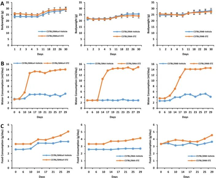
(3) 234. Seunghyun Lee et al.. Statistical analysis. All results expressed as mean±standard deviation (SD) and analyzed by one-way analysis of variance (ANOVA) followed by Newman-Keuls multiple range test (parametric). The acceptable level of significance was established at P<0.05. Results Changes in body weight, water and food consumption after final administration of streptozotocin. In mice treated with streptozotocin, the changes of body weight showed no significant difference compared with those of respective vehicle-treated mice during experimental period (Figure 1A). Significant increase in water intake were observed from 10 days after last dose. of streptozotocin in C57BL/6NKorl and C57BL/6NB mice, and from 14 days in C57BL/6NA mice (Figure 1B). Food consumption of mice were slightly increased in streptozotocin-treated mice compared with those of respective vehicle-treated mice (Figure 1C). Changes in serum glucose concentration and relative organ weight in streptozotocin-induced diabetic mice. Mice with greater than 300 mg/dL of blood glucose levels were considered diabetic in streptozotocin-induced model [18]. As shown in Figure 2A, serum glucose concentration of streptozotocin-treated mice was more than 300 mg/dL and it was successfully established in mice of all origins. The liver-to-body weight ratios (Figure 2B) were significantly increased 29, 42, and 21% in streptozotocin-treated C57BL/6NKorl, C57BL/. Figure 1. Changes in (A) body weight, (B) water and (C) food consumption after final administration of streptozotocin. Mice were given daily intraperitoneal injection with vehicle or 50 mg/kg body weight of streptozotocin (STZ) for 5 days. Changes were measured at least once a week after last dosing of streptozotocin during 4 weeks. Lab Anim Res | December, 2018 | Vol. 34, No. 4.
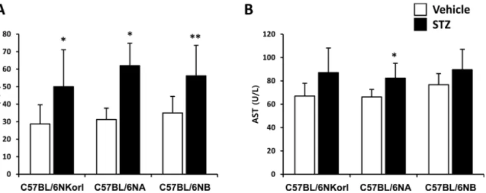
(4) Characterization of C57BL/6NKorl in streptozotocin-induced diabetic model. 235. Figure 2. Changes of (A) serum glucose level, and relative ratio of (B) liver-to-body and (C) kidney-to-body weight in streptozotocin-treated mice. Mice were given daily intraperitoneal injection with vehicle or 50 mg/kg body weight of streptozotocin (STZ) for 5 days. Changes were examined 4 weeks after final administration. ***Significantly different from the corresponding vehicle-treated mice (ANOVA followed by Newman-Keuls multiple range test, P<0.001, respectively).. Figure 3. Effect of streptozotocin on the activities of (A) ALT and (B) AST. Mice were given daily intraperitoneal injection with vehicle or 50 mg/kg body weight of streptozotocin (STZ) for 5 days. Activities of ALT and AST were examined 4 weeks after final administration. * **Significantly different from the corresponding vehicle-treated mice (ANOVA followed by Newman-Keuls multiple range test, P<0.05, 0.01, respectively). ,. 6NA, C57BL/6NB, respectively. The weight of the kidney (Figure 2C) was not changed in the streptozotocintreated mice. Effect of streptozotocin on the activities of ALT and AST Serum activity of ALT (Figure 3A) was significantly increased by the treatment of streptozotocin (C57BL/ 6NKorl, 1.74 fold; C57BL/6NA, 1.98 fold; C57BL/ 6NB, 1.60 fold). AST activity in serum showed slight increase with significance only in C57BL/6NA mice treated with streptozotocin (Figure 3B). There was no difference among the source of mice in the activities of both ALT and AST, suggesting that streptozotocin treatment affects liver function of diabetic mice. Changes in serum total cholesterol and HDL-, LDLcholesterol in streptozotocin-induced diabetic mice. There was no significant change in serum total cholesterol (Figure 4A) and HDL-cholesterol (Figure. 4B) by streptozotocin administration. LDL-cholesterol was slightly increased with significance only in C57BL/ 6NB mice (vehicle, 7.92±0.60 mg/dL; streptozotocin, 9.59±0.64 mg/dL) (Figure 4C). Changes in serum metabolites in streptozotocininduced diabetic mice. As shown Figure 5, many kinds of metabolites in serum were changed by streptozotocin treatment. The concentration of aspartate, citrulline, methionine, serine, threonine, glutamate, taurine, lysine, glycine, and tryptophan were decreased in streptozotocin-induced diabetic mice. Whereas, phenylalanine, ornithine, leucine, valine, and isoleucine were increased in streptozotocininduced diabetic mice. There was no changes in the serum levels of tyrosine, histidine, alanine, asparagine, and arginine streptozotocin treatment. The changed pattern of serum metabolites was no significant difference among the source of mice.. Lab Anim Res | December, 2018 | Vol. 34, No. 4.
(5) 236. Seunghyun Lee et al.. Figure 4. Changes in (A) total cholesterol, (B) HDL-cholesterol, and (C) LDL-cholesterol in streptozotocin-treated mice. Mice were given daily intraperitoneal injection with vehicle or 50 mg/kg body weight of streptozotocin (STZ) for 5 days. Changes were examined 4 weeks after final administration. *Significantly different from the corresponding vehicle-treated mice (ANOVA followed by Newman-Keuls multiple range test, P<0.05, respectively).. Figure 5. Heat map of changes in serum metabolites between vehicle- and streptozotocin (STZ)-treated mice. Mice were given daily intraperitoneal injection with vehicle or 50 mg/kg body weight of streptozotocin (STZ) for 5 days. Serum metabolites were determined 4 weeks after final administration. Each column represents one mouse sample and each row represents a different metabolite.. Discussion A number of animal models have been used to elucidate the pathophysiology of varying diseases and to provide insight into potential targets and approaches for therapeutic intervention. Although various alternatives to animal testing have been proposed to help overcome Lab Anim Res | December, 2018 | Vol. 34, No. 4. potential drawbacks related to animal experiments and avoid ethical issues, they are still considered critical to the drug development process for treating human diseases. In particular, the study of metabolic diseases (eg, diabetes and obesity), requires the use of animal models to monitor whole-body physiology [19]. One of the most widely used laboratory mouse strains in.
(6) Characterization of C57BL/6NKorl in streptozotocin-induced diabetic model. metabolic research is the inbred C57BL/6 mouse which is commonly referred to as “Black 6”, “B6”, or “C57 Black” [19-21]. Inbred stains including the C57BL6 mouse are commonly used for biomedical and behavioral experiments, and data resulting from these studies have contributed substantially to the field’s understanding of complicated biological processes [22-24]. In accordance with this, NIFDS has established its own inbred line of C57BL6/N named “C57BL6/NKorl” in the Guidelines for Nomenclature of Mouse Strains and been providing the characteristic information for animal researchers [25]. A diabetogenic effect of streptozotocin was first reported by Junod et al. who found that its effect resulted from selective destruction of pancreatic islet β-cells [26,27]. Several animal species are sensitive to the cytotoxic effect of streptozotocin and it is currently most often used to induce diabetes in rats and mice [28]. Specifically, multiple, lose-dose treatments of streptozotocin is increasingly being used to produce type-1 diabetic animal models which closely mimic symptoms seen in humans. In this study, a type-1 diabetic model of mice was induced by chronic injection of low dose streptozotocin and the response of C57BL/6NKorl mice was characterized by changes in basic physiology and serum metabolites. Furthermore, it was compared with the response of other mice obtained from two different commercially available sources. No significantly different responses between the streptozotocin-induced type-1 diabetic mouse models used in this study were observed. When comparing control and diabetic mice, increases in liver weight and disturbances in serum amino acids levels of diabetic mice were most remarkable. It has been suggested that increased triglyceride accumulation leads to an increase in the weight of livers from diabetic mouse model organisms because of the increased import of fatty acids into the liver induced by hypoinsulinemia and the low excretion of lipoprotein from the liver due to deficiency in the synthesis of apolipoprotein B [29-32]. In agreement with these reports, an important physiological change in this study is a significant increase in liver weight of streptozotocininduced diabetic mice. This change is accompanied by increased activities of both ALT and AST suggesting that type-1 diabetes may induce abnormal liver functions. Insulin leads to a decrease in serum amino acid levels through the stimulus of protein synthesis and the. 237. inhibition of proteolysis, which would explain the negative correlation between insulin dosage and plasma level of the amino acids in patients with diabetes [33]. It has been experimentally proven that insulin deficiency causes functional changes in the liver and muscle, which subsequently inhibits protein synthesis and promotes protein degradation. Interestingly, evidence is accumulating that there is a positive association between type-2 diabetes and circulating concentrations of BCAAs (eg, valine, leucine, isoleucine) [34-36]. Of note, the serum levels of many amino acids in the type-1 diabetic mice used in this study were decreased, but the serum concentration of BCAA was increased, which is similar to that observed in type-2 diabetes. Although the relationship between type-1 diabetes and BCAA has not been elucidated in this study, the results revealing a characteristic increase in diabetic mice of all origin are considered to be worthy of further study. In conclusion, the responses of C57BL/6NKorl mice following streptozotocin treatment (eg, physiological, biochemical, metabolic) are not significantly different among mice of different orgins. Thus, our results suggest the usefulness of C57BL/6NKorl mice in preclinical testing of antidiabetic substances and those aiming to better elucidate the mechanisms of type-1 diabetes progression in humans.. Acknowledgments This project was supported by a grant of NLAR (National Laboratory Animal Resources) from Ministry of Food and Drug Safety in 2017. Conflict of interests The authors declare that there is no financial conflict of interests to publish these results.. References 1. Maritim AC, Sanders RA, Watkins JB 3rd. Diabetes, oxidative stress, and antioxidants: a review. J Biochem Mol Toxicol 2003; 17(1): 24-38. 2. American Diabetes Association. Diagnosis and Classification of Diabetes Mellitus. Diabetes Care 2014; 37 Suppl 1: S81-90. 3. Chao KC, Chao KF, Fu YS, Liu SH. Islet-like clusters derived from mesenchymal stem cells in Wharton's Jelly of the human umbilical cord for transplantation to control type 1 diabetes. PLoS One 2008; 3(1): e1451. 4. Diabetes Prevention Trial--Type 1 Diabetes Study Group. Effects of insulin in relatives of patients with type 1 diabetes mellitus. N Engl J Med 2002; 346(22): 1685-1691. 5. van Belle TL, Coppieters KT, von Herrath MG. Type 1 diabetes: etiology, immunology, and therapeutic strategies. Physiol Rev Lab Anim Res | December, 2018 | Vol. 34, No. 4.
(7) 238. Seunghyun Lee et al.. 2011; 91(1): 79-118. 6. Knip M, Veijola R, Virtanen SM, Hyöty H, Vaarala O, Akerblom HK. Environmental triggers and determinants of type 1 diabetes. Diabetes 2005; 54 Suppl 2: S125-136. 7. Skyler JS, Cefalu WT, Kourides IA, Landschulz WH, Balagtas CC, Cheng SL, Gelfand RA. Efficacy of inhaled human insulin in type 1 diabetes mellitus: a randomised proof-of-concept study. Lancet 2001; 357(9253): 331-335. 8. Polat K, Güneş S. An expert system approach based on principal component analysis and adaptive neuro-fuzzy inference system to diagnosis of diabetes disease. Digital Signal Processing 2007; 17(4): 702-710. 9. Oelze M, Knorr M, Schuhmacher S, Heeren T, Otto C, Schulz E, Reifenberg K, Wenzel P, Münzel T, Daiber A. Vascular dysfunction in streptozotocin-induced experimental diabetes strictly depends on insulin deficiency. J Vasc Res 2011; 48(4): 275-284. 10. Lenzen S. The mechanisms of alloxan- and streptozotocininduced diabetes. Diabetologia 2008; 51(2): 216-226. 11. Szkudelski T. The mechanism of alloxan and streptozotocin action in B cells of the rat pancreas. Physiol Res 2001; 50(6): 537-546. 12. Eleazu CO, Eleazu KC, Chukwuma S, Essien UN. Review of the mechanism of cell death resulting from streptozotocin challenge in experimental animals, its practical use and potential risk to humans. J Diabetes Metab Disord 2013; 12(1): 60. 13. Kolb H. Mouse models of insulin dependent diabetes: low-dose streptozocin-induced diabetes and nonobese diabetic (NOD) mice. Diabetes Metab Rev 1987; 3(3): 751-778. 14. Like AA, Rossini AA. Streptozotocin-induced pancreatic insulitis: new model of diabetes mellitus. Science 1976; 193(4251): 415417. 15. Wu KK, Huan Y. Diabetic atherosclerosis mouse models. Atherosclerosis 2007; 191(2): 241-249. 16. Weide LG, Lacy PE. Low-dose streptozocin-induced autoimmune diabetes in islet transplantation model. Diabetes 1991; 40(9): 1157-1162. 17. REITMAN S, FRANKEL S. A colorimetric method for the determination of serum glutamic oxalacetic and glutamic pyruvic transaminases. Am J Clin Pathol 1957; 28(1): 56-63. 18. Motyl K, McCabe LR. Streptozotocin, type I diabetes severity and bone. Biol Proced Online 2009; 11: 296-315. 19. Fontaine DA, Davis DB. Attention to Background Strain Is Essential for Metabolic Research: C57BL/6 and the International Knockout Mouse Consortium. Diabetes 2016; 65(1): 25-33. 20. Collins S, Martin TL, Surwit RS, Robidoux J. Genetic vulnerability to diet-induced obesity in the C57BL/6J mouse: physiological and molecular characteristics. Physiol Behav 2004; 81(2): 243-248. 21. Winzell MS, Ahrén B. The high-fat diet-fed mouse: a model for studying mechanisms and treatment of impaired glucose tolerance and type 2 diabetes. Diabetes 2004; 53 Suppl 3: S215-219. 22. Mashimo T, Serikawa T. Rat resources in biomedical research. Curr Pharm Biotechnol 2009; 10(2): 214-220. 23. Croy BA. The 1999 Reginald Thomson Lecture. Custom-built mice: unique discovery tools in biomedical research. Can Vet J. Lab Anim Res | December, 2018 | Vol. 34, No. 4. 2000; 41(3): 201-206. 24. Ardaillou R. [Transgenic mice: a major advance in biomedical research]. Bull Acad Natl Med 2009; 193(8): 1773-1782. 25. Sundberg JP, Schofield PN. Commentary: mouse genetic nomenclature. Standardization of strain, gene, and protein symbols. Vet Pathol 2010; 47(6): 1100-1104. 26. Junod A, Lambert AE, Stauffacher W, Renold AE. Diabetogenic action of streptozotocin: relationship of dose to metabolic response. J Clin Invest 1969; 48(11): 2129-2139. 27. Junod A, Lambert AE, Orci L, Pictet R, Gonet AE, Renold AE. Studies of the diabetogenic action of streptozotocin. Proc Soc Exp Biol Med 1967; 126(1): 201-205. 28. Furman BL. Streptozotocin-Induced Diabetic Models in Mice and Rats. Curr Protoc Pharmacol 2015; 70: 5.47.1-20. 29. Habibuddin M, Daghriri HA, Humaira T, Al Qahtani MS, Hefzi AA. Antidiabetic effect of alcoholic extract of Caralluma sinaica L. on streptozotocin-induced diabetic rabbits. J Ethnopharmacol 2008; 117(2): 215-220. 30. Lee SI, Kim JS, Oh SH, Park KY, Lee HG, Kim SD. Antihyperglycemic effect of Fomitopsis pinicola extracts in streptozotocin-induced diabetic rats. J Med Food 2008; 11(3): 518-524. 31. Merzouk H, Madani S, Chabane Sari D, Prost J, Bouchenak M, Belleville J. Time course of changes in serum glucose, insulin, lipids and tissue lipase activities in macrosomic offspring of rats with streptozotocin-induced diabetes. Clin Sci (Lond) 2000; 98(1): 21-30. 32. Ohno T, Horio F, Tanaka S, Terada M, Namikawa T, Kitoh J. Fatty liver and hyperlipidemia in IDDM (insulin-dependent diabetes mellitus) of streptozotocin-treated shrews. Life Sci 2000; 66(2): 125-131. 33. Grill V, Björkman O, Gutniak M, Lindqvist M. Brain uptake and release of amino acids in nondiabetic and insulin-dependent diabetic subjects: important role of glutamine release for nitrogen balance. Metabolism 1992; 41(1): 28-32. 34. Newgard CB, An J, Bain JR, Muehlbauer MJ, Stevens RD, Lien LF, Haqq AM, Shah SH, Arlotto M, Slentz CA, Rochon J, Gallup D, Ilkayeva O, Wenner BR, Yancy WS Jr, Eisenson H, Musante G, Surwit RS, Millington DS, Butler MD, Svetkey LP. A branchedchain amino acid-related metabolic signature that differentiates obese and lean humans and contributes to insulin resistance. Cell Metab 2009; 9(4): 311-326. 35. Shah SH, Crosslin DR, Haynes CS, Nelson S, Turer CB, Stevens RD, Muehlbauer MJ, Wenner BR, Bain JR, Laferrère B, Gorroochurn P, Teixeira J, Brantley PJ, Stevens VJ, Hollis JF, Appel LJ, Lien LF, Batch B, Newgard CB, Svetkey LP. Branchedchain amino acid levels are associated with improvement in insulin resistance with weight loss. Diabetologia 2012; 55(2): 321330. 36. McCormack SE, Shaham O, McCarthy MA, Deik AA, Wang TJ, Gerszten RE, Clish CB, Mootha VK, Grinspoon SK, Fleischman A. Circulating branched-chain amino acid concentrations are associated with obesity and future insulin resistance in children and adolescents. Pediatr Obes 2013; 8(1): 52-61..
(8)
수치

관련 문서
