311
Focal Cerebral Ischemia Induces Decrease of Astrocytic Phosphoprotein PEA-15 in Brain Tissue and HT22 Cells
Phil-Ok Koh*
Department of Anatomy, College of Veterinary Medicine and Research Institute of Life Sciences, Gyeongsang National University, Jinju, Korea
PEA-15 is a small phosphoprotein (15 kDa) that is enriched in brain astrocytes. PEA-15 acts as an important modulator of cellular function including apoptosis and signal integration. This study investigated the expression of PEA-15 in focal cerebral ischemic injury. Cerebral ischemia was surgically induced in adult male rats by middle cerebral artery occlusion (MCAO), and brains were collected 24 hr after MCAO. A proteomic approach demonstrated decreases of PEA-15 protein spots in MCAO-operated animals in comparison to sham-operated animals. Western blot analysis clearly demonstrated that MCAO induces decreases in PEA-15 levels. We previously showed that glutamate toxicity induces cell death in a hippocampus-derived cell line (HT22). Glutamate exposure induces decreases of PEA-15 levels in HT22 cells. The results of this study suggest that focal cerebral ischemia induces cell death through down- regulation of PEA-15 protein.
Key words: HT22 cells, middle cerebral artery occlusion, PEA-15
Received 13 July 2010; Revised version received 31 July 2010; Accepted 7 September 2010
Stroke is a serious neurodegenerative disorder and is a major cause of morbidity and mortality. Middle cerebral artery occlusion (MCAO) is an important experimental model of stroke (Longa et al., 1989; Ferrer and Planas, 2003; Koh et al., 2010). The focal cerebral ischemia that is caused by MCAO results in the development of infarct lesions and leads to extensive neuronal damage in the cerebral cortex (Ferrer and Planas, 2003). We confirmed in a previous study that MCAO induces serious damage to the cerebral tissue (Koh, 2010). Cerebral ischemic injury induces down- and up- regulation of specific proteins that mediate neuronal cell death (Koh, 2010).
PEA-15 is a small acidic phosphoprotein (15 kDa) that is abundantly expressed in astrocytes (Araujo et al., 1993;
Danziger et al., 1995). PEA-15 is a multifunctional protein that is involved in the regulation of the cell cycle and apoptosis (Danziger et al., 1995; Renault et al., 2003; Krueger et al., 2005). Cerebral ischemia mediates apoptotic signaling and
survival signaling pathways and results in neuronal cell death (Ferrer and Planas, 2003). We previously reported that focal cerebral ischemia induces neuronal cell death by altering the expression levels of several proteins (Koh, 2010). However, the process of neuronal cell death in cerebral ischemia complex is poorly known. At present, little information is available regarding changes of protein expression that occur in damaged neurons. In this study, we detected decreases in PEA-15 proteins using a proteomic approach in an animal model of focal cerebral ischemia. We investigated the expressions of PEA-15 proteins in cerebral ischemic brain injury and hippocampal neuron culture conditions.
All animal experiments were performed according to a protocol approved by the Committee for Animal Experimentation at the Gyeongsang National University. Male Sprague-Dawley rats (225-250 g, n=20) were purchased from Samtako Co.
(Osan, Korea) and were randomly divided into two groups:
sham-operated group and MCAO-operated group (n=10 per group). Animals were maintained under controlled temperature (25
oC) and lighting (14L:10D), and allowed free access to commercial food and purified water.
MCAO was performed to induce focal cerebral ischemic injury (Longa et al., 1989). Animals were anesthetized with sodium pentobarbital (100 mg/kg). A piece of 4/0 mono-
*Corresponding author: Phil Ok Koh, Department of Anatomy, College of Veterinary Medicine, Gyeongsang National University, 900 Gajwa-dong, Jinju, Gyeongnam 660-701, Korea
Tel: +82-55-751-5809 Fax: +82-55-751-5803 E-mail: [email protected]
Lab. Anim. Res. 2010: 26(3), 311-314
Letter
312 Phil-Ok Koh
Lab. Anim. Res. | September, 2010 | Vol. 26, No. 3
filament nylon suture with its tip slightly rounded by gentle heating was inserted through the right internal carotid artery to the base of the right middle cerebral artery. Animals were sacrified 24 hr after MCAO for the investigation in late stage of apoptosis. The right cerebral cortex was removed. The tissues were homogenized in lysis buffer (8 M urea, 4% CHAPS, 0.5% ampholytes, and 40 mM Tris-HCl) and centrifuged at 16,000 g for 20 min at 4
oC. The total protein concentration was determined using the Bradford method (Bio-Rad, Hercules, CA, USA) according to the manufacturer’s protocol.
The two-dimensional (2D) gel electrophoresis was performed by the following steps. The IPG strips (IPG, pH 4-7, 17 cm, Bio-Rad) were re-hydrated in rehydration buffer at room temperature (8 M urea, 2% CHAPS, 20 mM DTT, 0.5% IPG buffer, a trace of bromophenol blue) that contained lysed proteins for 13 hr. The protein samples were focused on Protean isoelectric focusing (IEF) Cell (Bio-Rad) at 20
oC in three steps: 250 V (15 min), 10,000 V (3 hr), and then 10,000 V to 50,000 V. For the second dimension, gradient gels (7.5- 17.5%) were prepared. After equilibration of the IEF strips, 2D gel electro-phoresis was carried out at 10 mA/gel using Protein-II XI electrophoresis equipment (Bio-Rad) when the dye reached end of the gels. For the silver stain, the gels were fixed in a solution (12% acetic acid, 50% methanol).
The fixed gels stained with a sliver solution (0.2% silver nitrate, 0.75 mL/L formaldehyde). The images of stained gels were visualized by Agfar ARCUS 1200
TM(Agfar-Gevaert, Mortsel, BEL). The scanned gel images were analyzed using a standard protocol for PDQuest software (Bio-Rad). Differentially expressed proteins were identified in sham-operated and MCAO-operated animals. The spots of interest were cut and distained. The gel pieces were rehydrated with reduction solution (20 mM DTT in 0.1 M NH
4HCO
3) and reacted in alkylation solution for 30 min. The gel particles were incubated with trypsin-containing digestion buffer (Promegea, Madison, WI, USA). The mass analysis of extract peptides was performed on a Voyager-DE
TMSTR biospectrometry workstation (Applied Biosystem, Forster, CA, USA) for MALDI-TOF mass spectrometry.
Proteins were identified using search programs MS-Fit and ProFound program for identification. SWISS-PROT and NCBI were used as the protein sequence databases.
A mouse hippocampal cell line (HT22) was propagated in Dulbecco’s modified Eagle’s medium (without
L- glutamine), supplemented with 10% fetal bovine serum, streptomycin (100 µg/mL), and penicillin (100 unit/mL) (Gibco BRL, Gaithersburg, MD, USA). The cells were maintained at 37
oC in a humidified chamber with 5% CO
2atmosphere.
HT22 cells were seeded on 60-mm culture dishes at 100,000 cells per dish. Cell density was closely monitored to prevent
excessive growth and was maintained 70% or less confluence as described previously (Maher and Davis, 1996; Koh, 2007).
Glutamate (Sigma, St. Louis, MO, USA) was diluted to a final concentration of 5 mM in culture medium and cells were exposed for 24 hr. Ethanol was used at a final concentration of 0.1% as a vehicle control. This concentration of ethanol had no effect on cell viability or glutamate toxicity.
Western blot analysis was performed as previously described method (Koh, 2008). Equal amounts of protein (30 µg) per lane were loaded to 10% SDS-polyacrylamide gels and separated by electrophoresis. Proteins were transferred to a poly-vinylidene fluoride membrane (Millipore, Billerica, MA, USA). Blots were washed in Tris-buffered saline containing 0.1% Tween-20 and then incubated with the following antibodies: anti-PEA-15 and anti-actin antibodies (diluted 1:1,000, Santa Cruz Biotechnology, Santa Cruz, CA, USA) as primary antibodies. And the membrane was incubated with secondary antibody (1:5,000, Pierce, Rockford, IL, USA) and the ECL Western blot analysis system (Amersham Pharmacia Biotech, Piscataway, NJ, USA). The intensity analysis was carried out using SigmaGel 1.0 (Jandel Scientific, San Rafael, CA, USA) and SigmaPlot 4.0 (SPSS Inc., Point Richmond, CA, USA). All data are expressed as mean±S.E.M.
The results in each group were compared by one-way analysis of variance (ANOVA) followed by Student’s t-test.
Figure 1 illustrates PEA-15 protein spots identified in the
Figure 1. PEA-15 protein spots identified by MALDI-TOF.Arrows indicate the protein spots. The intensity of spots was measured using PDQuest software. The ratio of intensity is described as spots intensity of MCAO-operated animal to spots intensity of sham-operated animal. Data are shown as mean±
S.E.M. *P<0.05 (vs. Sham). Mw and IP indicate molecular weight and isoelectrical point, respectively.
The decreases of PEA-15 in brain injury 313
Lab. Anim. Res. | September, 2010 | Vol. 26, No. 3
cerebral cortexes of MCAO-operated and sham-operated rats.
The peptide mass of PEA-15 is 5/36 and the sequence of this protein is 37%. These protein levels decreased in MCAO- operated animals compared to sham-operated animals.
Western blot analysis clearly demonstrated that MCAO induces decreases in PEA-15 levels (Figure 2). The levels of PEA-15 were 1.09±0.04 and 0.48±0.03 in the cerebral cortexes of sham-operated and MCAO-operated animals, respectively.
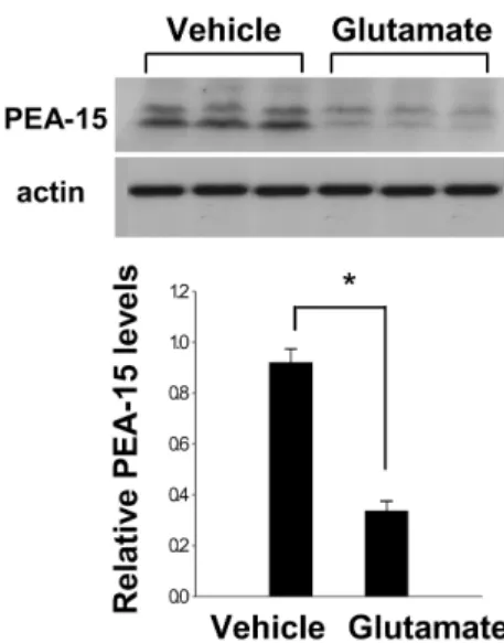
We previously showed that glutamate toxicity induces cell death in HT22 cells (Koh, 2007). Figure 3 indicates that glutamate exposure induces decreases of PEA-15 levels in HT22 cells. The levels of PEA-15 were 0.91±0.03 in the vehicle-treated group, and 0.35±0.03 in the glutamate-treated group, respectively (Figure 3).
Previous studies have established that MCAO leads to focal cerebral cortex ischemia and causes neuronal cell death through the apoptotic signaling pathway (Li et al., 1997; Ferrer and Planas, 2003). We previously identified differentially expressed proteins following ischemic brain injury using a proteomic approach (Koh, 2010). These proteins include 60 kDa heat shock protein, dehydropyrimidinase-related protein 2, thioredoxin, peroxiredoxin-2, stathmin, and ubiquitin carboxy-terminal hydrolase L1. Cerebral ischemic injury induces down- and up-regulation of these proteins. In the present study, we additionally identified the down-regulation of PEA-15 protein after MCAO-induced brain injury in an animal model.
PEA-15 is an abundant phosphoprotein in brain astrocytes
and plays a major role in modulating signal pathways that regulate apoptosis and cell proliferation (Araujo et al., 1993;
Danziger et al., 1995; Renault et al., 2003; Krueger et al., 2005). PEA-15 inhibits tumor-necrosis factor-α (TNF-α)- induced apoptosis, blocks activation of caspase, and regulates apoptosis (Renault et al., 2003; Sharif et al., 2003). Focal cerebral ischemia induces TNF-α expression, while inhibition of TNF-α mediates neuroprotection (Dawson et al., 1996).
PEA-15 binds to the Fas associated death domain and mediates apoptosis pathway (Krueger et al., 2005). Cerebral ischemia induces Fas-mediated apoptosis and leads to neuronal cell death (Wetzel et al., 2008; Jia et al., 2009). Moreover, PEA- 15 binds extracellular signal-regulated kinase and regulates mitogen-activated protein kinase signaling (Krueger et al., 2005). We identified decreases in PEA-15 protein spots in MCAO-induced brain injury. Western blot analysis confirmed that ischemic brain injury significantly decreases PEA-15 levels.
It is accepted that glutamate induces oxidative stress and causes neuronal cell death. Oxidative toxicity induces the disruption of redox homeostasis and results in cell death.
Thus, glutamate has been used as the inducer of ischemic condition in vitro study. Glutamate exposure induces a significant reduction in PEA-15 levels in neurons. Focal cerebral ischemia induces decreases in PEA-15 and leads to neuronal cell death. The results of this study demonstrate that PEA- 15 decreases in MCAO-induced injury and glutamate-exposed HT22 cells. PEA-15 mediates anti-apoptotic function and cell proliferation. However, decrease of PEA-15 in cerebral
Figure 2. Western blot analysis of PEA-15 in the cerebralcortex from sham-operated and MCAO-operated animals.
Each lane represents an individual experimental animal.
Densitometric analysis of PEA-15 levels is represented as intensity of PEA-15 to intensity of actin. Data are shown as mean±S.E.M. *P<0.05 (vs. Sham).
Figure 3. Western blot analysis of PEA-15 in HT22 cells.
Vehicle or glutamate (5 mM) was exposed to HT22 cells for 24 hr. Each lane represents an individual experimental animal.
Densitometric analysis of PEA-15 levels is represented as intensity of PEA-15 to intensity of actin. Data are shown as mean±S.E.M. *P<0.05 (vs. Vehicle).
314 Phil-Ok Koh
Lab. Anim. Res. | September, 2010 | Vol. 26, No. 3
ischemia is not yet reported. Although further studies are required to explain the mechanism of PEA-15 in ischemic brain injury, this study can demonstrate that decrease of PEA- 15 in cerebral ischemia inhibits anti-apoptotic function and induces neuronal cell death. Thus, the results of this study suggest that decrease in PEA-15 mediates neuronal cell death in brain injury.
Acknowledgments
This research was supported by Basic Science Research Program through the National Research Foundation of Korea (NRF) funded by the Ministry of Education, Science and Technology (2009-0070571).
References
Araujo, H., Danziger, N., Cordier, J., Glowinski, J. and Chneiweiss, H. (1993) Characterization of PEA-15, a major substrate for protein kinase C in astrocytes. J. Biol. Chem. 268(8), 5911- 5920.
Danziger, N., Yokoyama, M., Jay, T., Cordier, J., Glowinski, J. and Chneiweiss, H. (1995) Cellular expression, developmental regulation, and phylogenic conservation of PEA-15, the astrocytic major phosphoprotein and protein kinase C substrate. J. Neurochem. 64(3), 1016-1025.
Dawson, D.A., Martin, D. and Hallenbeck, J.M. (1996) Inhibition of tumor necrosis factor-alpha reduces focal cerebral ischemic injury in the spontaneously hypertensive rat. Neurosci. Lett.
218(1), 41-44.
Ferrer, I. and Planas, A.M. (2003) Signaling of cell death and cell survival following focal cerebral ischemia: life and death
struggle in the penumbra. J. Neuropathol. Exp. Neurol. 62(4), 329-339.
Jia J., Guan D., Zhu W., Alkayed N.J., Wang M.M., Hua Z. and Xu Y. (2009) Estrogen inhibits Fas-mediated apoptosis in experimental stroke. Exp. Neurol. 215(1), 48-52.
Koh, P.O. (2007) 17Beta-estradiol prevents the glutamate-induced decrease of Akt and its downstream targets in HT22 cells. J.
Vet. Med. Sci. 69(3), 285-288.
Koh, P.O. (2008) Melatonin prevents the injury-induced decline of Akt/forkhead transcription factors phosphorylation. J. Pineal Res. 45(2), 199-203.
Koh, P.O. (2010) Proteomic analysis of focal cerebral ischemic injury in male rats. J. Vet. Med. Sci. 72(2), 181-185.
Krueger, J., Chou, F.L., Glading, A., Schaefer, E. and Ginsberg, M.H. (2005) Phosphorylation of phosphoprotein enriched in astrocytes (PEA-15) regulates extracellular signal-regulated kinase-dependent transcription and cell proliferation. Mol.
Biol. Cell. 16(8), 3552-3561.
Li Y., Chopp, M., Powers, C. and Jiang, N. (1997) Apoptosis and protein expression after focal cerebral ischemia in rat. Brain Res. 765(2), 301-312.
Longa, E.Z., Weinstein, P.R., Carlson, S. and Cummins, R. (1989) Reversible middle cerebral artery occlusion without craniectomy in rats. Stroke 20(1), 84-91.
Maher, P. and Davis, J.B. (1996) The role of monoamine metabolism in oxidative glutamate toxicity. J. Neurosci.
16(20), 6394-6401.
Renault, F., Formstecher, E., Callebaut, I., Junier, M.P. and Chneiweiss, H. (2003) The multifunctional protein PEA-15 is involved in the control of apoptosis and cell cycle in astrocytes. Biochem. Pharmacol. 66(8), 1581-1588.
Sharif, A., Canton, B., Junier, M.P. and Chneiweiss, H. (2003) PEA-15 modulates TNF alpha intracellular signaling in astrocytes. Ann. N. Y. Acad. Sci. 1010, 43-50.
Wetzel, M., Li, L., Harms, K.M., Roitbak, T., Ventura, P.B., Rosenberg, G.A., Khokha, R. and Cunningham, L.A. (2008) Tissue inhibitor of metalloproteinases-3 facilitates Fas- mediated neuronal cell death following mild ischemia. Cell Death Differ. 15(1), 143-151.