• 검색 결과가 없습니다.

Endothelial Ca

N/A
N/A
Protected

Academic year: 2022

Share "Endothelial Ca"

Copied!
12
0
0

로드 중.... (전체 텍스트 보기)

전체 글

(1)

Author contributions: K.S.H. and M.G.L. conceptualized the review study.

K.S.H. and M.G.L. contributed to writing and editing of the manuscript.

This is an Open Access article distributed under the terms of the Creative Commons Attribution Non-Commercial License, which permits unrestricted non-commercial use, distribution, and reproduction in any medium, provided the original work is properly cited.

Copyright © Korean J Physiol Pharmacol, pISSN 1226-4512, eISSN 2093-3827

INTRODUCTION

The vascular endothelium is defined as a single layer of endo- thelial cells (ECs) that line the lumen of blood vessels and are me- chanically and metabolically dynamic organs. The endothelium, which consists of 1 – 6 × 10

13

individual ECs, is the largest organ in the body and exceeds 1,000 m

2

of estimated surface area [1-3].

This important organ is involved in a variety of physiological and pathological functions including blood supply, nutrient delivery,

immune cell adhesion, vasopermeability, angiogenesis, thrombo- genesis, and vascular tone [4-7].

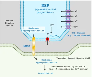
Endothelium-dependent vasodilation is largely determined by alterations in endothelial intracellular Ca

2+

concentrations in response to mechanical stimuli (e.g., shear stress, membrane stretch) or endogenous agonists (e.g., bradykinin, ATP, or reactive oxygen species [ROS]). Increased intracellular Ca

2+

levels produc- es nitric oxide (NO) and prostacyclin (PGI

2

) that are traditionally considered as endothelium-derived relaxing factors (EDRFs) [8,9].

Review Article

Endothelial Ca 2+ signaling-dependent vasodilation through transient receptor potential channels

Kwang-Seok Hong 1 and Man-Gyoon Lee 2, *

1

Department of Physical Education, College of Education, Chung-Ang University, Seoul 06974,

2

Sports Medicine and Science, Graduate School of Physical Education, Kyung Hee University, Yongin 17104, Korea

ARTICLE INFO

Received February 4, 2020 Revised April 3, 2020 Accepted April 14, 2020

*Correspondence Man-Gyoon Lee E-mail: [email protected] Key Words

Calcium signaling Endothelium Gap Junctions Ion channels Microcirculation Vasodilation

ABSTRACT Ca

2+

signaling of endothelial cells plays a critical role in controlling blood

flow and pressure in small arteries and arterioles. As the impairment of endothelial

function is closely associated with cardiovascular diseases (e.g., atherosclerosis,

stroke, and hypertension), endothelial Ca

2+

signaling mechanisms have received sub-

stantial attention. Increases in endothelial intracellular Ca

2+

concentrations promote

the synthesis and release of endothelial-derived hyperpolarizing factors (EDHFs, e.g.,

nitric oxide, prostacyclin, or K

+

efflux) or directly result in endothelial-dependent hy-

perpolarization (EDH). These physiological alterations modulate vascular contractility

and cause marked vasodilation in resistance arteries. Transient receptor potential

(TRP) channels are nonselective cation channels that are present in the endothe-

lium, vascular smooth muscle cells, or perivascular/sensory nerves. TRP channels are

activated by diverse stimuli and are considered key biological apparatuses for the

Ca

2+

influx-dependent regulation of vasomotor reactivity in resistance arteries. Ca

2+

-

permeable TRP channels, which are primarily found at spatially restricted microdo-

mains in endothelial cells (e.g., myoendothelial projections), have a large unitary or

binary conductance and contribute to EDHFs or EDH-induced vasodilation in concert

with the activation of intermediate/small conductance Ca

2+

-sensitive K

+

channels. It

is likely that endothelial TRP channel dysfunction is related to the dysregulation of

endothelial Ca

2+

signaling and in turn gives rise to vascular-related diseases such as

hypertension. Thus, investigations on the role of Ca

2+

dynamics via TRP channels in

endothelial cells are required to further comprehend how vascular tone or perfusion

pressure are regulated in normal and pathophysiological conditions.

(2)

In addition, it has been well documented that changes in global or localized EC Ca

2+

signaling stimulate Ca

2+

-sensitive K

+

channels and elicit the membrane hyperpolarization of ECs and vascular smooth muscle cells (VSMCs) in a sequential manner [10]. This discovery contributes to the development of a novel concept, endothelium-dependent hyperpolarization (EDH), which is a pri- mary mechanism of vasodilation in small resistance arteries.

It is no exaggeration to state that the elucidation of EC Ca

2+

signaling has been accomplished by the progression of sophisti- cated imaging techniques and fast/high-affinity fluorescent Ca

2+

indicators, including a genetically modified mouse specifically expressing a Ca

2+

indicator (e.g., GCaMP2) in ECs, high-quality (i.e., high speed and resolution) confocal microscopy, and total in- ternal reflection fluorescent (TIRF) microscopy [11]. These tech- nological advances have led investigators to explore complicated Ca

2+

dynamics in ECs including Ca

2+

release from the endoplas- mic reticulum (ER; i.e., propagated Ca

2+

wave [12], Ca

2+

pulsars [13], Ca

2+

wavelets [14]) and Ca

2+

entry from the extracellular space (i.e., Ca

2+

sparklets [15]) that result in vasodilation. Howev- er, the molecular mechanisms underlying the Ca

2+

influx in ECs and their regulations have been poorly defined. In light of this, the identification of transient receptor potential (TRP) channels has provided new insights into Ca

2+

mobilization in ECs that is required for EDH(F) and vasodilation. Thus, this review focuses on describing the contribution of TRP channels to fundamental Ca

2+

signals (i.e., serve as a crucial means of altering intracellular Ca

2+

levels) in the ECs of resistance arteries.

Ca 2+ SIGNALING AND EDH

The discovery of NO and PGI

2

generated from ECs has pro- vided insight into the novel paradigm that the endothelium is an organ that does not merely cover the inner wall of blood vessels;

it also controls the vascular tone and blood flow [10]. In addi- tion, succeeding investigations have shown that EC-dependent VSMC hyperpolarization (caused by releasing factors from the ECs: endothelium-dependent hyperpolarizing factors [EDHFs]) in response to muscarinic receptor activation elicits vasodila- tion by inhibiting voltage-dependent Ca

2+

channels (VDCCs) in VSMCs [16,17]. Chen and colleagues [17] sought to directly assess the effects of acetylcholine (ACh) on the membrane potential of VSMCs in the aorta and main pulmonary artery of rats. ACh- induced VSMC hyperpolarization was still detected even in the presence of inhibitors of NO or guanylyl cyclase, suggesting that EDHFs are distinct from EDRFs. Importantly, that study dem- onstrated that K

+

efflux is a key component of EDHFs [17]. Since then, the previous findings on EDHFs have evolved into a new concept of EDH; additionally, such studies have seminally identi- fied intermediate/small conductance Ca

2+

-sensitive K

+

channels and microdomain structures (e.g., myoendothelial projections [MEPs] and myoendothelial gap junctions [MEGJs]) that allow

the movement of hyperpolarizing currents from ECs to VSMCs [18].

In the Garland and McPherson study [19], although VSMC hy- perpolarization in response to NO was significantly attenuated by a K

+

channel blocker (i.e., glibenclamide), ACh-stimulated hyper- polarization was not affected by the same blocker. The hyperpo- larization and vasorelaxation induced by ACh were not sensitive to NO synthase (NOS) or cyclooxygenase (COX) inhibitors (i.e., L-NNA or indomethacin, respectively) in rat small mesenteric arteries [19]. These results suggest that ACh-mediated VSMC hy- perpolarization leads to vasorelaxation that occurs independently of NO or prostaglandins. Further, subsequent works have demon- strated that ACh-induced hyperpolarization and vasorelaxation are associated with the activation of Ca

2+

-sensitive IK

Ca

and SK

Ca

channels distributed in ECs [20-22]. This finding implies that muscarinic receptor activation evokes inositol trisphosphate (IP

3

)- mediated Ca

2+

release from the ER and activates IK

Ca

/SK

Ca

chan- nels. Another investigation in rat hepatic arteries showed that K

+

efflux from ECs through IK

Ca

/SK

Ca

channels acts as an EDHF by activating inwardly rectifying K

+

(K

ir

) channels and Na

+

-K

+

ATPase in adjacent VSMCs [23]. However, since blockers of K

ir

channels and Na

+

-K

+

ATPase (Ba

2+

and ouabain, respectively) did not entirely abolish vasorelaxation in rat small mesenteric arter- ies, EC hyperpolarization might be directly transmitted to the VSMCs through MEGJs [23].

It is generally accepted that the vasorelaxation or dilation of small resistance arteries primarily depends on EDH rather than on EDRFs (e.g., NO, PGI

2

) or EDHFs [18]. MEPs are defined as extensions of ECs that project through the fenestration of the internal elastic lamina (IEL) to adjacent VSMCs. The MEGJs lo- cated at the tip of the MEP are intercellular pores that allow the spread of second messengers (e.g., IP

3

) or charged ions for hetero- cellular (EC-VSMC) communication [24]. IK

Ca

and SK

Ca

channels that are indispensable for EDH-mediated vasodilation in resis- tance arteries are activated by the binding of the Ca

2+

-calmodulin complex to the C-terminus of those channels [25]. K

+

loss via the Ca

2+

-sensitive K

+

channels elicits hyperpolarization that is con- ducted to VSMCs through MEGJs, which leads to vasodilation by inhibiting VDCCs (Fig. 1). Thus, it is strongly suggested that EC Ca

2+

signaling, in particular at MEPs, plays a critical role in EDH- dependent vasodilation. Indeed, since it was first discovered that an alteration in intracellular Ca

2+

concentrations is a major deter- minant of vascular reactivity [26], considerable strides have been made in understanding the role of EC Ca

2+

signaling in EC-de- pendent vasodilation in resistance arteries. According to previous studies, cation-permeable TRP ion channels that allow for Ca

2+

entry into ECs have been shown to induce EDH-mediated vaso-

dilation in small arteries [13-15,27,28]. Thus, EC TRP channel-

dependent Ca

2+

signaling will be discussed in this review.

(3)

Ca 2+ INFLUX THROUGH TRP CHANNELS AND SUBSEQUENT VASODILATION

The TRP superfamily is encoded by 28 genes and is classified according to DNA and protein sequence homology. Consequent- ly, TRP channels are categorized into six subfamilies: TRPA (an- kyrin), TRPC (canonical), TRPM (melastatin), TRPML (muco- liptin), TRPP (polycystin), and TRPV (vanilloid) channels. TRP channels are generally comprised of seven hydrophobic domains.

Six (S1–S6) of the domains span the plasma membrane, and the seventh domain intracellularly exists with the COOH- and NH

2

-termini of TRP channels [29-31]. The ion-conducting pore region is located between the fifth and sixth domains (S5 and S6 domains). The TRP box, a conserved 25-amino acid element, is formed immediately after the S6 domain in the subfamilies of TRPC, TRPM, and TRPV channels [32]. Although the TRP domain has been incompletely examined, it has been elucidated to date that the conserved box serves as a platform for a phospha- tidylinositol 4,5-bisphosphate (PIP

2

) binding site or ion channel heteromeric assembly [33,34]. The intracellular C- and N-termini of TRP channels exist for multiple regulatory elements, interac- tion sites, and enzymatic domains [32].

TRP channels are postulated to participate in diverse physi-

ological processes (e.g., mechanotransduction, cellular excit- ability, protein trafficking, and vascular contractility) by regulat- ing cation influx across the cell membrane [32]. TRP channels physiologically control the membrane potential and regulate Ca

2+

signaling in ECs and VSMCs. Among TRP channel subfamilies, Na

+

-permeable TRPM4 channels (equally permeant to K

+

, but not Ca

2+

-permeable) are activated by globally or locally increased intracellular Ca

2+

levels in VSMCs [35]. The activated channels primarily elicit inward currents of Na

+

in cerebral VSMCs, induce membrane depolarization, and subsequently activate VDCCs [36]. In addition to membrane potential regulation, Ca

2+

signal- ing that is required for vasoconstriction or dilation is controlled by Ca

2+

-permeable TRP channels in a direct or indirect (i.e., metabotropic) manner. For example, Ca

2+

influx via the TRPV4 and TRPA1 channels in ECs that are directly stimulated by shear stress, endogenous lipid arachidonic acid, or exogenous agonists (e.g., GSK1016790A or AITC) triggers the vasodilation of cerebral or mesenteric arteries [15,37-39].

Localized Ca

2+

signaling through endothelial non-selective cation TRP channels primarily plays a vital role in the generation of EDH via the downstream activation of IK

Ca

/SK

Ca

channels and vasodilation in small-sized resistance arteries. It is well docu- mented that vascular peripheral resistance is a major determinant of arterial pressure and blood flow to organs and tissues. Further- more, vascular resistance is regulated by EDH and subsequent endothelium-dependent vasodilation in the microcirculation.

Indeed, impaired EDH and endothelium-dependent vasodilation in microvascular beds is found to cause cardiovascular diseases including hypertension. Thus, this review mainly focused on TRP channel-mediated EDH in resistance arteries as they are physiologically significant in the regulation of vascular resistance, intravascular pressure, and blood flow.

TRPC1

TRPC1 channels are non-selectively permeable to Ca

2+

(a unitary conductance of ~5pS) [40]. Although TRPC1 channel is known to be expressed in the endothelium, it is debatable that TRPC1 channels show homomeric channel in intact endothelial cells. In contrast, it is definitely elucidated that TRPC1 chan- nels form heteromeric channels with TRPC3 [41], TRPC4 [40], TRPC5 [40], and TPRV4 [42,43] channels. With regard to the role of TRPC1 channels in endothelial intracellular Ca

2+

signaling, the activation of Ca

2+

-sensing receptor (CaSR) stimulates Ca

2+

influx through TRPC1 channels and leads to NO production in the en- dothelium [44]. Furthermore, once CaSR is activated by the eleva- tion of extracellular Ca

2+

concentration, TRPC1 channels that are co-localized with TRPV4 channels showed vasorelaxation, which was significantly abrogated in the presence of TRPC1 and TRPV4 inhibitors [42]. Therefore, it is suggested that the heteromeric TRPC1/TRPV4 channels stimulated by CaSR activation induce NO-mediated vasorelaxation.

Fig. 1. Transient receptor potential (TRP) channel-mediated endo- thelium-dependent hyperpolarization (EDH) and vasodilation. TRP channels (e.g., representatively TRPV4 channels) are placed myoendo- thelial projections (MEPs) that are extensions of endothelial cells (ECs) through internal elastic lamina to contact adjacent vascular smooth muscle cells (VSMCs). Localized Ca

2+

entry through endothelial TRP channels stimulate intermediate/small conductance Ca

2+

-sensitive po- tassium (IK

Ca

/SK

Ca

) channels and in turn results in EDH via K

+

ion efflux.

EDH is transmitted to adjacent VSMCs through myoendothelial gap

junctions (MEGJs) comprising of two hemi-channels, which initiates

VSMC hyperpolarization. The altered membrane potential of VSMCs

suppresses voltage-dependent Ca

2+

channels (VDCCs) and evokes va-

sodilation.

(4)

TRPC3

TRPC3 channels are expressed in arteriolar myocytes and contribute to membrane depolarization-induced vasoconstriction (i.e., permeability: Ca

2+

= Na

+

> K

+

) [45-48]. Meanwhile, growing evidence has shown that TRPC3 cation channels are expressed in ECs and are responsible for EC hyperpolarization-mediated vasodilation. The vasoreactivity of rat small mesenteric arteries was considerably blunted in response to flow or bradykinin in the treatment of TRPC3 antisense oligonucleotide [49]. Subsequent pharmacological studies with a specific inhibitor of TRPC3 chan- nels (i.e., Pyr3) have consistently demonstrated that the ACh- induced vasodilation of human internal mammary arteries or rat mesenteric arteries is markedly reduced with Pyr3 pretreatment [50,51], implying that EC GPCR activation by shear stress or exogenous agonists is coupled to TRPC3-induced downstream signaling for vasodilation. Senadheera and colleagues used ultra- structural immunohistochemistry to show that TRPC3 channels are spatially restricted at MEPs (i.e., five-fold higher expression compared to non-MEPs) in resistance mesenteric arteries [51].

This investigation also revealed that TRPC3 inhibition with Pyr3 causes a significant reduction in EC hyperpolarization in response to ACh [51]. More recently, studies have shown that EC TRPC3 channels in cerebral arteries are translocated to the plasma membrane following purinergic receptor activation with ATP and are then involved in EDH-mediated vasodilation [52].

Collectively, it is speculated that second messengers (e.g., DAG) produced by GPCR activation stimulate Ca

2+

influx via TRPC3 channels and in turn lead to EDH-mediated vasodilation in resis- tance small arteries.

TRPC4

TRPC4 channels preferably allow Ca

2+

influx in the endothe- lium through store-operated Ca

2+

entry and are stimulated by GPCR downstream signaling [32,53]. Freichel and colleagues [54]

demonstrated that endothelium-dependent vasodilation occurs through the activation of TRPC4 channels in mouse aorta. The specific molecular mechanism underlying the vasodilation is at- tributed to endothelial NO synthase (eNOS) activation and NO production followed by the activation of muscarinic acetylcholine receptors [54,55]. Even though subsequent studies failed to show that endothelial store-operated Ca

2+

entry is mediated by TRPC4 channels [53,56], it is noteworthy to understand that TRPC4 channels could be involved in endothelium-dependent vasodila- tion in conduit arteries, but not small-sized resistance arteries.

TRPC5

TRPC5 channels show a unitary conductance of 40–60 pS and are more permeable to Ca

2+

than Na

+

(P

Ca

:P

Na

= 1.8–9.5) [57,58].

Although the role of TRPC5 channels in endothelial function has

not been clearly identified to date, previous investigations found that TRPC5 channels are distributed in the endothelium from mouse aorta, human coronary, and cerebral arteries [59-61]. Mori and colleagues revealed that Ca

2+

influx through endothelial TRPC5 channel is regulated by post-translational modifications.

Specifically, S-nitrosylation, the covalent attachment of an NO to cysteine groups, enhances the activity of TRPC5 channels and Ca

2+

influx in cultured ECs [62]. However, the alteration of vasodilatory response to ACh was not monitored in mouse aortic rings originated from TRPC5 knockout models [60]. This sug- gests that NO-mediated vasodilation in the presence of ACh was not augmented by TRPC5 channels, which is inconsistent with previous studies [62]. Thus, it is necessary in future studies to explore if Ca

2+

entry via TRPC5 channels elicits vasodilation and via what supporting mechanisms.

TRPM2

TRPM2 channels that have a similar permeability of Ca

2+

and Na

+

ions display the unitary conductance of 58–76 pS [63].

The subtype of TRPM2 channels is gated by several activators including ADP-ribose, arachidonic acid, hydrogen peroxide [64-66]. The Ca

2+

influx of TRPM2 channels is also clarified to induce endothelium-dependent vasodilation. It was found in rat cremaster resistance arteries that exogenous hydrogen peroxide application leads to Ca

2+

entry and subsequent vasodilation [67].

However, those Ca

2+

signaling and vascular reactivity were mark- edly diminished by the treatments of TRPM2 antibody or IK

Ca

/ SK

Ca

channel inhibitors. These results indicate that hydrogen peroxide-mediated Ca

2+

entry through TRPM2 channels evokes endothelial vasodilation in the manner dependent of EDH [67].

TRPV1

TRPV1 nonselective cation channels are expressed in arte- rial ECs [68,69] and are favorably permeable to Ca

2+

compared with Na

+

(P

Ca

:P

Na

≈ 9.6) [32]. Capsaicin, endocannabinoids, and noxious heat activate TRPV1 channels [70,71]. Anandamide- evoked TRPV1 activation increased NO in the endothelium of rat mesenteric arteries [72]. A synthetic cannabinoid compound (VSN16) induced vasorelaxation of the mesenteric arteries, which was markedly attenuated by capsazepine (a TRPV1 antagonist), L-NNA (an NOS inhibitor), charybdotoxin (an IK

Ca

/BK

Ca

chan- nel inhibitor), or apamin (a SK

Ca

blocker) [73]. In gracilis skeletal muscle arterioles, the capsaicin-mediated stimulation of TRPV1 channels produced vasodilation that was substantially attenuated by L-NAME (an NOS inhibitor), which suggests that EC TRPV1- mediated vasodilation occurs in an NO-dependent manner [74].

Interestingly, NO-mediated vasorelaxation/dilation following

TRPV1 activation differs from vasodilatory mechanisms of other

TRP subfamilies (i.e., TRPV4, TRPA1) that display EDH-depen-

dent vasodilation. In accordance with previous studies showing a

(5)

relationship between EC TRPV1 activation and NO production, capsaicin treatment increased intracellular EC Ca

2+

levels and elicited the phosphorylation of eNOS in cultured mouse aortic ECs [75]. Additionally, the chronic administration of capsaicin profoundly increased eNOS phosphorylation at Ser

1177

, NO gen- eration, and protein kinase A (PKA) activation in mouse mes- enteric arteries; these changes were absent in TRPV1 knockout mice [75]. As expected, the chronic activation of TRPV1 channels was coupled to the enhancement of EC-dependent vasorelax- ation in freshly isolated mesenteric arteries [75]. Surprisingly, this study showed convincing clinical evidence that chronic capsaicin application significantly reduces blood pressure in spontane- ously hypertensive rats. This finding implies that TRPV1 chan- nels participate in the regulation of vascular tone and arterial pressure. The following study in bovine aortic ECs found that capsaicin-induced Ca

2+

entry via TRPV1 channels stimulates Akt-calmodulin-dependent protein kinase II (CaMKII)-eNOS pathway; in turn, phosphorylated eNOS physically interacts with TRPV1 channels to produce NO [76]. However, despite consistent findings, a recent study elucidated that large renal arteries in mice do not respond to a relatively higher concentration of capsaicin (i.e., a 100-fold higher level of EC

50

is required for significant va- sodilation), whereas mesenteric small arteries showed a marked vasodilation in response to 25 nM of capsaicin [77]. Thus, the magnitude of TRPV1-dependent vasoreactivity is specific to vas- cular beds.

TRPV3

TRPV3 channels display a large unitary conductance (≈

150–200 pS) [78]. Dietary monoterpenoid compounds including carvacrol, eugenol, and thymol stimulate TRPV3 channels in the oral/nasal epithelium [79]. With regard to the roles of TRPV3 in EC-dependent vasodilation, Earley and co-workers revealed for the first time that carvacrol that is typically found in the Medi- terranean diet, induces vasodilation in rat cerebral arteries [80].

Increased endothelial Ca

2+

concentrations and TRPV3-like cur- rents have been found in the presence of carvacrol [80]. Moreover, TRPV3-dependent cerebral vasodilation was found to result from VSMC hyperpolarization that was inhibited by TRAM- 34, apamin, or BaCl

2

(an inhibitor of inwardly rectifying K

+

(K

ir

) channels), but not by L-NNA or indomethacin. This suggests that TRPV3 activation-induced K

+

efflux through IK

Ca

or SK

Ca

leads to the K

ir

-mediated hyperpolarization of VSMCs and cerebral vasodilation [80]. Recently, it was demonstrated that EC TRPV3 Ca

2+

sparklets (i.e., localized Ca

2+

influx through EC TRPV3 channels) are directly and optically detected with carvacrol in the ECs of rat cerebral parenchymal arterioles [27]. The unitary Ca

2+

events in ECs were significantly attenuated by a selective TRPV3 antagonist, isopentenyl pyrophosphate (IPP). This study also noted that TRPV3 sparklet-mediated vasodilation is not sensitive to NOS or COX inhibitors. By contrast, the inhibition of IK

Ca

or

SK

Ca

channels markedly reduced the dietary compound-induced vasodilation. Importantly, the authors also suggested that the amplitude of TRPV3 channels (∆F/F

0

= 0.20; peak fluorescence [F] is normalized to baseline fluorescence [F

0

]) is greater than that of TRPA1 (∆F/F

0

= 0.13) and TRPV4 (∆F/F

0

= 0.06) channels.

TRPV3 Ca

2+

sparklets were implicated to play a key role in EC Ca

2+

signaling in cerebral resistance arterioles [27]. However, to the best of our knowledge, there have been few observations on the influence of TRPV3 channels on vasomotor reactivity. Thus, further observations are required for a better understanding of the contribution of EC TRPV3 for the modulation of vascular function.

TRPV4

TRPV4 channels were shown to have a specific permeability to Ca

2+

, Na

+

, and Mg

2+

(P

Mg

:P

Na

≈ 2–3:1; P

Ca

:P

Na

≈ 6–10:1) [68,81,82].

TRPV4 channels are stimulated by a variety of endogenous activa- tors or synthetic agonists including 4α-phorbol 12,13-didecano- ate (4α-PDD; 200 nM of EC

50

; [83]), GSK1016790A (18 nM of EC

50

; [84]), RN-1747 (4 μM of EC

50

; [85]), or arachidonic acid metabolites (e.g., 5,6-EET; 130 nM of EC

50

; [86]). When Ca

2+

- permeable TRPV4 channels were pharmacologically activated, increased whole-cell currents or Ca

2+

influx were observed in isolated ECs [87-90]. Previous investigations demonstrated that in small arteries, EC TRPV4-mediated alterations in Ca

2+

levels following the activation of muscarinic or purinergic receptors in ECs contribute to marked vasodilation. ACh fails to dilate mes- enteric arteries with a genetic deletion of TRPV4 channels [90]. In this study, ACh-mediated Ca

2+

influx and NO production were also significantly blunted in TRPV4

-/-

mice. Similarly, a potent TRPV4 antagonist (RN-1734) pretreatment considerably disrupt- ed the ACh-evoked vasodilation of radial uterine arteries [91].

Sullivan et al. [92] and Sonkusare et al. [15] marked the begin- ning of a new area of endothelial Ca

2+

influx through TRPV4 channels, called TRPV4 Ca

2+

sparklets, by using improved and sophisticated optical imaging techniques including high-speed/

resolution confocal or TIRF microscopy. The Sonkusare and

Nelson group sought to delineate the properties of elementary

TRPV4 Ca

2+

sparklets spatially identified at MEPs. TRPV4 chan-

nels were revealed to exhibit quantal events of Ca

2+

entry, and the

quantal amplitudes were shown to be dependent on the electro-

chemical gradient. Surprisingly, three to eight TRPV4 sparklet

sites per EC was considered firmly sufficient for maximal vasodi-

lation [15,92]. Extensive monitoring also showed that Ca

2+

signal-

ing through EC TRPV4 channels is explicitly coupled to IK

Ca

and

SK

Ca

activation-mediated hyperpolarization at MEPs and vasodi-

lation [15]. Further, K

+

efflux via IK

Ca

and SK

Ca

channels enhanced

vasodilation by stimulating K

ir

channels in ECs and VSMCs and

by amplifying the hyperpolarization of both cells [23,93]. Next,

one of the novel characteristics of TRPV4 sparklets is channel

cooperative behavior [94]. Ca

2+

-permeable TRPV4 channels are

(6)

interestingly designed with a four-channel (tetramer) metastruc- ture. The clustered TRPV4 channels locally distributed at MEPs are cooperatively gated, which is determined by protein kinase C- anchoring protein 150 (AKAP150) and intracellular Ca

2+

[94]. It is conceivable that opening only one single TRPV4 channel may induce a minimal Ca

2+

influx and activate a small number of IK

Ca

and/or SK

Ca

channels at the microdomains. However, increased cooperativity of the clustered TRPV4 channels (i.e., quantal events) gives rise to augmented Ca

2+

amplitudes and enhanced hyperpolarization [94]. Angiotensin II-induced hypertension was shown to impair AKAP150-dependent cooperative activity of TRPV4 channels by disrupting the scaffolding protein expression at MEPs [94]. It is speculated that EC TRPV4 and AKAP150 play a critical role in preventing pathophysiological circumstances in the cardiovascular system.

With respect to the roles of TRPV channels in mechanotrans- duction, since ECs directly interact with shear stress induced by blood flow velocity and blood viscosity due to their unique ana- tomical position, considerable attention was given to flow (shear stress)-mediated EC TRPV4 activation and subsequent vasodila- tion in small resistance arteries. Further, because most previous studies of TRPV4 Ca

2+

imaging were implemented in the presence of chemical agonist compounds (e.g., 4α-PDD, GSK1016790A), the impact of physiological modulators including shear stress or intravascular pressure on EC TRPV4 activation and downstream signaling for vasodilation requires further exploration. TRPV4 channels activated by shear stress resulted in increased Ca

2+

entry in the ECs of mesenteric arteries and vasodilation that was sig- nificantly suppressed in TRPV4-deficient mice [38]. Pharmaco- logical inhibition to NOS entirely suppressed shear stress (10 dyn cm

-2

)-mediated vasodilation in mesenteric arteries [38], which was consistently shown in gracilis muscle arterioles [88]. These studies suggest that the mechanoactivation of TRPV4 channels exerts vasodilation through NO production; however, it is not completely clear as to how the channels are directly or indirectly activated by shear stress or metabolites (e.g., epoxyeicosatrienoic acids [EETs]) produced by shear stress. Conversely, the perfusion of 4α-PDD and 11,12-EET still evoked vasodilation in mesenteric arteries even in the presence of NOS and COX inhibitors [72,95].

It is implied that different physiological or pharmacological forms of activation of TRPV4 channels may result in disparate downstream signaling for vasodilation [88], presumably due to the unique polymodal nature of TRPV4 channels. Further, it was delineated in human coronary arteries that flow-dependent activation of TRPV4 channels generates mitochondrial ROS (i.e., hydrogen peroxide and superoxide) that play an important role in vasodilation in human coronary arteries [96].

As mentioned above, although VSMCs are constantly ex- posed to intraluminal pressure, biological processes in ECs are mechanically affected by shear stress. Nonetheless, EC TRPV4 channel activity has been demonstrated to be modulated by low intravascular pressure (i.e., below 50 mmHg) in rat cremaster or

mesenteric arterioles [97]. Specifically, the frequency of EC Ca

2+

events via EC TRPV4 channels was further augmented at a low intraluminal pressure (two- to three-fold higher than those at 60–80 mmHg of arterial intraluminal pressure). Both pharma- cological (i.e., selective TRPV4 agonists) and physiological (i.e., intraluminal pressure) TRPV4 activations were markedly dimin- ished by RN1734 or HC067047 [97]. Although they revealed that TRPV4, IK

Ca

, and SK

Ca

channels are clustered at perforations in the IEL, the inhibition of IK

Ca

with TRAM-34 (but not the in- hibition of SK

Ca

with apamin) considerably enhanced the basal myogenic tone in cremaster arterioles. Additionally, the blockade of PLC and the IP

3

receptor with U-73122 and xestospongin fully abrogated the low pressure-mediated increase in Ca

2+

sparklet events. Thus, Ca

2+

influx via EC TRPV4 channels in response to low intraluminal pressure stimulates IK

Ca

channels through PLC and IP

3

R signaling, which in turn leads to EC/VSMC hyperpolar- ization-dependent vasodilation [97].

Arachidonic acid and its metabolites are potential physiological modulators for the activation of TRPV4 channels in ECs. Arachi- donic acid is converted to EETs such as 5,6-EET, 8,9-EET, 11,12- EET, and 14,15-EET through cytochrome P450 (CYP) epoxygen- ase [86]. It has been well known that EETs have anti-hypertensive/

inflammatory effects and contribute to vasodilation as a prevail- ing EDHF [98-100]. Watanabe and colleagues [86] determined that EETs stimulate TRPV4 channels in aortic ECs and may in- duce vasodilation. A subsequent study elucidated that exogenous 11,12-EET application in mesenteric arteries elicited vasodilation via hyperpolarization in VSMCs, and this vasomotor activity was substantially abrogated by TRPV4 knockout or the inhibition of Ca

2+

-sensitive K

+

channels, but not by the blockade of NOS and COX [95]. More recently, pre-constricted human coronary arter- ies with endothelin-1 exhibited vasodilation in the presence of arachidonic acid, which was inhibited by RN-1734 [101]. In addi- tion, the arachidonic acid-induced increase in Ca

2+

concentration and vasodilation was profoundly reduced by the combination of TRAM-34 and apamin (inhibitors of IK

Ca

and SK

Ca

channels, respectively), the removal of extracellular Ca

2+

, or pretreatment with CYP inhibitors, suggesting that CYP-dependent metabolite generation from arachidonic acid increases Ca

2+

influx (but not Ca

2+

release from the sarcoplasmic reticulum) through EC TRPV4 channels and subsequently requires IK

Ca

and SK

Ca

-mediated hy- perpolarization for vasodilation in human coronary arteries [101].

As most studies on TRPV4 channels have been performed

under ex vivo preparations, many investigators have raised ques-

tions regarding the regulatory effect of TRPV4 channels on the

cardiovascular system. The administration of GSK1016790A (a

selective agonist for TRPV4 channels) interestingly decreased

mean arterial pressure [102], which implies that the systemic

activation of TRPV4 channels may contribute to the attenua-

tion of peripheral resistance [32]. Consistently, there are studies

showing that TRPV4-deficient mice did exhibit a higher mean

arterial pressure compared with that of control mice [103] . More-

(7)

over, in the presence of the concurrent administration of an NOS inhibitor (L-NAME) and angiotensin II, TRPV4 knockout mice showed a much greater mean arterial pressure [104], indicating that TRPV4 may provide an alternative compensatory pathway in pathophysiological circumstances to prevent cardiovascular diseases (e.g., hypertension) by reducing peripheral resistance or inducing vasodilation [105].

TRPA1

TRPA1 channels are nonselective cation (i.e., Na

+

, Ca

2+

) chan- nels (but specifically permeable to Ca

2+

compared with Na

+

, P

Ca

:P

Na

≈ 7.9) that have a large unitary conductance (≈ 98 pS) [106,107]. As the name of the ion channel implies, TRPA1 chan- nels have a distinguished structure that exhibits approximately 14–19 ankyrin repeat domains in the intracellular N-terminus of the ion channels [108]. The ankyrin repeat domains participate in protein-protein interactions and modulate channel activity in response to agonists or heat [109,110]. It is well established that diverse pungent dietary compounds such as allyl isothiocyanate (AITC) from mustard, allicin from garlic, eugenol from cloves, or cinnamaldehyde from cinnamon [111] activate TRPA1 channels by exerting covalent modifications on cysteine residues of the N- terminus of the channels [112,113]. Along with the dietary activa- tors, oxidative stress and specific metabolites of lipid peroxidation act as physiological regulators for TRPA1 channels. Previous studies determined that endogenous aldehydes, 4-hydroxy-2-non- enal (4-HNE), 4-oxononenal (4-ONE), or 4-hydroxyhexenal (4- HHE) derived from the peroxidation of ω6 polyunsaturated fatty acids in the plasma membrane activate TRPA1 channels [114-118].

Further, hydrogen peroxide (H

2

O

2

) resulted in the oxidative mod- ification of cysteine residues and subsequently activated TRPA1 channels in neuron cells [114,116]. Therefore, oxidative stress me- tabolites play a vital role in stimulating TRPA1 channels.

In light of the physiological consequences of TRPA1 activation, it was first noted that AITC application leads to vasorelaxation in rat mesenteric arteries by activating the target ion channels in perivascular nerves [119]. Interestingly, the AITC-mediated relax- ant response was markedly attenuated after the pretreatment of the calcitonin gene-related peptide (CGRP) receptor antagonist (i.e., CGRP 8–37). Previous findings demonstrated that the di- etary TRPA1 activators caused cultured trigeminal neurons to release CGRP, which was abolished by a TRPA1 channel blocker [120]. Therefore, it is likely that CGRP released from perivascular nerve endings elicits vasodilation. Subsequent studies further showed that cinnamaldehyde-mediated vasodilation in mesen- teric arteries is considerably abrogated in TRPA1-deficient mice, whereas endothelial denudation has minor effects on vasodila- tion [121]. This suggests that pungent dietary activators of TRPA1 channels elicit vasodilation by inducing Ca

2+

influx-dependent CGRP release from perivascular nerves and subsequently bind- ing the peptide to CGRP receptors in VSMCs. Recently, it was

reported that TRPA1 channels regulate the cold-induced vascular response [122]. Specifically, sequential vasoconstriction and vaso- dilation occur following local cold exposure to prevent cutaneous tissues from suffering from cold injury. Although TRPA1 chan- nel-induced superoxide generation provoked vasoconstriction through α

2

-adrenoreceptor activation and myosin light chain phosphorylation immediately after environmental cold exposure, neuropeptide CGRP released from sensory nerves evoked TRPA1 channel-dependent vasodilation for the restoration of reduced blood flow [122].

Meanwhile, TRPA1 channels are identified in ECs and in perivascular or sensory nerves. However, the distribution of EC TRPA1 channels is limited to only the cerebral arteries, not the mesenteric, coronary, or renal arteries [28,37]. The direct influ- ence of EC TRPA1 channels on the vasomotor response following the treatment of an electrophilic TRPA1 activator derived from mustard oil (e.g., AITC) was first assessed in pressurized cere- bral arteries [37]. AITC concentration-dependent vasodilation of cerebral arteries was observed, which was accompanied by a significant reduction in the intracellular Ca

2+

levels of cerebral ar- terial myocytes. Because VDCCs are a key determinant for Ca

2+

dynamics in VSMCs, TRPA1 activation was suggested to induce VSMC hyperpolarization and vasodilation. TRPA1 channel- mediated vasodilation was diminished in EC-denuded arteries or after TRPA1 channel blocker (HC-030031) pretreatment [37], indicating that AITC-dependent vasodilation in cerebral arteries is EC-dependent and distinct from CGRP-induced vasorelax- ation in mesenteric arteries [119]. Moreover, in intact ECs, TRPA1 channels are detected at MEPs where IK

Ca

channels, but not SK

Ca

channels, are locally expressed [37]. Pharmacological approaches

have substantiated that although TRPA1-induced cerebral vaso-

dilation is not affected by the inhibition of NOS and COX, the

application of Ca

2+

-sensitive K

+

channel blockers or a K

ir

channel

inhibitor markedly suppresses AITC-induced vasodilation. Thus,

EC TRPA1-dependent Ca

2+

entry (called TRPA1 Ca

2+

sparklets)

elicits a K

+

efflux via IK

Ca

/SK

Ca

channels and provokes vasodila-

tion through EC and VSMC hyperpolarization, which is ampli-

fied by K

ir

channels in cerebral arteriolar myocytes [37]. Earley

and co-workers extended their novel hypotheses on TRPA1

channel-induced vasodilation and revealed innovative cerebral

dilatory mechanisms. They found that EC TRPA1 channels fre-

quently display a binary channel gating behavior (i.e., two TRPA1

channels open simultaneously) [28]. Their study focused on the

ROS-mediated activation of TRPA1 channels in cerebral arteries

[28]. NADPH (nicotinamide adenine dinucleotide phosphate)

oxidase (NOX) was shown to generate ROS [123,124], and TRPA1

channels in neurons are stimulated by ROS or ROS-derived me-

tabolites [114,116,125]. In cerebral arteries, the administration of

exogenous NADPH-induced vasodilation that was profoundly

decreased in the presence of HC-030031 or TRAM-34 [28]. Con-

sistent with their prior study [37], the combination of L-NNA and

indomethacin did not affect NADPH-induced vasodilation [28].

(8)

Collectively, NADPH-NOX downstream signaling was shown to stimulate Ca

2+

entry via EC TRPA1 channels and subsequently led to IK

Ca

channel-dependent vasodilation. To investigate spe- cific signaling pathways, diverse pharmacological interventions including apocynin (an NOX inhibitor), catalase (a hydrogen peroxide-metabolizing enzyme), and deferoxamine (an inhibitor for the hydroxyl radical and lipid peroxidation) were employed in that study [28]. As a result, NADPH-mediated vasodilation was completely abolished in the presence of those inhibitors, suggest- ing that NOX-derived H

2

O

2

causes the production of lipid per- oxidation metabolites, which stimulate EC TRPA1 Ca

2+

sparklets and provoke IK

Ca

activation-dependent vasodilation in cerebral arteries [28]. Therefore, since NOX-derived ROS production was reported to be much greater in the cerebral vasculature than in other vascular beds [126], it is strongly noted that ROS and lipid peroxidation products may serve as endogenous modulators of TRPA1 channels and physiological regulators for vasomotor reac- tivity in the cerebral circulation.

CONCLUSIONS AND PERSPECTIVES

Impaired endothelial function has been documented as a hall- mark of cardiovascular disorders including hypertension, vaso- spasm, atherosclerosis, and coronary/cerebral ischemia. The en- dothelium plays an important role in the modulation of vascular contractility, which is regulated by alterations in endothelial Ca

2+

signaling. Despite the significance of Ca

2+

dynamics in ECs, the mechanisms underlying endothelium-dependent Ca

2+

influx and hyperpolarization remain uncertain. In light of this, TRP chan- nels have drawn attention to the regulation of Ca

2+

mobilization and vascular tone via EDH-mediated vasorelaxation/dilation, as have advances in optical Ca

2+

imaging techniques and highly sen- sitive Ca

2+

-detecting molecules (e.g., fluo-4, GCaMP2 biosensor).

As a result, previous groundbreaking studies have demonstrated that TRP channels participate in Ca

2+

entry and in turn induce EDH-mediated vasodilation in small resistance arteries. However, because most of those works have utilized pharmacological ago- nists/antagonists to explore physiological outcomes and biophysi- cal properties of the TRP channels under ex vivo preparation, the endogenous modulators for TRP channel-dependent endothelial Ca

2+

signaling must be identified and the effects of biological stimuli (e.g., shear stress, intravascular pressure, stretch/tension of plasma membrane) on the activation of TRP channels in the ECs of resistance arteries must be studied. It is strongly believed that such studies will contribute to an exact understanding of the complexity of EC Ca

2+

signaling through TRP channels and the discovery of therapeutic targets for cardiovascular disease treat- ments.

ACKNOWLEDGEMENTS

This work was supported by the Ministry of Education of the Republic of Korea and the National Research Foundation of Ko- rea (NRF-2017S1A5B8067020).

CONFLICTS OF INTEREST

The authors declare no conflicts of interest.

REFERENCES

1. Augustin HG, Kozian DH, Johnson RC. Differentiation of endo- thelial cells: analysis of the constitutive and activated endothelial cell phenotypes. Bioessays. 1994;16:901-906.

2. Fishman AP. Endothelium: a distributed organ of diverse capabili- ties. Ann N Y Acad Sci. 1982;401:1-8.

3. Müller MM, Griesmacher A. Markers of endothelial dysfunction.

Clin Chem Lab Med. 2000;38:77-85.

4. Cook-Mills JM, Deem TL. Active participation of endothelial cells in inflammation. J Leukoc Biol. 2005;77:487-495.

5. Lum H, Malik AB. Regulation of vascular endothelial barrier func- tion. Am J Physiol. 1994;267(3 Pt 1):L223-L241.

6. Sandoo A, van Zanten JJ, Metsios GS, Carroll D, Kitas GD. The endothelium and its role in regulating vascular tone. Open Cardio- vasc Med J. 2010;4:302-312.

7. Sullivan MN, Earley S. TRP channel Ca

2+

sparklets: fundamental signals underlying endothelium-dependent hyperpolarization. Am J Physiol Cell Physiol. 2013;305:C999-C1008.

8. Bredt DS, Snyder SH. Isolation of nitric oxide synthetase, a calmod- ulin-requiring enzyme. Proc Natl Acad Sci U S A. 1990;87:682-685.

9. Brotherton AF, Hoak JC. Role of Ca

2+

and cyclic AMP in the regu- lation of the production of prostacyclin by the vascular endothe- lium. Proc Natl Acad Sci U S A. 1982;79:495-499.

10. Garland CJ, Hiley CR, Dora KA. EDHF: spreading the influence of the endothelium. Br J Pharmacol. 2011;164:839-852.

11. Pires PW, Earley S. No static at all: tuning into the complexities of Ca

2+

signaling in the endothelium. Circ Res. 2016;118:1042-1044.

12. Tallini YN, Brekke JF, Shui B, Doran R, Hwang SM, Nakai J, Salama G, Segal SS, Kotlikoff MI. Propagated endothelial Ca

2+

waves and arteriolar dilation in vivo: measurements in Cx40BAC GCaMP2 transgenic mice. Circ Res. 2007;101:1300-1309.

13. Ledoux J, Taylor MS, Bonev AD, Hannah RM, Solodushko V, Shui B, Tallini Y, Kotlikoff MI, Nelson MT. Functional architecture of inositol 1,4,5-trisphosphate signaling in restricted spaces of myo- endothelial projections. Proc Natl Acad Sci U S A. 2008;105:9627- 9632.

14. Tran CH, Taylor MS, Plane F, Nagaraja S, Tsoukias NM, Solo- dushko V, Vigmond EJ, Furstenhaupt T, Brigdan M, Welsh DG.

Endothelial Ca

2+

wavelets and the induction of myoendothelial feedback. Am J Physiol Cell Physiol. 2012;302:C1226-C1242.

15. Sonkusare SK, Bonev AD, Ledoux J, Liedtke W, Kotlikoff MI, Hep-

pner TJ, Hill-Eubanks DC, Nelson MT. Elementary Ca

2+

signals

(9)

through endothelial TRPV4 channels regulate vascular function.

Science. 2012;336:597-601.

16. Bolton TB, Lang RJ, Takewaki T. Mechanisms of action of nor- adrenaline and carbachol on smooth muscle of guinea-pig anterior mesenteric artery. J Physiol. 1984;351:549-572.

17. Chen G, Suzuki H, Weston AH. Acetylcholine releases endotheli- um-derived hyperpolarizing factor and EDRF from rat blood ves- sels. Br J Pharmacol. 1988;95:1165-1174.

18. Garland CJ, Dora KA. EDH: endothelium-dependent hyperpolari- zation and microvascular signalling. Acta Physiol (Oxf). 2017;219:

152-161.

19. Garland CJ, McPherson GA. Evidence that nitric oxide does not mediate the hyperpolarization and relaxation to acetylcholine in the rat small mesenteric artery. Br J Pharmacol. 1992;105:429-435.

20. Adeagbo AS, Triggle CR. Varying extracellular [K

+

]: a functional approach to separating EDHF- and EDNO-related mechanisms in perfused rat mesenteric arterial bed. J Cardiovasc Pharmacol.

1993;21:423-429.

21. Holzmann S, Kukovetz WR, Windischhofer W, Paschke E, Graier WF. Pharmacologic differentiation between endothelium-depen- dent relaxations sensitive and resistant to nitro-L-arginine in coro- nary arteries. J Cardiovasc Pharmacol. 1994;23:747-756.

22. Waldron GJ, Garland CJ. Contribution of both nitric oxide and a change in membrane potential to acetylcholine-induced relaxation in the rat small mesenteric artery. Br J Pharmacol. 1994;112:831- 836.

23. Edwards G, Dora KA, Gardener MJ, Garland CJ, Weston AH. K

+

is an endothelium-derived hyperpolarizing factor in rat arteries. Na- ture. 1998;396:269-272.

24. Kerr PM, Tam R, Ondrusova K, Mittal R, Narang D, Tran CH, Welsh DG, Plane F. Endothelial feedback and the myoendothelial projection. Microcirculation. 2012;19:416-422.

25. Ledoux J, Werner ME, Brayden JE, Nelson MT. Calcium-activated potassium channels and the regulation of vascular tone. Physiology (Bethesda). 2006;21:69-78.

26. Lückhoff A, Pohl U, Mülsch A, Busse R. Differential role of extra- and intracellular calcium in the release of EDRF and prostacyclin from cultured endothelial cells. Br J Pharmacol. 1988;95:189-196.

27. Pires PW, Sullivan MN, Pritchard HA, Robinson JJ, Earley S.

Unitary TRPV3 channel Ca

2+

influx events elicit endothelium-de- pendent dilation of cerebral parenchymal arterioles. Am J Physiol Heart Circ Physiol. 2015;309:H2031-H2041.

28. Sullivan MN, Gonzales AL, Pires PW, Bruhl A, Leo MD, Li W, Ou- lidi A, Boop FA, Feng Y, Jaggar JH, Welsh DG, Earley S. Localized TRPA1 channel Ca

2+

signals stimulated by reactive oxygen species promote cerebral artery dilation. Sci Signal. 2015;8:ra2.

29. Adamian L, Liang J. Prediction of transmembrane helix orienta- tion in polytopic membrane proteins. BMC Struct Biol. 2006;6:13.

30. Clapham DE, Runnels LW, Strübing C. The TRP ion channel fam- ily. Nat Rev Neurosci. 2001;2:387-396.

31. Vannier B, Zhu X, Brown D, Birnbaumer L. The membrane topol- ogy of human transient receptor potential 3 as inferred from glyco- sylation-scanning mutagenesis and epitope immunocytochemistry.

J Biol Chem. 1998;273:8675-8679.

32. Earley S, Brayden JE. Transient receptor potential channels in the vasculature. Physiol Rev. 2015;95:645-690.

33. García-Sanz N, Fernández-Carvajal A, Morenilla-Palao C, Planells-

Cases R, Fajardo-Sánchez E, Fernández-Ballester G, Ferrer-Montiel A. Identification of a tetramerization domain in the C terminus of the vanilloid receptor. J Neurosci. 2004;24:5307-5314.

34. Rohács T, Lopes CM, Michailidis I, Logothetis DE. PI(4,5)P

2

regulates the activation and desensitization of TRPM8 channels through the TRP domain. Nat Neurosci. 2005;8:626-634.

35. Earley S. TRPM4 channels in smooth muscle function. Pflugers Arch. 2013;465:1223-1231.

36. Gonzales AL, Yang Y, Sullivan MN, Sanders L, Dabertrand F, Hill- Eubanks DC, Nelson MT, Earley S. A PLCγ1-dependent, force- sensitive signaling network in the myogenic constriction of cerebral arteries. Sci Signal. 2014;7:ra49.

37. Earley S, Gonzales AL, Crnich R. Endothelium-dependent cerebral artery dilation mediated by TRPA1 and Ca

2+

-activated K

+

channels.

Circ Res. 2009;104:987-994.

38. Mendoza SA, Fang J, Gutterman DD, Wilcox DA, Bubolz AH, Li R, Suzuki M, Zhang DX. TRPV4-mediated endothelial Ca

2+

influx and vasodilation in response to shear stress. Am J Physiol Heart Circ Physiol. 2010;298:H466-H476.

39. Vriens J, Owsianik G, Fisslthaler B, Suzuki M, Janssens A, Voets T, Morisseau C, Hammock BD, Fleming I, Busse R, Nilius B.

Modulation of the Ca

2

permeable cation channel TRPV4 by cy- tochrome P450 epoxygenases in vascular endothelium. Circ Res.

2005;97:908-915.

40. Strübing C, Krapivinsky G, Krapivinsky L, Clapham DE. TRPC1 and TRPC5 form a novel cation channel in mammalian brain. Neuron.

2001;29:645-655.

41. Strübing C, Krapivinsky G, Krapivinsky L, Clapham DE. Forma- tion of novel TRPC channels by complex subunit interactions in embryonic brain. J Biol Chem. 2003;278:39014-39019.

42. Greenberg HZE, Carlton-Carew SRE, Khan DM, Zargaran AK, Ja- han KS, Vanessa Ho WS, Albert AP. Heteromeric TRPV4/TRPC1 channels mediate calcium-sensing receptor-induced nitric oxide production and vasorelaxation in rabbit mesenteric arteries. Vascul Pharmacol. 2017;96-98:53-62.

43. Ma X, Cheng KT, Wong CO, O'Neil RG, Birnbaumer L, Ambudkar IS, Yao X. Heteromeric TRPV4-C1 channels contribute to store- operated Ca

2+

entry in vascular endothelial cells. Cell Calcium.

2011;50:502-509.

44. Qu YY, Wang LM, Zhong H, Liu YM, Tang N, Zhu LP, He F, Hu QH. TRPC1 stimulates calcium-sensing receptor-induced store-op- erated Ca

2+

entry and nitric oxide production in endothelial cells.

Mol Med Rep. 2017;16:4613-4619.

45. Albert AP, Pucovsky V, Prestwich SA, Large WA. TRPC3 proper- ties of a native constitutively active Ca

2+

-permeable cation channel in rabbit ear artery myocytes. J Physiol. 2006;571(Pt 2):361-369.

46. Kamouchi M, Philipp S, Flockerzi V, Wissenbach U, Mamin A, Raeymaekers L, Eggermont J, Droogmans G, Nilius B. Properties of heterologously expressed hTRP3 channels in bovine pulmonary artery endothelial cells. J Physiol. 1999;518 Pt 2:345-358.

47. Large WA, Saleh SN, Albert AP. Role of phosphoinositol 4,5-bis- phosphate and diacylglycerol in regulating native TRPC channel proteins in vascular smooth muscle. Cell Calcium. 2009;45:574- 48. Peppiatt-Wildman CM, Albert AP, Saleh SN, Large WA. Endothe- 582.

lin-1 activates a Ca

2+

-permeable cation channel with TRPC3 and

TRPC7 properties in rabbit coronary artery myocytes. J Physiol.

(10)

2007;580(Pt3):755-764.

49. Liu CL, Huang Y, Ngai CY, Leung YK, Yao XQ. TRPC3 is involved in flow- and bradykinin-induced vasodilation in rat small mesen- teric arteries. Acta Pharmacol Sin. 2006;27:981-990.

50. Gao G, Bai XY, Xuan C, Liu XC, Jing WB, Novakovic A, Yang Q, He GW. Role of TRPC3 channel in human internal mammary ar- tery. Arch Med Res. 2012;43:431-437.

51. Senadheera S, Kim Y, Grayson TH, Toemoe S, Kochukov MY, Abramowitz J, Housley GD, Bertrand RL, Chadha PS, Bertrand PP, Murphy TV, Tare M, Birnbaumer L, Marrelli SP, Sandow SL.

Transient receptor potential canonical type 3 channels facilitate endothelium-derived hyperpolarization-mediated resistance artery vasodilator activity. Cardiovasc Res. 2012;95:439-447.

52. Kochukov MY, Balasubramanian A, Abramowitz J, Birnbaumer L, Marrelli SP. Activation of endothelial transient receptor potential C3 channel is required for small conductance calcium-activated potassium channel activation and sustained endothelial hyperpo- larization and vasodilation of cerebral artery. J Am Heart Assoc.

2014;3:e000913.

53. Abdullaev IF, Bisaillon JM, Potier M, Gonzalez JC, Motiani RK, Trebak M. Stim1 and Orai1 mediate CRAC currents and store- operated calcium entry important for endothelial cell proliferation.

Circ Res. 2008;103:1289-1299.

54. Freichel M, Suh SH, Pfeifer A, Schweig U, Trost C, Weissgerber P, Biel M, Philipp S, Freise D, Droogmans G, Hofmann F, Flockerzi V, Nilius B. Lack of an endothelial store-operated Ca

2+

current im- pairs agonist-dependent vasorelaxation in TRP4

-/-

mice. Nat Cell Biol. 2001;3:121-127.

55. Freichel M, Vennekens R, Olausson J, Hoffmann M, Müller C, Stolz S, Scheunemann J, Weissgerber P, Flockerzi V. Functional role of TRPC proteins in vivo: lessons from TRPC-deficient mouse models. Biochem Biophys Res Commun. 2004;322:1352-1358.

56. Shinde AV, Motiani RK, Zhang X, Abdullaev IF, Adam AP, González-Cobos JC, Zhang W, Matrougui K, Vincent PA, Trebak M. STIM1 controls endothelial barrier function independently of Orai1 and Ca

2+

entry. Sci Signal. 2013;6:ra18.

57. Jung S, Mühle A, Schaefer M, Strotmann R, Schultz G, Plant TD.

Lanthanides potentiate TRPC5 currents by an action at extracellu- lar sites close to the pore mouth. J Biol Chem. 2003;278:3562-3571.

58. Schaefer M, Plant TD, Obukhov AG, Hofmann T, Gudermann T, Schultz G. Receptor-mediated regulation of the nonselective cation channels TRPC4 and TRPC5. J Biol Chem. 2000;275:17517-17526.

59. Amer MS, McKeown L, Tumova S, Liu R, Seymour VA, Wilson LA, Naylor J, Greenhalgh K, Hou B, Majeed Y, Turner P, Sedo A, O'Regan DJ, Li J, Bon RS, Porter KE, Beech DJ. Inhibition of endo- thelial cell Ca

2+

entry and transient receptor potential channels by Sigma-1 receptor ligands. Br J Pharmacol. 2013;168:1445-1455.

60. Li Z, Guo G, Wang H, Si X, Zhou G, Xiong Y, Li S, Dai R, Yang C. TRPC5 channel modulates endothelial cells senescence. Eur J Pharmacol. 2017;802:27-35.

61. Yip H, Chan WY, Leung PC, Kwan HY, Liu C, Huang Y, Michel V, Yew DT, Yao X. Expression of TRPC homologs in endothelial cells and smooth muscle layers of human arteries. Histochem Cell Biol.

2004;122:553-561.

62. Yoshida T, Inoue R, Morii T, Takahashi N, Yamamoto S, Hara Y, Tominaga M, Shimizu S, Sato Y, Mori Y. Nitric oxide activates TRP channels by cysteine S-nitrosylation. Nat Chem Biol. 2006;2:596-

63. Togashi K, Hara Y, Tominaga T, Higashi T, Konishi Y, Mori Y, 607.

Tominaga M. TRPM2 activation by cyclic ADP-ribose at body tem- perature is involved in insulin secretion. EMBO J. 2006;25:1804- 1815.

64. Hara Y, Wakamori M, Ishii M, Maeno E, Nishida M, Yoshida T, Ya- mada H, Shimizu S, Mori E, Kudoh J, Shimizu N, Kurose H, Okada Y, Imoto K, Mori Y. LTRPC2 Ca

2+

-permeable channel activated by changes in redox status confers susceptibility to cell death. Mol Cell. 2002;9:163-173.

65. Kolisek M, Beck A, Fleig A, Penner R. Cyclic ADP-ribose and hy- drogen peroxide synergize with ADP-ribose in the activation of TRPM2 channels. Mol Cell. 2005;18:61-69.

66. Perraud AL, Fleig A, Dunn CA, Bagley LA, Launay P, Schmitz C, Stokes AJ, Zhu Q, Bessman MJ, Penner R, Kinet JP, Scharenberg AM. ADP-ribose gating of the calcium-permeable LTRPC2 chan- nel revealed by Nudix motif homology. Nature. 2001;411:595-599.

67. Chidgey J, Fraser PA, Aaronson PI. Reactive oxygen species facili- tate the EDH response in arterioles by potentiating intracellular endothelial Ca

2+

release. Free Radic Biol Med. 2016;97:274-284.

68. Inoue R, Jensen LJ, Shi J, Morita H, Nishida M, Honda A, Ito Y.

Transient receptor potential channels in cardiovascular function and disease. Circ Res. 2006;99:119-131.

69. Yao X, Garland CJ. Recent developments in vascular endothelial cell transient receptor potential channels. Circ Res. 2005;97:853- 863.

70. Baylie RL, Brayden JE. TRPV channels and vascular function. Acta Physiol (Oxf). 2011;203:99-116.

71. Caterina MJ, Schumacher MA, Tominaga M, Rosen TA, Levine JD, Julius D. The capsaicin receptor: a heat-activated ion channel in the pain pathway. Nature. 1997;389:816-824.

72. Poblete IM, Orliac ML, Briones R, Adler-Graschinsky E, Huidobro- Toro JP. Anandamide elicits an acute release of nitric oxide through endothelial TRPV1 receptor activation in the rat arterial mesen- teric bed. J Physiol. 2005;568(Pt 2):539-551.

73. Hoi PM, Visintin C, Okuyama M, Gardiner SM, Kaup SS, Bennett T, Baker D, Selwood DL, Hiley CR. Vascular pharmacology of a novel cannabinoid-like compound, 3-(5-dimethylcarbamoyl-pent- 1-enyl)-N-(2-hydroxy-1-methyl-ethyl)benzamide (VSN16) in the rat. Br J Pharmacol. 2007;152:751-764.

74. Kark T, Bagi Z, Lizanecz E, Pásztor ET, Erdei N, Czikora A, Papp Z, Edes I, Pórszász R, Tóth A. Tissue-specific regulation of microvas- cular diameter: opposite functional roles of neuronal and smooth muscle located vanilloid receptor-1. Mol Pharmacol. 2008;73:1405- 1412.

75. Yang D, Luo Z, Ma S, Wong WT, Ma L, Zhong J, He H, Zhao Z, Cao T, Yan Z, Liu D, Arendshorst WJ, Huang Y, Tepel M, Zhu Z.

Activation of TRPV1 by dietary capsaicin improves endothelium- dependent vasorelaxation and prevents hypertension. Cell Metab.

2010;12:130-141.

76. Ching LC, Kou YR, Shyue SK, Su KH, Wei J, Cheng LC, Yu YB, Pan CC, Lee TS. Molecular mechanisms of activation of endothe- lial nitric oxide synthase mediated by transient receptor potential vanilloid type 1. Cardiovasc Res. 2011;91:492-501.

77. Chen L, Kaβmann M, Sendeski M, Tsvetkov D, Marko L, Michal-

ick L, Riehle M, Liedtke WB, Kuebler WM, Harteneck C, Tepel

M, Patzak A, Gollasch M. Functional transient receptor potential

(11)

vanilloid 1 and transient receptor potential vanilloid 4 channels along different segments of the renal vasculature. Acta Physiol ( Oxf). 2015;213:481-491.

78. Chung MK, Lee H, Mizuno A, Suzuki M, Caterina MJ. 2-aminoe- thoxydiphenyl borate activates and sensitizes the heat-gated ion channel TRPV3. J Neurosci. 2004;24:5177-5182.

79. Xu H, Delling M, Jun JC, Clapham DE. Oregano, thyme and clove- derived flavors and skin sensitizers activate specific TRP channels.

Nat Neurosci. 2006;9:628-635.

80. Earley S, Gonzales AL, Garcia ZI. A dietary agonist of transient re- ceptor potential cation channel V3 elicits endothelium-dependent vasodilation. Mol Pharmacol. 2010;77:612-620.

81. Randhawa PK, Jaggi AS. TRPV1 and TRPV4 channels: potential therapeutic targets for ischemic conditioning-induced cardiopro- tection. Eur J Pharmacol. 2015;746:180-185.

82. Strotmann R, Harteneck C, Nunnenmacher K, Schultz G, Plant TD. OTRPC4, a nonselective cation channel that confers sensitivity to extracellular osmolarity. Nat Cell Biol. 2000;2:695-702.

83. Watanabe H, Davis JB, Smart D, Jerman JC, Smith GD, Hayes P, Vriens J, Cairns W, Wissenbach U, Prenen J, Flockerzi V, Droogmans G, Benham CD, Nilius B. Activation of TRPV4 chan- nels (hVRL-2/mTRP12) by phorbol derivatives. J Biol Chem.

2002;277:13569- 3577.

84. Thorneloe KS, Sulpizio AC, Lin Z, Figueroa DJ, Clouse AK, Mc- Cafferty GP, Chendrimada TP, Lashinger ES, Gordon E, Evans L, Misajet BA, Demarini DJ, Nation JH, Casillas LN, Marquis RW, Votta BJ, Sheardown SA, Xu X, Brooks DP, Laping NJ, et al. N-((1S)-1-{[4-((2S)-2-{[(2,4-dichlorophenyl)sulfonyl]amino}- 3-hydroxypropanoyl)-1-piperazinyl]carbonyl}-3-methylbutyl)- 1-benzothiophene-2-carboxamide (GSK1016790A), a novel and po- tent transient receptor potential vanilloid 4 channel agonist induces urinary bladder contraction and hyperactivity: part I. J Pharmacol Exp Ther. 2008;326:432-442.

85. Vincent F, Acevedo A, Nguyen MT, Dourado M, DeFalco J, Gus- tafson A, Spiro P, Emerling DE, Kelly MG, Duncton MA. Identifi- cation and characterization of novel TRPV4 modulators. Biochem Biophys Res Commun. 2009;389:490-494.

86. Watanabe H, Vriens J, Prenen J, Droogmans G, Voets T, Nilius B.

Anandamide and arachidonic acid use epoxyeicosatrienoic acids to activate TRPV4 channels. Nature. 2003;424:434-438.

87. Hartmannsgruber V, Heyken WT, Kacik M, Kaistha A, Grgic I, Harteneck C, Liedtke W, Hoyer J, Köhler R. Arterial response to shear stress critically depends on endothelial TRPV4 expression.

PLoS One. 2007;2:e827.

88. Köhler R, Heyken WT, Heinau P, Schubert R, Si H, Kacik M, Busch C, Grgic I, Maier T, Hoyer J. Evidence for a functional role of en- dothelial transient receptor potential V4 in shear stress-induced vasodilatation. Arterioscler Thromb Vasc Biol. 2006;26:1495-1502.

89. Watanabe H, Vriens J, Suh SH, Benham CD, Droogmans G, Nilius B. Heat-evoked activation of TRPV4 channels in a HEK293 cell ex- pression system and in native mouse aorta endothelial cells. J Biol Chem. 2002;277:47044-47051.

90. Zhang DX, Mendoza SA, Bubolz AH, Mizuno A, Ge ZD, Li R, Warltier DC, Suzuki M, Gutterman DD. Transient receptor poten- tial vanilloid type 4-deficient mice exhibit impaired endothelium- dependent relaxation induced by acetylcholine in vitro and in vivo.

Hypertension. 2009;53:532-538.

91. Senadheera S, Bertrand PP, Grayson TH, Leader L, Murphy TV, Sandow SL. Pregnancy-induced remodelling and enhanced endo- thelium-derived hyperpolarization-type vasodilator activity in rat uterine radial artery: transient receptor potential vanilloid type 4 channels, caveolae and myoendothelial gap junctions. J Anat.

2013;223:677-686.

92. Sullivan MN, Francis M, Pitts NL, Taylor MS, Earley S. Optical recording reveals novel properties of GSK1016790A-induced vanil- loid transient receptor potential channel TRPV4 activity in primary human endothelial cells. Mol Pharmacol. 2012;82:464-472.

93. Sonkusare SK, Dalsgaard T, Bonev AD, Nelson MT. Inward recti- fier potassium (Kir2.1) channels as end-stage boosters of endotheli- um-dependent vasodilators. J Physiol. 2016;594:3271-3285.

94. Sonkusare SK, Dalsgaard T, Bonev AD, Hill-Eubanks DC, Kot- likoff MI, Scott JD, Santana LF, Nelson MT. AKAP150-dependent cooperative TRPV4 channel gating is central to endothelium-de- pendent vasodilation and is disrupted in hypertension. Sci Signal.

2014;7:ra66.

95. Earley S, Pauyo T, Drapp R, Tavares MJ, Liedtke W, Brayden JE.

TRPV4-dependent dilation of peripheral resistance arteries influ- ences arterial pressure. Am J Physiol Heart Circ Physiol. 2009;297:

H1096-H1102.

96. Bubolz AH, Mendoza SA, Zheng X, Zinkevich NS, Li R, Gutterman DD, Zhang DX. Activation of endothelial TRPV4 channels medi- ates flow-induced dilation in human coronary arterioles: role of Ca

2+

entry and mitochondrial ROS signaling. Am J Physiol Heart Circ Physiol. 2012;302:H634-H642.

97. Bagher P, Beleznai T, Kansui Y, Mitchell R, Garland CJ, Dora KA.

Low intravascular pressure activates endothelial cell TRPV4 chan- nels, local Ca

2+

events, and IK

Ca

channels, reducing arteriolar tone.

Proc Natl Acad Sci U S A. 2012;109:18174-18179.

98. Campbell WB, Gebremedhin D, Pratt PF, Harder DR. Identi- fication of epoxyeicosatrienoic acids as endothelium-derived hyperpolari zing factors. Circ Res. 1996;78:415-423.

99. Fisslthaler B, Popp R, Kiss L, Potente M, Harder DR, Fleming I, Busse R. Cytochrome P450 2C is an EDHF synthase in coronary arteries. Nature. 1999;401:493-497.

100. Zhao X, Yamamoto T, Newman JW, Kim IH, Watanabe T, Ham- mock BD, Stewart J, Pollock JS, Pollock DM, Imig JD. Soluble ep- oxide hydrolase inhibition protects the kidney from hypertension- induced damage. J Am Soc Nephrol. 2004;15:1244-1253.

101. Zheng X, Zinkevich NS, Gebremedhin D, Gauthier KM, Nishijima Y, Fang J, Wilcox DA, Campbell WB, Gutterman DD, Zhang DX.

Arachidonic acid-induced dilation in human coronary arterioles:

convergence of signaling mechanisms on endothelial TRPV4- mediated Ca

2+

entry. J Am Heart Assoc. 2013;2:e000080.

102. Willette RN, Bao W, Nerurkar S, Yue TL, Doe CP, Stankus G, Turner GH, Ju H, Thomas H, Fishman CE, Sulpizio A, Behm DJ, Hoffman S, Lin Z, Lozinskaya I, Casillas LN, Lin M, Trout RE, Votta BJ, Thorneloe K, et al. Systemic activation of the transient receptor potential vanilloid subtype 4 channel causes endothelial failure and circulatory collapse: part 2. J Pharmacol Exp Ther.

2008;326:443-452.

103. Suzuki M, Mizuno A, Kodaira K, Imai M. Impaired pressure sen- sation in mice lacking TRPV4. J Biol Chem. 2003;278:22664-22668.

104. Nishijima Y, Zheng X, Lund H, Suzuki M, Mattson DL, Zhang DX.

Characterization of blood pressure and endothelial function in

수치

Fig. 1. Transient receptor potential (TRP) channel-mediated endo- endo-thelium-dependent hyperpolarization (EDH) and vasodilation

참조

관련 문서

(d) displays the CA curves of the Ni2P2O7 electrocatalyst at an applied potential of 1.47 V (vs. • The steady-state CA curve acquired by the Ni 2 P 2 O 7 microsheets

Most of the plastic strain is localized in narrow shear bands, which form approximately on the planes of maximum resolved shear stress. The inhomogeneous flow in

Shear and elongational viscosity Normal

 For an ideal column (no eccentricity or residual stress) buckling will occur at the tangent modulus load, given by.. ( E t

- Turbulence is generated primarily by friction effects at solid boundaries or by the interaction of fluid streams that are moving past each other with different velocities

State or other Participating States, are increasing in such a manner as to cause, or threaten to cause, serious injury to domestic industry that produces like or

Taking account of these results, in this work, it looks that chlorogenic acid depresses the CA secretory effect revealed by cholinergic secretagogues as well as Ang II

• Bending moments in the thin-walled beams are accompanied by transverse shear force → give rise to shear flow distribution.. - evaluated by introducing the axial flow,