Analysis of midpalatal miniscrew-assisted maxillary molar distalization patterns with simultaneous use of fixed appliances: A preliminary study
Skeletal anchorage-assisted upper molar distalization has become one of the standard treatment modalities for the correction of Class II malocclusion.
The purpose of this study was to analyze maxillary molar movement patterns according to appliance design, with the simultaneous use of buccal fixed orthodontic appliances. The authors devised two distinct types of midpalatal miniscrew-assisted maxillary molar distalizers, a lingual arch type and a pendulum type. Fourteen patients treated with one of the two types of distalizers were enrolled in the study, and the patterns of tooth movement associated with each type were compared. Pre- and post-treatment lateral cephalograms were analyzed. The lingual arch type was associated with relatively bodily upper molar distalization, while the pendulum type was associated with distal tipping with intrusion of the upper molar. Clinicians should be aware of the expected tooth movement associated with each appliance design. Further well designed studies with larger sample sizes are required.
[Korean J Orthod 2016;46(1):55-61]
Key words: Distalizing, Orthodontic mini-implant, Class II Su-Jung Maha
Ji-Eun Kima Eun Jin Ahnb Jong-Hyun Namc Ji-Young Kimd Yoon-Goo Kangb
aDepartment of Orthodontics, Dental Hospital, Kyung Hee University Hospital at Gangdong, Seoul, Korea
bDepartment of Orthodontics, College of Dentistry, Kyung Hee University, Seoul, Korea
cPrivate Practice, Seoul, Korea
dPrivate Practice, Seoul, Korea
Received August 20, 2015; Revised October 21, 2015; Accepted November 17, 2015.
Corresponding author: Yoon-Goo Kang.
Associate Professor, Department of Orthodontics, College of Dentistry, Kyung Hee University, 26 Kyungheedae-ro, Dongdaemun-gu, Seoul 02447, Korea.
Tel +82-2-440-6273 e-mail [email protected]
©
2016 The Korean Association of Orthodontists.The authors report no commercial, proprietary, or financial interest in the products or companies described in this article.
This is an Open Access article distributed under the terms of the Creative Commons Attribution Non-Commercial License (http://creativecommons.org/licenses/by-nc/4.0) which permits unrestricted non-commercial use, distribution, and reproduction in any medium, provided the original work is properly cited.
pISSN 2234-7518 • eISSN 2005-372X http://dx.doi.org/10.4041/kjod.2016.46.1.55
INTRODUCTION
Maxillary molar distalization is one of the most frequently used orthodontic treatment modalities for the correction of a Class II molar relationship and/or space gaining. Appliances including headgear, pendulums, Jones jigs, and distal jets have conventionally been used for maxillary molar distalization.
1-4However, distalization is often associated with adverse effects such as anchor loss and uncontrolled tipping. Currently, skeletal anchorage, including orthodontic miniscrew-assisted upper molar distalization is the standard treatment modality. It is not limited by patient compliance, and it can prevent dental anchor loss.
There are numerous clinical skeletal anchorage options available for upper molar distalization. One method utilizes miniplates inserted into the buccal maxillary, zygomatic, and palatal bones.
5-7The utilization of miniscrews inserted into the buccal interdental alveolar bone, maxillary tuberosity, and palatal area for the distalization of upper molars has also been reported.
8-10Furthermore, combinations of conventional maxillary molar distalizing appliances and skeletal anchorages such as miniscrew-assisted pendulum appliances have been proposed.
11-13All of these treatment modalities produce similar but different dental effects, with potentially varying levels of convenience for both clinicians and patients. Clinicians are required to acquire a full understanding of the effects of each appliance prior to treatment selection; therefore, the effects of each distalizing appliance require analysis.
A treatment system using midpalatal miniscrews has previously been reported.
8,14Among the proposed biomechanical options, maxillary molar distalization can be obtained using two distinct appliance designs, lingual arches and pendulum arms. The aim of this current study was to compare the patterns of tooth movement associated with these two different designs, using midpalatal miniscrews with the simultaneous use of buccal fixed orthodontic appliances.
Maxillary molar distalization with two midpalatal miniscrews
Midpalatal miniscrew placement
A multi-purpose miniscrew-supported biomechanical system to control maxillary dentition three-dimensionally has previously been described.
8This system utilizes two miniscrews placed in the midpalatal area, each with a 0.0215 × 0.0250-inch rectangular slot on its head. Miniscrews are inserted 2−3 mm lateral to the midpalatal suture on both the left and right sides, and in the sagittal position around the line connecting the right and left first upper molar. Placement is performed using a conventional miniscrew insertion technique.
Under local anesthesia, miniscrews are inserted with a speed-reduction contra-angle hand-piece under saline irrigation. Through the insertion of various foms of wire into the miniscrews’ rectangular slots, different types of maxillary tooth movement, including distalization, can be achieved.
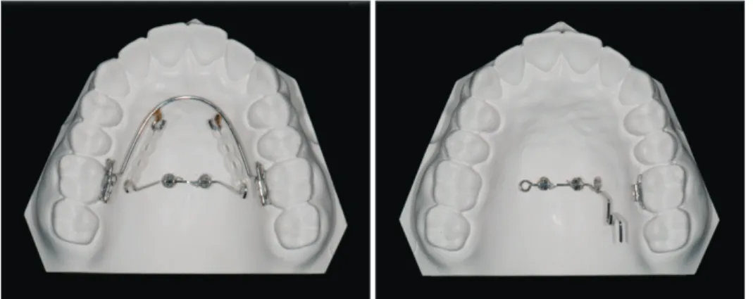
Lingual arch type appliance for upper molar distalization The lingual arch type of biomechanical system utilizes a lingual arch (0.8−0.9 mm) connecting the right and left first molars with hooks soldered onto the mesial part (Figure 1). A rectangular wire with hooked ends is inserted into the miniscrews’ rectangular slots to connect two midpalatal miniscrews (Figure 1). Elastomeric chain or coil springs are engaged between the lingual arch hook and midpalatal miniscrew connecting wires to deliver a distalizing force to the molars. By adjusting the vertical length of the midpalatal miniscrew connecting wires, the maxillary molar distalizing pattern can be controlled: shorter lengths (hooks placed near the palatal roof) can provide greater root distalizing movement, while longer lengths (hooks placed near the tooth crown) can provide greater crown distalizing movement.
In all of the cases included in the present study, the midpalatal miniscrew connecting wires and the lingual arches were adjusted to direct the distalizing force vector
Figure 1. Midpalatal minisc-
rew assisted maxillary molar
distalizer. Right, lingual arch-
type appliance; Left, pendu-
lum type appliance
through the furcation of the maxillary first molar and parallel to the occlusal plane.
Pendulum type appliance for maxillary molar distaliza tion The pendulum type of midpalatal miniscrew-supported maxillary molar distalizer adopts the wire design of the conventional Hilgers pendulum appliance.
15A stainless steel 0.0215 × 0.0250 inch midpalatal-miniscrew connecting wire with a helix and a horizontal loop is utilized, with its end inserted into the lingual sheath of the first molar (Figure 1). The activation mode is the same as that for the conventional pendulum appliance:
the helix is activated to distalize the whole pendulum arm and then inserted into the lingual sheath of the first molar.
MATERIALS AND METHODS
To investigate the molar distalization patterns associated with lingual arch type and pendulum type appliances, 14 patients who each received one of the two appliances were enrolled into the study. All patients exhibited a bilateral or unilateral Class II molar relationship at the start of treatment. Seven patients (14 molars, age 19.2±4.4 years) were treated with the lingual arch type appliance, and seven (12 molars, age 20.9±7.6 years) were treated with the pendulum type appliance.
All patients received full fixed orthodontic appliances.
With brief regard to treatment mechanics, alignment and leveling was performed after placing full fixed
appliances until 0.016 × 0.022-inch stainless steel wire could be engaged passively. Molar distalization was performed until a super Class I relationship was achieved.
Molar distalizing force was subsequently decreased to hold the position while retracting premolars, canines, and incisors.
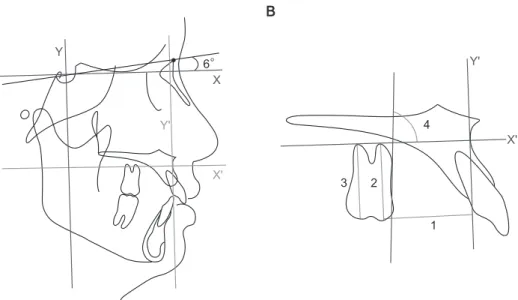
Initial (pre-orthodontic treatment) and final (post- orthodontic treatment) lateral cephalograms were obtained and analyzed. Maxillary molar movement was analyzed using measurements described by Cha and Ngan.
16The X-Y coordinate was constructed using a horizontal rotating sella-nasion line in a downward direction at 6
o(X-axis) and a vertical line perpendicular to the horizontal line passing through the sella point (Y-axis). Using the X-Y coordinate, a 0−0 point was set at point A and a new X´-Y´ coordinate was constructed (Figure 2A). Measurements recorded included the Y´ line to maxillary first molar mesial cusp tip distance (mm), X´ line to maxillary first molar mesial cusp tip distance (mm), X´ line to maxillary first molar distal cusp tip distance (mm), and angle (
o) between the X´ line and a line starting from the mesial point of the maxillary first molar crown tangent to the root (Figure 2B).
Statistical evaluation
The Mann-Whitney U test was used to compare measurements between the two groups (lingual arch versus pendulum), while for intragroup comparisons (pre- and post-treatment), the Wilcoxon signed-rank test was used.
A B
Y
X
Y'
X' 6
X' Y'
1 2
3
4
Figure 2. Cephalometric measurements. A, The X-Y coordinate was constructed using a horizontal rotating sella-nasion
line in a downward direction at 6° (X-axis) and a vertical line perpendicular to the horizontal line passing through
the sella point (Y-axis). Using the X-Y coordinate, a 0−0 point was set at point A, and a new X´-Y´ coordinate was
constructed. B, 1: Y´ line to maxillary first molar mesial cusp tip distance (mm). 2: X´ line to maxillary first molar mesial
cusp tip distance (mm). 3: X´ line to maxillary first molar distal cusp tip distance (mm). 4: Angle (
o) between X´ line and a
line starting from the mesial point of the maxillary first molar crown tangent to the root.
RESULTS
Pre-treatment measurements did not differ statistically significantly between the two groups (Table 1). Post- treatment, first molar angulation differed significantly between the groups (Table 1). Post-treatment, the pendulum appliance group showed greater distal angulation than the lingual arch appliance group.
In the lingual arch appliance group, pre- and post- treatment comparisons yielded a mean distalization of 2.4 mm (Table 2). The vertical position exhibited slight intrusion (0.3 mm); however, this was not statistically significant. A statistically significant reduction in angulation to 0.8 mm indicated the occurrence of crown mesial tipping or root distal tipping.
For the pendulum type appliance, pre- and post- treatment comparisons yielded a mean distalization of 1.8 mm, and intrusion of the first molar differed statistically significantly (Table 2). The amount of intrusion differed between the mesial cusp and the distal cusp: distal cusp intrusion (X´-distal cusp distance
−1.1 ± 0.4 mm) was greater than mesial cusp intrusion (X´-mesial cusp distance −0.8 ± 0.5 mm). Additionally, angulation changed significantly to +1.5
o± 1.3
o, indicating the occurrence of distal tipping during distalization.
DISCUSSION
The utilization of midpalatal miniscrews as an absolute anchorage mechanism offers several advantages, and can aid in the achievement of optimal treatment outcomes.
The most essential advantage is the low failure rate.
The midpalatal area lacks critical anatomical structures such as large sized vessels and nerves, and dental roots which are reportedly responsible for increasing the risk of miniscrew failure when they are implanted in close proximity.
17,18In contrast, the midpalatal area has an
abundance of keratinized gingiva with an excellent quality of cortical bones, favoring the stability of miniscrews.
19However, the utilization of midpalatal miniscrews is not as popular as interdental miniscrews because the midpalatal area is far from maxillary dentition. Several articles describing the biomechanics of midpalatal miniscrew utilization for the control of maxillary dentition have been published.
8,14,20The current report analyzes maxillary molar distalizing patterns as a follow-up study.
Distalizing force vector was adjusted to pass through furcation of the upper first molar for the lingual arch type appliance group. As expected, the results showed almost bodily distal movement: mean 0.8
omesial crown tipping or root distal tipping occurred, while a mean distal movement of 2.4 mm was achieved. This equates to approximately 0.3
oof mesial crown tipping per 1 mm of distal movement, and is clinically negligible. The clinical advantage of the lingual arch appliance is the control of molar tipping; however, it is disadvantaged by its complex design (two wires in the palate) which can increase patient discomfort. Furthermore, it can only be applied in bilateral distalizing cases.
The pendulum type appliance produced significant distal tipping of the maxillary molars during distali- zation. This was anticipated based on previous studies investigating the effects of the conventional pendulum appliance, which included distal tipping of the maxillary molars during distalization.
3,4,21Mean distal crown tipping of 1.5
ooccurred during a mean distalization of 1.8 mm, which equates to approximately 0.8
odistal tipping per 1 mm of distalization. The extent of distal tipping was lower than that reported in previous studies investigating the pendulum appliance; however, direct comparison is not possible as total distalization was much smaller in the present study. In addition, the movement pattern of the upper first molars measured in the present study was actually the result of a Table 1. Pre and post treatment measurements comparison between the appliance groups using Mann-Whitney U-test
Pretreatment Group
difference (p-value)
Posttreatment Group
difference (p-value) Lingual arch
type (n = 14) Pendulum
type (n = 12) Lingual arch
type (n = 14) Pendulum type (n = 12) Sagittal measurement (mm)
Y´-Mx6 26.5 ± 3.2 30.5 ± 4.5 0.231 29.1 ± 3.9 32.6 ± 4.3 0.820
Vertical measurement (mm)
X´-mesial cusp 17.9 ± 3.0 16.8 ± 3.7 0.667 17.5 ± 2.8 16.0 ± 3.4 0.432
X´-distal cusp 15.4 ± 3.7 14.8 ± 4.7 0.899 15.0 ± 3.5 13.8 ± 4.4 0.527
Angulation (°) 84.2 ± 5.3 80.2 ± 8.9 0.403 85.3 ± 4.8 78.6 ± 8.5 0.046*
Values are presented as mean ± standard deviation.
*p < 0.05.