Different Location of Triaxial Accelerometer and Different Energy Expenditures
Do Yoon Kim,
1Yoo-Suk Jung,
1Rae-Woong Park,
2and Nam-Seok Joo
31Fit. Life Inc., Suwon; Departments of 2Medical Informatics and 3Family Practice and Community Health, Ajou University School of Medicine, Suwon, Korea.
Received: March 25, 2013 Revised: November 19, 2013 Accepted: November 25, 2013
Corresponding author: Dr. Nam-Seok Joo, Department of Family Practice and Community Health,
Ajou University School of Medicine, 164 World cup-ro, Yeongtong-gu, Suwon 443-380, Korea.
Tel: 82-31-219-5324, Fax: 82-31-219-5218 E-mail: [email protected]
∙ The authors have no financial conflicts of interest.
© Copyright:
Yonsei University College of Medicine 2014 This is an Open Access article distributed under the terms of the Creative Commons Attribution Non- Commercial License (http://creativecommons.org/
licenses/by-nc/3.0) which permits unrestricted non- commercial use, distribution, and reproduction in any medium, provided the original work is properly cited.
Purpose: We performed a study to determine the best appropriate wearing site of a
triaxial accelerometer at different exercise speeds. Materials and Methods: We con- ducted an observational study with 66 healthy Korean adults (26 men and 40 wom- en). Resting metabolic rate (RMR) before exercise, physical activity-related energy expenditure (PAEE) by cardiorespiratory gas analyzer and Signal Vector Magnitude (SVM) were measured while wearing four triaxial accelerometers on four different sites (wrist, waist, upper arm, and ankle) at exercise speeds from 2--10 km/h. Results:
The mean RMR was 4.03 mL/kg/min and Actual METs (oxygen consumption at dif- ferent exercise speeds divided by individual RMR) compared with the calculated METs (oxygen consumption divided by 3.5 mL/kg/min) showed relatively low value.
The overall correlation between PAEE and SVM was highest when the accelerometer was worn on the wrist at low exercise speed (r=0.751, p<0.001), waist at a moderate speed (r=0.821, p<0.001), and ankle at a high speed (r=0.559, p<0.001). Using regres- sion analysis, it was shown that the ankle at a low speed (R
2=0.564, p<0.001), high speed (R
2=0.559, p<0.001), and the waist at a moderate speed (R
2=0.821, p<0.001) were the best appropriate sites. Conclusion: When measuring the PAEE and SVM at different exercise speeds, the ankle in low and high exercise speed, and waist in mod- erate speed are the most appropriate sites for an accelerometer.
Key Words:
Physical activity, energy expenditure, Signal Vector Magnitude, tri- axial accelerometer
INTRODUCTION
Maintaining proper physical activity or exercise is an important factor in reducing
metabolic risks
1as well as obesity.
2In the modern global society, many people live
under circumstances where physical activity or exercise is limited due to a sedentary
lifestyle or daily job-related burdens, even though they recognize the importance of
health and physical activity/exercise. Moderate-intensity cardiorespiratory exercise
training (≥30 min per day, ≥5 days per week, for a total ≥150 minutes per week),
vigorous-intensity cardiorespiratory exercise training (≥20 min per day, ≥3 days per
week, for a total ≥75 minutes per week), or a combination of moderate- and vigor-
ous-intensity exercise to achieve a total energy expenditure of ≥500--1000 MET/
and arrhythmia or cardiomegaly in 12-lead electrocardiog- raphy (EKG). All participants received a medical interview and all EKGs were normal. The Institutional Review Board of Ajou University Hospital approved this study (AJIRB- DEV-DE2-10-298).
Study design
All measurements were conducted by a trained research nurse and research staff in the clinical trial center in Ajou University Hospital. All participants were requested to mea- sure resting metabolic rate (RMR, mL/kg/min) for 5 min rest before exercise by cardiorespiratory gas analyzer. PAEE calculated by oxygen consumption also measured at differ- ent speeds on the treadmill adjusted by individual RMR.
Oxygen consumption was measured by cardiorespiratory gas analyzer during exercise (walk or running, as subjects’
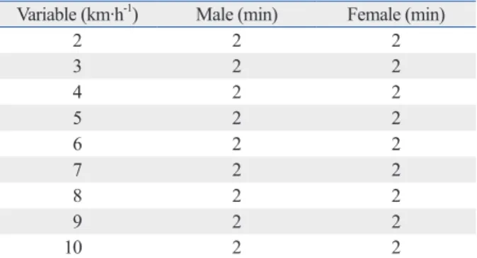
habitual exercise) with different speeds (2, 3, 4, 5, 6, 7, 8, 9, and 10 km/h). Each was continued for 2 min, for a total of 18 min of exercise (Table 1). During exercise, the activity of exercise was measured as SVM by simultaneously apply- ing four triaxial accelerometers on the wrist, ankle, upper arm, and waist. Accelerometer data and energy expenditure data were synchronized under the control of a computer sys- tem before measurement, which could control time set up.
To acquire stable exercise data for each speed, the data of the first 50 sec and the last 10 sec in each 2 min exercise period were eliminated, since the posture of exercise during the first 50 sec and last 10 sec could be changed or influ- enced by the speed change, with the remaining 60 sec at each exercise speed being analyzed. All participants completed the study after 5 min rest under the close observation by the re- searchers. There were no adverse events during the study.
Measurement of RMR, PAEE, and SVM
RMR in mL/kg/min of each participant was measured us- ing an Ultima PFX cardiorespiratory gas analyzer (Medical Graphic, St. Paul, MN, USA) after 5 min rest without fast- min/wk is recommended to maintain general health.
3Addi-
tionally, the number of studies assessing cost-effectiveness of exercise intervention in various diseases is still limited.
4Therefore, the exact measurement of physical activity is im- portant. On the other hand, objective measurement of physi- cal activity is difficult due to the fact that the exact descrip- tion of movement that is robust to location on the human body itself is difficult to acquire.
5Triaxial accelerometers have been used to monitor physi- cal activity or exercise in many clinical fields,
6,7partly be- cause quantitative monitoring of physical activity in daily life has an important role. A triaxial accelerometer is a small device that can be carried or worn on the wrist, upper arm, waist, ankle, and other body sites. It permits an objective measurement of physical activity by Signal Vector Magni- tude (SVM, expressed as cm/s
2) and convert those measured values into physical activity-related energy expenditure (PAEE, expressed as kcal/kg/min).
8,9However, the past lit- erature is unclear concerning the best body site to evaluate physical activity or PAEE by the use of a triaxial acceler- ometer, even though some accelerometer products were de- veloped to wear on sites including the wrist, upper arm, waist, ankle, and hip. Because the exact monitoring of physi- cal activity has a role in the behavior modification for the maintenance of proper physical activity or exercise,
10the body location of a triaxial accelerometer during exercise at different speeds is an important consideration. Therefore, this study was undertaken to determine and recommend the most appropriate site to wear a triaxial accelerometer dur- ing different exercise speeds in healthy Korean adults, with the goal of determining the best relationship between PAEE determined by cardiorespiratory gas analyzer and SVM measured by triaxial accelerometer.
MATERIALS AND METHODS
Participants
This study was conducted in the Department of Family Practice and Community Health, Ajou University Hospital, Suwon, Gyeonggi-do, Republic of Korea. Sixty six healthy adults (26 men and 40 women), 20--49 years of age volun- teered for the study and were enrolled after providing in- formed consent. Exclusion criteria included pregnancy, cur- rent breast feeding in women, past and/or current history of heart disease or chronic lung diseases, dyspnea or chest pain by simple exercise, an evident myocardial ischemia,
Table 1. Testing Protocol for All Activities
Variable (km∙h-1) Male (min) Female (min)
2 2 2
3 2 2
4 2 2
5 2 2
6 2 2
7 2 2
8 2 2
9 2 2
10 2 2
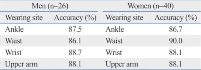
ed body sites were 86.1--90.0%, which was considered high enough to represent the PAEE in different sites (Table 2).
Other measurements
Body weight and height were measured with a test gown using an automatic height-weight meter before each exer- cise test. Results were described by 0.1 cm and 0.1 kg, re- spectively. Body mass index (BMI) was calculated as weight (kg)/height (m)
2.
Statistical analyses
All measured values showed normal distribution. General characteristics, calculated METs, Actual METs, PAEE, and SVM were expressed as mean±standard deviation. Actual METs were divided by three exercise speed categories: low (<3.00 MET), moderate (3.00≤MET<6.00), and high (MET
≥6.00). The corresponding actual exercise speed of low METs was 2--4 km/h, moderate was 5--7 km/h, and high was 8--10 km/h. After the division of actual exercise speed, we used the partial correlation method to evaluate the asso- ciation of PAEE using oxygen consumption with measured SVM in different wearing sites in the speed category after age, sex, and BMI adjustments. Finally, to determine the proper wearing site of triaxial accelerometer for the match- ing of PAEE with SVM at the different speeds, linear re- gression analysis was used. Data were analyzed using SPSS 18.0 (SPSS Inc., Chicago, IL, USA) with p<0.05 were con- sidered statistically significant.
RESULTS
Clinical characteristics of study subjects
The mean age of participants in this study was 36.0±10.3 ing. This gas analyzer measures energy expenditure using
the ratio of O
2to CO
2. Pulmonary Exercise Ultima PF is in- direct calorimeter that calculates respiratory exchange ratio (CO
2·O
2-1). Each participant wore the Ultima PF, while per- forming each activity and throughout the rest periods. The Ultima PF is a gas exchange system that measures ventila- tion, expired concentration of oxygen and carbon dioxide, which then estimates energy expenditure. The Ultima PF is small in size and lightweight, 36×33×36 cm and 12 kg. It has a range of ±18 L·sec
-1, accuracy ±3% or 50 mL, which- ever is greater (meets or exceeds ATS/ERS clinical perfor- mance standards), and resolution 8.64 mL·sec
-1. For the RMR measurements, we used the latter half of the exercise period (2.5 min) to get the stable data. The reason for the omission of fasting was our view that the actual RMR with- out fasting is more important in daily life and is based on the oxygen consumption at rest, which is different from the basal metabolic rate. In addition to measuring RMR, we obtained two kinds of activity-related oxygen consumption (METs): Calculated METs and Actual METs. To calculate Actual MET, the measured oxygen consumption at a partic- ular speed was divided by the individual RMR. For the Cal- culated MET, the measured oxygen consumption at each speed was divided by 3.5 mL/kg/min. Calculation of PAEE using oxygen consumption was based on the assumption that 1 liter of oxygen produces 5 kcal. Simultaneously, SVM was measured using a Fitmeter triaxial accelerometer (Fit. Life, Suwon, Korea), which was sum of total accelera- tion values of all three axes (x, y, z cm/s
2). Usually, the ac- celerometer is worn on a belt positioned around the partici- pants’ ankle, waist, wrist, and upper arm. The Fitmeter accelerometer was positioned on the ankle, waist, wrist, and upper arm and the Actical accelerometer was posi- tioned on the ankle, waist, and wrist. The Fitmeter acceler- ometer is a small (35×35×12 mm) and lightweight (13.5 g) devices that uses triaxial vector data in activity that mea- sures accelerations in the range of -8--+8 G. These values correspond to the range in which most human activities are performed. Samples at a rate of 32 Hz and these values are then summed over a specified time period (epoch). The Fit- meter was worn on the ankle, waist, wrist, and humerus sites in nylon pouch that was secured to a belt provided by the manufacturer. The device was initialized using 1/32 sec epochs and converted to 1-min epoches for the data analy- sis. The results were downloaded directly to a PC compati- ble computer using a USB cable. The predictive accuracy of the algorithm in calculating Actual METs in the four select-
Table 2. Predictive Accuracy of Algorithm at Each Wearing Site on the Body
Men (n=26) Women (n=40)
Wearing site Accuracy (%) Wearing site Accuracy (%)
Ankle 87.5 Ankle 86.7
Waist 86.1 Waist 90.0
Wrist 88.7 Wrist 88.1
Upper arm 88.1 Upper arm 88.1
PAEE, physical activity-related energy expenditure; SVM, Signal Vector Magnitude.
Each accuracy at different triaxial accelerometer wearing sites represent the relationship between actual physical activity (METs or kcal) and predic- tive physical activity (METs or kcal) using PAEE and SVM measured using Fitmeter accelerometers.
years, mean BMI was 23.4±3.0 kg/m
2, and the measured RMR by cardiorespiratory gas analyzer after 5 min rest was 4.03±0.60 mL/kg/min (Table 3).
Comparison of METs, PAEE, and SVM at each exercise speed
Mean actual METs of study subjects at exercise speed of 2, 3, 4, 5, 6, 7, 8, 9, and 10 km/h was 2.14±0.29, 2.56±0.38, 2.92±0.51, 3.56±0.72, 4.58±0.94, 5.96±1.08, 7.11±1.09, 7.77±1.19, and 8.40±1.17, respectively. For each exercise speed (values from each MET divided by 3.5 mL/kg/min), the Actual MET values were lowered than Calculated MET values. Therefore, the use of Calculated MET may overesti- mate the real exercise intensity. Mean calculated PAEE us- ing oxygen consumption at exercise speeds of 2, 3, 4, 5, 6, 7, 8, 9, and 10 km/h was 1.45±0.40, 1.98±0.50, 2.43±0.61, 3.22±0.83, 4.51±1.14, 6.22±1.28, 7.68±1.42, 8.60±1.62, and 9.51±1.78 kcal/min, respectively. Measured SVM (cm/
s
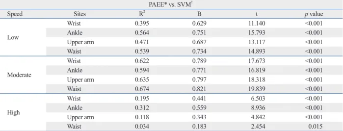
2) by the Fitmeter triaxial accelerometer were also deter- mined at the wrist, ankle, upper arm, and waist. All values increased as the exercise speed increased (Table 4). Overall relation between METs and SVM showed a high correla- tion with the ankle in low intensity (r=0.751, p<0.001), waist in moderate intensity (r=0.821, p<0.001), and ankle in high intensity (r=0.559, p<0.001) after adjustments with age, sex, and BMI.
Regression analysis of PAEE with SVM at different wearing sites for the exercise speed categories
Linear regression analysis was used to determine the most appropriate site to represent the association between PAEE using oxygen consumption and SVM measured using triaxi- al accelerometers during exercise at the designated speeds.
Similar to the correlation analysis, results also showed prom- inent significant relation between PAEE and SVM. At low speed, moderate speed, and high speed, the most significant site was the ankle (R
2=0.564, p<0.001), waist (R
2=0.674,
Table 3. Clinical Characteristics of Study SubjectsVariables Mean (SD)
Numbers of subjects (women) 66 (40)
Age (yrs) 36.0 (10.3)
Body weight (kg) 64.3 (11.9)
Height (cm) 165.3 (9.1)
BMI (kg/m2) 23.4 (3.0)
Measured RMR (mL/kg/min) 4.03 (0.60) BMI, body mass index; RMR, resting metabolic rate.
RMR measured by Cardiorespiratory Gas Analyzer. Table 4. Mean Values of Exercise Intensity (METs), PAEE, and SVM in the Different Speed and Wearing Sites Exercise speed 2 km/h3 km/h4 km/h5 km/h6 km/h7 km/h8 km/h9 km/h10 km/h METs (calculated)* 2.44 (0.43) 2.93 (0.49) 3.33 (0.59) 4.04 (0.72) 5.20 (0.89) 6.73 (0.81) 8.04 (0.73) 8.79 (0.83) 9.62 (0.85) † METs (actual) 2.14 (0.29) 2.56 (0.38) 2.92 (0.51) 3.56 (0.72) 4.58 (0.94) 5.96 (1.08) 7.11 (1.09) 7.77 (1.19) 8.40 (1.17) ‡ PAEE 1.45 (0.40) 1.98 (0.50) 2.43 (0.61) 3.22 (0.83) 4.51 (1.14) 6.22 (1.28) 7.68 (1.42) 8.60 (1.62) 9.51 (1.78) § SVM Wrist 8190.6 (3384.7) 13090.6 (7312.5) 18124.2 (11832.6) 25466.3 (16095.9) 38977.9 (24797.1) 66351.3 (30547.8) 96905.4 (18909.5)106136.9 (19522.9) 115522.1 (21094.4) Ankle20164.6 (5244.0)31837.6 (5259.8)41433.6 (6238.6)50536.6 (6880.5)59879.1 (7600.0)72059.9 (7958.7)85472.8 (7698.0)94452.3 (7105.2)103577.1 (7758.9) Upper arm 7212.4 (2197.6)10666.7 (3594.0)13981.8 (6993.2) 20152.6 (13187.8) 31214.4 (19145.3) 52564.6 (22326.5) 77607.6 (12479.6) 83625.4 (12527.7) 88768.7 (12768.4) Waist 7476.6 (1773.1)11981.0 (2296.2)17329.7 (4115.5) 25618.1 (11320.9) 37025.2 (14647.7) 54097.6 (16401.2) 70498.0 (11643.3) 74852.1 (10823.2) 78335.9 (11069.6) All values are mean (standard deviation). *Calculated oxygen consumption (Calculated METs, mL/kg/min) measured by Cardiorespiratory Gas Analyzer at each speed divided by 3.5 mL/kg/min. † Actual oxygen consumption (actual METs, mL/kg/min) measured by Cardiorespiratory Gas Analyzer at each speed divided by individual resting metabolic rate. ‡ Physical activity-related energy expenditure (PAEE), represents activity energy expenditure (kcal/kg/min) at each speed, which calculated from the equation ‘Oxygen consumption by exercise--Resting Metabolic Rate’, and then transformed under the assumption ‘1 liter of oxygen produce 5 kcal’. §2 Signal Vector Magnitude (SVM) is the total value of vector of three axes (x, y, z cm/s) measured using Fitmeter triaxial accelerometers.
treadmill walking at self-selected speeds
16or correlation of moderate-to-vigorous physical activity in different racial/
ethnic groups, which might require culturally tailored strat- egies.
17In one study, to obtain optimal results with acceler- ometers in clinical trials, the authors recommended detailed protocols for monitor use, calibration of monitors and vali- dation of data quality and use of validated equations for analysis.
18However, there has been no recommendation of the wearing site of triaxial accelerometer during exercise with different speeds or exercise intensities. In a study re- porting the validity of three accelerometers during treadmill walking and motor vehicle travel,
19the researchers followed the recommendations of each accelerometer user guideline;
in which the location of the accelerometer was the anterior thigh, ankle, and waist. Moreover, most clinical trials have been conducted based on the recommendations from the manufacturer and all participants were instructed to wear the device at certain locations. For example, consistent with the anatomical location of the anterior iliac spine, with the accelerometer is placed in a vertical position and the acceler- ometer worn on the belt or waistband of the clothing. Alter- nately, subjects were provided with a belt to secure the accel- erometer to the proper location at the waist in the event that it could not be attached properly to their clothing,
20which seemed to be the standard wearing site. But, a triaxial accel- erometer can be worn on various sites, such as wrist, ankle, upper arm, and waist using a band or other accessories.
p<0.001), and ankle (R2
=0.312, p<0.001), respectively, even though other sites also showed a significant relation- ship (Table 5).
DISCUSSION
In this trial to determine the best appropriate body location of a triaxial accelerometer, at low and high exercise speed, the ankle was the best site to represent that association. For moderate exercise speed, the waist was the best site to rep- resent the association, even though other sites showed sig- nificant results.
Many studies have addressed the relationship between physical activity or PAEE and diseases such as osteoarthri- tis,
11early aerobic endurance training intervention in pa- tients with coronary artery diseases,
12and weight loss main- tenance behavioral intervention in a diverse population of high-risk patients,
13as well as an intervention study to eval- uate the changes in visceral fat in a group of subjects who exercised vigorously with restricted caloric intake.
14In many clinical trials, however, accurate measurement of physical activity or PAEE is essential to guarantee research quality and reduce measurement error.
With careful consideration, individual differences of physi- cal activity during exercise may exist, such as accelerometer wear time,
15measurement of physical activity in level-ground,
Table 5. Regression Analysis of PAEE of Different Wearing Sites with SVM in the Low, Moderate, High Speed Exercise Cat- egories
PAEE* vs. SVM†
Speed Sites R2 Β t p value
Low
Wrist 0.395 0.629 11.140 <0.001
Ankle 0.564 0.751 15.793 <0.001
Upper arm 0.471 0.687 13.117 <0.001
Waist 0.539 0.734 14.893 <0.001
Moderate
Wrist 0.622 0.789 17.673 <0.001
Ankle 0.594 0.771 16.819 <0.001
Upper arm 0.635 0.797 18.318 <0.001
Waist 0.674 0.821 19.839 <0.001
High
Wrist 0.195 0.441 6.503 <0.001
Ankle 0.312 0.559 8.936 <0.001
Upper arm 0.118 0.343 4.842 <0.001
Waist 0.034 0.183 2.454 0.015
PAEE, physical activity-related energy expenditure.
All values are from linear regression analysis. Low represents <3.00 METs, 2--4 km/h. Moderate represents 3.00≤METs<6.00 METs, 5--7 km/h. High repre- sents ≥6.00 METs, 8--10 km/h.
*PAEE, represents energy expenditure (kg/kcal/min) at each speed, which calculated from the equation ‘Oxygen consumption by exercise--Resting Meta- bolic Rate’, and then transformed under the assumption ‘1 liter of oxygen produce 5 kcal’.
†Signal Vector Magnitude (SVM) is the total value of vector of three axes (x, y, z cm/s2) measured using Fitmeter triaxial accelerometers.
individuals would occur. SVM might also vary with adop- tion of different postures during exercise at different speeds.
There were several strengths of the study. The study is the first to evaluate the relationship between PAEE and SVM at different body locations at different exercise speeds. This study design revealed the benefit of different accelerometer body sites at different exercise speeds for most accurate measurements. The study also determined that measuring PAEE and SVM at one site during the entire period of exer- cise can potentially produce a measurement error. There- fore, to best evaluate PAEE and SVM, the body site should be considered with respect to the intended speed or intensi- ty of the exercise, as well as proper adjustment of the wear- ing site. Furthermore, development of a proper algorism ac- cording to the wearing site is important. Actually, in real outdoor activity, wearing an accelerometer on the ankle may be inconvenient, therefore, wearing it on the waist for low and moderate speed exercise and the wrist for high speed exercise is recommended.
In conclusion, when measuring the PAEE and SVM at low (2--4 km/h or <3.00 METs) and high (8--10 km/h or
≥6.00 METs) exercise speeds, the ankle is the best site for an accelerometer. For moderate exercise speed (5--7 km/h or 3.00≤METs<6.00), the waist is the best site to represent the association between PAEE and SVM. Research de- signed to evaluate the PAEE and SVM at different speeds should consider the most appropriate site of wearing a tri- axial accelerometer under the exercise conditions.
ACKNOWLEDGEMENTS
This study was supported by a grant of the Korea Healthcare technology R&D Project, Ministry for Health, Welfare &
Family Affairs, Republic of Korea (A084120). We thank all participants in this clinical trial and research nurse Myung- Eun Son for her excellent assistance.
REFERENCES
1. Simmons RK, Griffin SJ, Steele R, Wareham NJ, Ekelund U; Pro- Active Research Team. Increasing overall physical activity and aer- obic fitness is associated with improvements in metabolic risk: co- hort analysis of the ProActive trial. Diabetologia 2008;51:787-94.
2. Goodpaster BH, Delany JP, Otto AD, Kuller L, Vockley J, South- Paul JE, et al. Effects of diet and physical activity interventions on weight loss and cardiometabolic risk factors in severely obese adults: a randomized trial. JAMA 2010;304:1795-802.
The benefits and drawbacks of each body location site of the accelerometer can be summarized as follows. The ac- celerometer can be worn at the waist by securing the device to a belt or waistband of the clothing, or carrying the device in a pocket of tight skirts, pants, or trousers. The benefits of this method are a relatively high accuracy due to the central location, irrespective of a subject’s sedentary behavior, and the option of hiding the accelerometer underneath the cloth- ing for subjects concerned with the appearance of the de- vice. An accelerometer positioned on the wrist or upper arm is likely to yield relatively low accuracy in a subject with a sedentary lifestyle because of an overestimation due to higher arm movement compared with whole body move- ment. However, a wrist or upper arm location carries the benefit of device use as an accessory or watch that makes it easy for a subject to monitor their calorie use and physical activity. Additionally, wearing an accelerometer on the up- per arm enables music during exercise, if the accelerometer provides MP3 function. An accelerometer positioned on an ankle is better for accurate recording of subjects with a sed- entary lifestyle, since leg movement is not typically as pro- nounced as arm movement in such subjects. But, an ankle location can be inconvenient to install and wear, which may lower the compliance of use.
Many clinical trials are likely to use the triaxial acceler- ometer worn on the waist or central core of the body. The present results indicate that this and other body locations are acceptable to represent the relationship between PAEE and SVM. Contrary to our expectation, however, the ankle was the most appropriate site for low speed and high speed exercise or intensity, and best reflected the relationship be- tween PAEE and SVM in these conditions. For moderate speed exercise or intensity, the waist proved to be the most appropriate site. Given that accelerometers are sensitive to the changes in treadmill speed,
21the possible influence of changing speed should be taken into account when select- ing an accelerometer site in terms of evaluating PAEE or SVM. Most of clinical trials are likely to compare physical activity or PAEE measured at a single accelerometer body location during the entire period of exercise, which creates the possibility of low correspondence with actual physical activity or PAEE, given the different speed measured by different wearing sites.
This study had some limitations. Study subjects were com-
prised of one ethnic group and healthy population. All par-
ticipants were requested to engage in their exercise as their
usual exercise posture or pattern, and so variations between
tive. Arthritis Care Res (Hoboken) 2010;62:1724-32.
12. Hansen D, Eijnde BO, Roelants M, Broekmans T, Rummens JL, Hensen K, et al. Clinical benefits of the addition of lower extremi- ty low-intensity resistance muscle training to early aerobic endur- ance training intervention in patients with coronary artery disease:
a randomized controlled trial. J Rehabil Med 2011;43:800-7.
13. Hollis JF, Gullion CM, Stevens VJ, Brantley PJ, Appel LJ, Ard JD, et al. Weight loss during the intensive intervention phase of the weight-loss maintenance trial. Am J Prev Med 2008;35:118-26.
14. Nicklas BJ, Wang X, You T, Lyles MF, Demons J, Easter L, et al.
Effect of exercise intensity on abdominal fat loss during calorie restriction in overweight and obese postmenopausal women: a randomized, controlled trial. Am J Clin Nutr 2009;89:1043-52.
15. Napolitano MA, Borradaile KE, Lewis BA, Whiteley JA, Longval JL, Parisi AF, et al. Accelerometer use in a physical activity inter- vention trial. Contemp Clin Trials 2010;31:514-23.
16. Hendrick P, Boyd T, Low O, Takarangi K, Paterson M, Claydon L, et al. Construct validity of RT3 accelerometer: a comparison of level-ground and treadmill walking at self-selected speeds. J Re- habil Res Dev 2010;47:157-68.
17. Kelly EB, Parra-Medina D, Pfeiffer KA, Dowda M, Conway TL, Webber LS, et al. Correlates of physical activity in black, Hispanic, and white middle school girls. J Phys Act Health 2010;7:184-93.
18. Jakicic JM, Gregg E, Knowler W, Kelley DE, Lang W, Miller GD, et al. Activity patterns of obese adults with type 2 diabetes in the look AHEAD study. Med Sci Sports Exerc 2010;42:1995-2005.
19. Maddocks M, Petrou A, Skipper L, Wilcock A. Validity of three accelerometers during treadmill walking and motor vehicle travel.
Br J Sports Med 2010;44:606-8.
20. Winkler EA, Gardiner PA, Clark BK, Matthews CE, Owen N, Healy GN. Identifying sedentary time using automated estimates of accelerometer wear time. Br J Sports Med 2012;46:436-42.
21. Jacobi D, Perrin AE, Grosman N, Doré MF, Normand S, Oppert JM, et al. Physical activity-related energy expenditure with the RT3 and TriTrac accelerometers in overweight adults. Obesity (Silver Spring) 2007;15:950-6.
3. Garber CE, Blissmer B, Deschenes MR, Franklin BA, Lamonte MJ, Lee IM, et al. American College of Sports Medicine position stand. Quantity and quality of exercise for developing and main- taining cardiorespiratory, musculoskeletal, and neuromotor fitness in apparently healthy adults: guidance for prescribing exercise.
Med Sci Sports Exerc 2011;43:1334-59.
4. Roine E, Roine RP, Räsänen P, Vuori I, Sintonen H, Saarto T.
Cost-effectiveness of interventions based on physical exercise in the treatment of various diseases: a systematic literature review.
Int J Technol Assess Health Care 2009;25:427-54.
5. Crouter SE, DellaValle DM, Haas JD, Frongillo EA, Bassett DR.
Validity of ActiGraph 2-regression model, Matthews cut-points, and NHANES cut-points for assessing free-living physical activi- ty. J Phys Act Health 2013;10:504-14.
6. Feinglass J, Lee J, Semanik P, Song J, Dunlop D, Chang R. The effects of daily weather on accelerometer-measured physical ac- tivity. J Phys Act Health 2011;8:934-43.
7. Camhi SM, Sisson SB, Johnson WD, Katzmarzyk PT, Tudor- Locke C. Accelerometer-determined lifestyle activities in US adults. J Phys Act Health 2011;8:382-9.
8. Ohkawara K, Oshima Y, Hikihara Y, Ishikawa-Takata K, Tabata I, Tanaka S. Real-time estimation of daily physical activity intensity by a triaxial accelerometer and a gravity-removal classification al- gorithm. Br J Nutr 2011;105:1681-91.
9. Sloane R, Snyder DC, Demark-Wahnefried W, Lobach D, Kraus WE. Comparing the 7-day physical activity recall with a triaxial accelerometer for measuring time in exercise. Med Sci Sports Ex- erc 2009;41:1334-40.
10. Rogers LQ, Hopkins-Price P, Vicari S, Markwell S, Pamenter R, Courneya KS, et al. Physical activity and health outcomes three months after completing a physical activity behavior change inter- vention: persistent and delayed effects. Cancer Epidemiol Bio- markers Prev 2009;18:1410-8.
11. Song J, Semanik P, Sharma L, Chang RW, Hochberg MC, Mysiw WJ, et al. Assessing physical activity in persons with knee osteo- arthritis using accelerometers: data from the osteoarthritis initia-