• 검색 결과가 없습니다.

2020 Aug https://doi.org/10.4070/kcj.2020.0016 pISSN 1738-5520·eISSN 1738-5555

N/A
N/A
Protected

Academic year: 2022

Share "2020 Aug https://doi.org/10.4070/kcj.2020.0016 pISSN 1738-5520·eISSN 1738-5555"

Copied!
2
0
0

로드 중.... (전체 텍스트 보기)

전체 글

(1)Korean Circ J. 2020 Aug;50(8):736-737 https://doi.org/10.4070/kcj.2020.0016 pISSN 1738-5520·eISSN 1738-5555. Images in Cardiovascular Medicine. Weeping Heart: Regrowing Thrombus at Basal Left Ventricle in Acute Stroke. Soon Myung Jung , MD1, Yong Hyun Park , MD, PhD1, Sang Hyun Lee , MD, PhD1, Soo Yong Lee , MD, PhD1, Kyung-Pil Park , MD, PhD2, and June Hong Kim, MD, PhD1 Division of Cardiology, Department of Internal Medicine, Pusan National University Yangsan Hospital, Yangsan, Korea 2 Department of Neurology, Pusan National University Yangsan Hospital, Yangsan, Korea 1. Received: Jan 13, 2020 Accepted: Jan 22, 2020 Correspondence to Yong Hyun Park, MD, PhD Division of Cardiology, Department of Internal Medicine, Pusan National University Yangsan Hospital, 20 Geumo-ro, Yangsan 50612, Korea. E-mail: [email protected] Copyright © 2020. The Korean Society of Cardiology This is an Open Access article distributed under the terms of the Creative Commons Attribution Non-Commercial License (https:// creativecommons.org/licenses/by-nc/4.0) which permits unrestricted noncommercial use, distribution, and reproduction in any medium, provided the original work is properly cited. ORCID iDs Soon Myung Jung https://orcid.org/0000-0003-0846-3133 Yong Hyun Park https://orcid.org/0000-0001-6122-214X Sang Hyun Lee https://orcid.org/0000-0001-7196-2643 Soo Yong Lee https://orcid.org/0000-0003-2616-1294 Kyung-Pil Park https://orcid.org/0000-0003-4952-3796 Conflict of Interest The authors have no financial conflicts of interest.. A 39-year-old male visited the emergency department 90 minutes after the sudden onset of right-sided weakness and aphasia following endoscopic examination for epigastric discomfort and dyspepsia. He had suffered congestive heart failure for years, but was lost to follow-up. Brain magnetic resonance imaging revealed acute embolic infarction at the right cerebrum and left cerebellum. Chest X-ray showed cardiomegaly. The electrocardiogram revealed sinus rhythm and left ventricular (LV) hypertrophy. Laboratory findings were non-specific. On transthoracic echocardiography (TTE), global hypo-kinesis and severe LV systolic dysfunction were found, but specific embolic sources were not evident (Figure 1A, Supplementary Video 1). After 3 days of medication with antiplatelet agents, transesophageal echocardiography found a mobile echogenic mass in the basal anterior wall of LV, which was not observed in the previous TTE (Figure 1B, Supplementary Video 2). Repeated TTE also found newly appeared mobile echogenic mass at the basal anterior wall of LV (Figure 1C, Supplementary Video 3). After a resolution of thrombus with 1 week of adequate anticoagulation therapy, TTE revealed a recess in the basal left ventricle, which had played a role of the nest for a nidus formation (Figure 1D, Supplementary Video 4). After 6 days of uneventful recovery, the patient was discharged with warfarin and antihypertensive therapy in good condition. In this case, the combination of decreased LV systolic function and a ventricular recess might be a necessary condition for thrombus formation. Thorough vigilance should be exerted throughout the LV from apex to base to find out the source of cardiac embolism.. SUPPLEMENTARY MATERIALS Supplementary Video 1 TTE demonstrates no visible embolic source in the LV. Click here to view. Supplementary Video 2 TEE showed newly appeared echogenic mobile mass on LV basal anterior wall. Click here to view. https://e-kcj.org. 736.

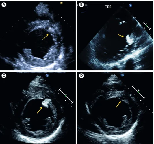
(2) Teardrop of the Heart. Author Contributions Conceptualization: Park YH, Lee SY, Park KP, Kim JH; Supervision: Park YH, Park KP, Kim JH; Writing - original draft: Jung SM; Writing - review & editing: Park YH, Lee SH, Lee SY, Kim JH.. A. B. C. D. TEE. Figure 1. Echocardiographic images. (A) TTE demonstrates no visible embolic source in the LV. (B) TEE showed newly appeared echogenic mobile mass on LV basal anterior wall. (C) TTE also showed newly appeared echogenic mobile mass not seen in previous TTE. (D) After 1 week of anticoagulation, TTE revealed a recess in the basal LV. LV = left ventricle; TEE = transthoracic echocardiography; TTE = transthoracic echocardiography.. Supplementary Video 3 TTE also showed newly appeared echogenic mobile mass not seen in previous TTE. Click here to view. Supplementary Video 4 After 1 week of anticoagulation, TTE revealed a recess in the basal LV. Click here to view. https://e-kcj.org. https://doi.org/10.4070/kcj.2020.0016. 737.

(3)

수치

Figure 1. Echocardiographic images. (A) TTE demonstrates no visible embolic source in the LV

참조

관련 문서

11:20 Preliminary Study on Conceptual Design Analysis of PCCS for SMART Hae Seong Lee, Soon Joon Hong, Yeon Joon Choo, and Jeong Hee Ha(FNC Tech.) Chun Tae Park, Young In Kim,

Chang Je Park, Kwoen Ho Kang, Sang Ho Na, Young Hee Kim, Ho Jin Ryu, Geun Il Park, and Kee Chan Song (KAERI). Geun-Suk Choi and

Dong Won Lee, Young Dug Bae, Suk Kwon Kim, Hee Yun Shin, Bong Guen Hong, Hyun Kyu Jung, Yang Il Jung, Jeong Yong Park, Byung Kwon Choi, and Yong Hwan Jeong(KAERI). P07B04

11:40 Fabrication of Nitride Coated U-Mo Powders for an Advanced Research Reactor Fuel Jae Soon Park, Yong Jin Jeong, Sang Oh Bae, Sun Chil Kwon, Eung Soo Kim, Se Jung Jang,

Sang-Keun Woo, Yong Jin Lee, WonHo Lee, Min Hwan Kim, Ji Ae Park, In Ok Ko, Jin Su Kim, Jong Guk Kim, Young Hoon Ji, Joo Hyun Kang, Gi Jeong Cheon, Chang Woon Choi, Sang Moo

Min Seok Ko, Bo An Lee, Sung Yong Lee, Yeong Jun Jang, Yoon Jeong Hong, and Sin Kim(JNU) TR-PIV Performance Test for a Flow Field Measurement in a Single Rod Test Section Ju

IL Soon Hwang, Myung Hyun Kim, Han Gyu Joo, Kyung Woo Yi, Bong Yoo, Moo Hwan Kim, Seung Rok Oh, Yoon Jae Kim, Jong Gye Shin, Kwang Myung Lee, Jae Yong

녹내장의 진행정도는 시야검사 상 평균 mean deviation (MD) 값이 –11.96 dB 으로 중기 녹내장 소견 을 보였고, 녹내장의 종류는 일차성개방우각녹내장 (primary