© 2017 The Korean Ophthalmological Society
This is an Open Access article distributed under the terms of the Creative Commons Attribution Non-Commercial License (http://creativecommons.org/licenses /by-nc/3.0/) which permits unrestricted non-commercial use, distribution, and reproduction in any medium, provided the original work is properly cited.
Original Article
Clinical Features of Ocular Ischemic Syndrome and Risk Factors for Neovascular Glaucoma
Yung Hui Kim, Mi Sun Sung, Sang Woo Park
Department of Ophthalmology and Research Institute of Medical Sciences, Chonnam National University Hospital, Chonnam National University Medical School, Gwangju, Korea
Purpose: We aimed to examine the clinical features and prognosis of ocular ischemic syndrome and to investi- gate the risk factors for the development of neovascular glaucoma (NVG).
Methods: The medical records from 25 patients (25 eyes) who were diagnosed with ocular ischemic syndrome were retrospectively analyzed. We recorded the length of time between symptom onset and diagnosis, visu- al acuity, intraocular pressure, clinical findings of the anterior and posterior segments of the eye, fluorescein angiography, systemic diseases, smoking history, and the extent of any ipsilateral carotid artery stenosis. The risk factors for NVG in patients with ocular ischemic syndrome were investigated.
Results: The mean age was 67.9 ± 12.5 years, and 21 men and 4 women were included in this study. At initial examination, the mean logarithm of the minimum angle of resolution (logMAR) was 2.02 ± 1.26, and the mean intraocular pressure was 21.0 ± 10.3 mmHg. Among 25 eyes of the 25 patients, NVG occurred in 17 eyes after a mean period of 12.6 ± 14.0 months. The length of time between symptom onset and diagnosis ( p = 0.025) and the extent of ipsilateral carotid artery stenosis (p = 0.032) were identified as significant risk factors for NVG. At the final follow-up, the mean logMAR visual acuity was 3.13 ± 1.24, showing a poor prognosis regard- less of whether NVG occurred.
Conclusions: Overall, the prognosis for ocular ischemic syndrome is very poor. The risk of NVG increases with the length of time between symptom onset and diagnosis, as well as with the severity of ipsilateral carotid ar- tery stenosis.
Key Words: Carotid stenosis, Glaucoma, Neovascular, Ocular ischemic syndrome, Risk factors
Ocular ischemic syndrome (OIS) is a disease character- ized by ischemia of the anterior and posterior segments of the eye due to reduced blood flow and is associated with a
variety of clinical symptoms. The incidence of this condi- tion increases with age and rarely occurs in people under 50 years old. Previous studies have shown that there are no ethnic differences in the incidence rate and that the condi- tion occurs twice as frequently in men as in women, re- flecting the higher rate of atherosclerosis in men. There is no difference in the incidence rate between the left and right eyes, and both eyes are simultaneously affected in approximately 20% of cases of OIS. Annually, 7.5 people
Received: June 27, 2016 Accepted: September 4, 2016
Corresponding Author: Sang Woo Park, MD, PhD. Department of
Ophthalmology and Research Institute of Medical Sciences, Chonnam
National University Hospital, Chonnam National University Medical
School, #42 Jebong-ro, Dong-gu, Gwangju 61469, Korea. Tel: 82-62-220-
6742, Fax: 82-62-227-1642, E-mail: [email protected]
per million are diagnosed with OIS [1-6]. However, the ac- tual prevalence is thought to be higher because OIS can be obscured by or misdiagnosed as retinal vein occlusion or diabetic retinopathy [2-4].
OIS occurs most frequently as a secondary condition to carotid artery atherosclerosis. As a result, it is necessary to examine the whole body when OIS is suspected [2]. More than 90% of patients with OIS have pre-existing ipsilateral carotid stenosis [3]. Additionally, occlusion of either the common or internal carotid artery is seen in the majority of cases. A complete occlusion of the carotid artery on the affected side is found in 50% of all patients with OIS, and 10% of patients show complete occlusion of the carotid ar- tery bilaterally [1]. However, even in cases of complete oc- clusion, OIS may not occur if the collateral circulation is well developed. According to Magargal et al. [3], occlusion of the ipsilateral ophthalmic artery alone can cause OIS.
They suggested that this possibility should be considered if OIS is suspected clinically, even if the test results for the carotid artery are normal. Generally, the prognosis for vi- sion is very poor in patients diagnosed with OIS, and 90%
or more of patients with neovascular glaucoma (NVG) will become legally blind.
Although some studies have been conducted in other ethnic groups to investigate the clinical features of OIS and the risk factors for NVG, there have been no such studies to date in a Korean population. In this study, we aimed to ex- amine the clinical features and prognosis of OIS and to in- vestigate the factors associated with the development of NVG.
Materials and Methods
We conducted a retrospective review of the medical re- cords of patients who had been diagnosed with OIS at Chonnam National University Hospital between January 2011 and December 2014. The inclusion criteria were an OIS diagnosis confirmed by slit lamp biomicroscopy, fluo- rescein angiography (FAG), and evaluation of the carotid artery, as well as a follow-up period of at least 6 months.
Patients were excluded if they had a history of intraocular surgery or other underlying ophthalmological diseases such as open angle glaucoma, closed angle glaucoma, sec- ondary glaucoma (including pigmentary glaucoma, exfoli- ation glaucoma, and uveitis glaucoma), or other chorioreti-
nal diseases (including macular degeneration and uveitis).
Patients with mild diabetic retinopathy or age-related cata- racts were included.
To diagnose OIS, the patients underwent a full ophthal- mic examination including best-corrected visual acuity as the mean logarithm of the minimum angle of resolution (logMAR), manifest refraction, slit lamp biomicroscopy, measurement of the intraocular pressure (IOP) using Gold- mann applanation tonometry, an anterior chamber angle ex- amination using gonioscopy, a fundus examination using fundus photography (Canon, Tokyo, Japan), and FAG using a Heidelberg HRA (Heidelberg Engineering, Heidelberg, Germany). The presence or absence of various comorbidi- ties, including hypertension, diabetes mellitus, dyslipidemia, cerebrovascular diseases, and other cardiovascular diseases, as well as smoking history, were also recorded. A diagno- sis of OIS was made from the carotid artery evaluation and FAG if there was either a prolonged arm-to-choroid (>15 seconds) and arm-to-retina circulation time (>18 seconds), or a prolonged retinal arteriovenous time (>11 seconds). We defined NVG in subjects as an IOP higher than 21 mmHg (measured by Goldmann tonometry), angle neovascular- ization observed using a gonioscopy lens, or neovascular- ization of the iris (NVI).
All patients were evaluated by the Department of Neu- rology for the extent of carotid artery stenosis and by the Department of Cardiology for coronary artery stenosis. The extent of carotid artery stenosis in all patients was assessed with non-invasive, contrast-enhanced magnetic resonance angiography and classified according to the criteria of the North American Symptomatic Carotid Endarterectomy Tri- al Collaborators (NASCET) [7] into the following groups:
grade 0 (stenosis of 29% or less), grade 1 (30%–49%), grade 2 (50%–69%), grade 3 (70%–80%), grade 4 (80%–90%), and grade 5 (90%–99%).
Statistical analysis was performed using PASW ver. 18.0 for Windows (SPSS Inc., Chicago, IL, USA). Patients with OIS were divided into two groups, those with and those without NVG. The two groups were compared using the Mann-Whitney U-test and Fisher exact test, as appropriate.
Logistic regression analysis was performed to examine the
risk factors for development of NVG. Pretreatment and
posttreatment visual function were compared using the
Wilcoxon signed-rank test. A p-value of less than 0.05 was
considered to have statistical significance.
Results
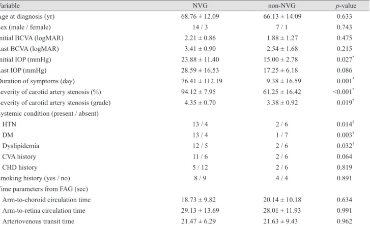
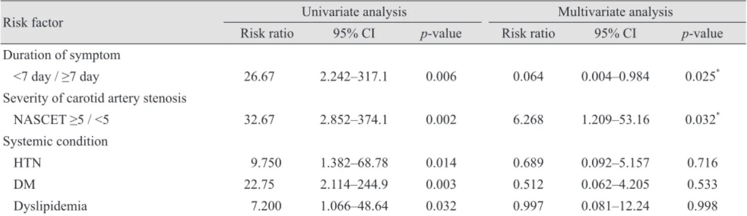
Among the 30 patients diagnosed with OIS, 25 patients (25 eyes) were included in this study. Of the five eyes (of five patients) excluded from the analysis, four eyes had not received follow-up care and one eye had a comorbidity of glaucoma. The mean age at diagnosis of the 25 patients was 67.9 ± 12.50 years (range, 40 to 91 years), and there were 21 men and 4 women. The mean duration of symp- toms was 54.96 ± 97.41 days. The most significant symp- toms found at the initial examination were decreased visu- al acuity in 23 eyes and ocular or periorbital pain in five eyes. At the initial examination, the mean visual acuity was 2.11 ± 0.99 logMAR; and the mean IOP was 21.04 ± 10.33 mmHg (Table 1). Stenosis of the internal carotid ar- tery was observed in all patients, and severe stenosis of the carotid artery of NASCET grade 4 or higher was diagnosed in 18 eyes (72%). Nine out of those 18 patients showed ste- nosis of 90% or more in the carotid artery that required surgical treatment, such as a carotid endarterectomy (Table 2). During a mean follow-up period of 12.60 ± 14.04 months, NVG developed in 17 eyes (68.0%). There were no significant differences in age, gender, visual acuity at initial examination, the incidence of comorbidities of cardiovas- cular and cerebrovascular diseases, smoking history, or time parameters of FAG between patients with and without NVG. However, there were significant differences in IOP at initial examination, the length of time between symp- tom occurrence and diagnosis, and the extent of ipsilateral carotid artery stenosis between the two groups (p = 0.027, p = 0.001, and p = 0.019, respectively). In addition, the inci- dence of hypertension, diabetes, and dyslipidemia was higher in the NVG group when compared with the non- NVG group (Table 3). The logistic regression analysis showed that the probability of NVG development was sig- nificantly higher if the time interval between the onset of symptoms and the initial hospital visit was longer than 7 days (p = 0.025; risk ratio, 0.064) or if the carotid artery stenosis was of NASCET grade 5 or higher (p = 0.032; risk ratio, 6.268) (Table 4).
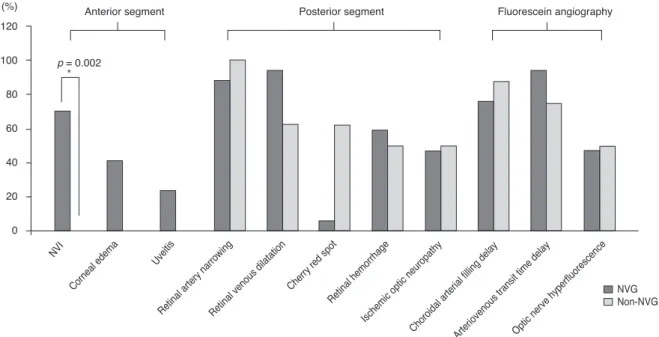
Various clinical features were found in patients with OIS, including NVI (48%), corneal edema (28%), narrow- ing of the retinal artery (92%), widening of the retinal vein (84%), macular cherry-red spots (16%), retinal hemorrhage (56%), and ischemic optic neuropathy (48%). In addition, delayed arterial filling (80%), delayed arteriovenous transit
time (88%), and excessive fluorescence of the optic nerve (4%) were found using FAG. These findings in the anterior segment and the fundus were compared between the two groups; only NVI had a significantly higher rate (p = 0.002) in the NVG group (Fig. 1).
Among the 17 eyes with NVG, 13 eyes underwent pan- retinal photocoagulation and seven eyes were additionally treated with an intracameral injection of a vascular endo-
Table 1. Baseline patient characteristics
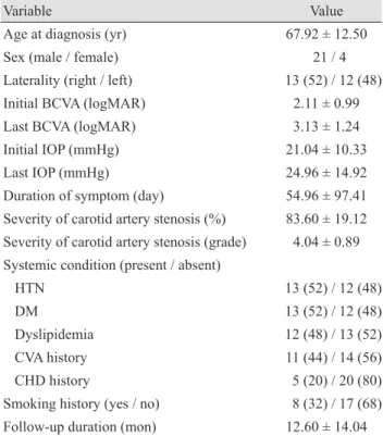
Variable Value
Age at diagnosis (yr) 67.92 ± 12.50
Sex (male / female) 21 / 4
Laterality (right / left) 13 (52) / 12 (48)
Initial BCVA (logMAR) 2.11 ± 0.99
Last BCVA (logMAR) 3.13 ± 1.24
Initial IOP (mmHg) 21.04 ± 10.33
Last IOP (mmHg) 24.96 ± 14.92
Duration of symptom (day) 54.96 ± 97.41 Severity of carotid artery stenosis (%) 83.60 ± 19.12 Severity of carotid artery stenosis (grade) 4.04 ± 0.89 Systemic condition (present / absent)
HTN 13 (52) / 12 (48)
DM 13 (52) / 12 (48)
Dyslipidemia 12 (48) / 13 (52)
CVA history 11 (44) / 14 (56)
CHD history 5 (20) / 20 (80)
Smoking history (yes / no) 8 (32) / 17 (68) Follow-up duration (mon) 12.60 ± 14.04 Values are presented as mean ± standard deviation or number (%).
BCVA = best-corrected visual acuity; logMAR = logarithm of the minimum angle of resolution; IOP = intraocular pressure;
HTN = hypertension; DM = diabetes mellitus; CVA = cerebro- vascular accident; CHD = coronary heart disease.
Table 2. NASCET grading in eyes with ocular ischemic syn- drome
NASCET criteria (%) Grade NVG Non-NVG Total
0–29 0 0 0 0
30–49 1 0 0 0
50–69 2 0 1 1
70–79 3 2 4 6
80–89 4 7 2 9
90–99 5 8 1 9
NASCET = North American Symptomatic Carotid Endarterec-
tomy Trial Collaborators; NVG = neovascular glaucoma.
thelial growth factor inhibitor (Avastin; Roche Pharma, Switzerland). Six eyes with uncontrolled IOP and NVG underwent surgical management. One eye underwent a trabeculectomy, and Ahmed valve implantation was per- formed on the other five eyes. Carotid endarterectomy was performed in nine eyes with OIS in which the carotid ar- tery was occluded. Despite these treatments, visual acuity was significantly decreased in any intervention groups (Table 5). At the last follow-up, the logMAR visual acuity was not significantly different between the NVG and non- NVG groups, and showed a poor prognosis in both groups.
Blindness with no light perception developed in ten eyes (58.8%) from the NVG group and three eyes (37.5%) from the non-NVG group (p = 0.411).
Discussion
OIS is a disease in which ischemia occurs in the anterior
or posterior segment of the eye due to reduced blood flow, and typically, 90% or more of the ipsilateral common ca- rotid artery or internal carotid artery is narrowed [3,8-12].
If ischemia of the anterior or posterior segment persists as a result of carotid artery stenosis, NVG can develop [4,13,14].
Occlusion of the carotid artery is the third most common cause of NVG, following central retinal vein occlusion and diabetes. According to Lazzaro [6], occlusion of the carotid artery is also found in approximately 50% of patients with retinal vein occlusion [5]. Therefore, the incidence of ca- rotid artery occlusion as a cause of NVG may increase if the diagnostic methods used for the carotid artery are im- proved. This is the first study analyzing the associations between clinical characteristics, including the extent of ca- rotid artery stenosis, of patients with OIS and the risk for NVG.
Decreased visual acuity is the most common symptom present at the initial hospital visit for OIS and is observed in more than 90% of cases [1]. Mizener et al. [2] reported
Table 3. Comparison of characteristics between the NVG group and the non-NVG group
Variable NVG non-NVG p-value
Age at diagnosis (yr) 68.76 ± 12.09 66.13 ± 14.09 0.633
Sex (male / female) 14 / 3 7 / 1 0.743
Initial BCVA (logMAR) 2.21 ± 0.86 1.88 ± 1.27 0.475
Last BCVA (logMAR) 3.41 ± 0.90 2.54 ± 1.68 0.215
Initial IOP (mmHg) 23.88 ± 11.40 15.00 ± 2.78 0.027
*Last IOP (mmHg) 28.59 ± 16.53 17.25 ± 6.18 0.086
Duration of symptoms (day) 76.41 ± 112.19 9.38 ± 16.59 0.001
*Severity of carotid artery stenosis (%) 94.12 ± 7.95 61.25 ± 16.42 <0.001
*Severity of carotid artery stenosis (grade) 4.35 ± 0.70 3.38 ± 0.92 0.019
*Systemic condition (present / absent)
HTN 13 / 4 2 / 6 0.014
†DM 13 / 4 1 / 7 0.003
†Dyslipidemia 12 / 5 2 / 6 0.032
†CVA history 11 / 6 2 / 6 0.064
CHD history 5 / 12 2 / 6 0.819
Smoking history (yes / no) 8 / 9 4 / 4 0.891
Time parameters from FAG (sec)
Arm-to-choroid circulation time 18.73 ± 9.82 20.14 ± 10.18 0.634
Arm-to-retina circulation time 29.13 ± 13.69 28.01 ± 11.93 0.991
Arteriovenous transit time 21.47 ± 6.29 21.63 ± 9.43 0.962
NVG = neovascular glaucoma; BCVA = best-corrected visual acuity; logMAR = logarithm of the minimum angle of resolution; IOP
= intraocular pressure; HTN = hypertension; DM = diabetes mellitus; CVA = cerebrovascular accident; CHD = coronary heart disease;
FAG = fluorescein angiography.
*
Mann-Whitney U-test;
†Fisher exact test.
that in 13% of the eyes with OIS, periorbital pain was also noted at the first hospital visit. In this study, four out of 17 patients with NVG (23.5%) and one out of eight patients without NVG (12.5%) complained of periorbital pain. Due to the relatively small sample size, the difference between the two groups did not reach statistical significance.
In this study, NVI was found in 12 out of 25 patients with OIS (48%). Ino-ue et al. [15] reported that NVI is a poor visual prognostic factor and that it is therefore essen- tial to recognize the early stages of OIS associated with di- abetes.
Posterior segment findings are more common than find- ings in the anterior segment of the eye. Both narrowing of the retinal artery and widening of the retinal vein are ob- served in almost all patients with OIS, and retinal hemor- rhage is observed in approximately 80% of patients, while a macular cherry-red spot is observed in approximately 12%
of patients [8]. In this study, narrowing of the retinal artery (92%) and widening of the retinal vein (84%) were observed in most patients, while a macular cherry-red spot (16%) was a less common finding.
The presence of an atheromatous plaque in the carotid artery is a major cause of OIS. According to a study by Sivalingam et al. [9], ischemic cardiovascular disease, a history of cerebrovascular accident, and peripheral arterial disease were observed in 48%, 27%, and 19% of patients, respectively, and hypertension and diabetes were observed in 73% and 56% of patients, respectively. According to a retrospective study by Brown and Magargal [10], hyper-
tension was observed in 56% of patients, diabetes in 43%, cardiovascular disease in 35%, and a history of cerebrovas- cular accident or transient ischemic attack in 26%. These results are consistent with the findings of our study. These comorbidities might be explained by the pathogenic simi- larity between OIS and the aforementioned systemic con- ditions. An atheromatous plaque in the carotid artery leads to continuous ocular ischemia, making the eyes more sus- ceptible to OIS.
In the present study, the extent of carotid artery stenosis was graded according to the NASCET criteria [7]. Carotid artery stenosis was significantly more severe in the NVG group compared with the non-NVG group, and a NASCET grade of 5 or higher was a significant risk factor for NVG.
Since carotid artery stenosis progresses slowly, severe ste- nosis of the carotid artery might result in decreased blood flow to the eye over a long period of time and therefore predispose eye to NVG [16-24]. The long-term decrease in blood flow could cause underlying structural changes, such as fibrovascular proliferation, in the anterior chamber an- gle.
The multivariate analysis also identified that a delay of 7 days or longer from the onset of symptoms to the patient seeking medical advice was a risk factor for developing NVG. As the length of time between symptom onset and the hospital visit increased, the incidence of NVG also in- creased. Karacostas et al. [19] reported a case of isolated OIS with no cerebral involvement and suggested that earli- er referral for a simple carotid artery work-up can prevent
Table 4. Risk factors for neovascular glaucoma in patients with ocular ischemic syndrome
Risk factor Univariate analysis Multivariate analysis
Risk ratio 95% CI p-value Risk ratio 95% CI p-value
Duration of symptom
<7 day / ≥7 day 26.67 2.242–317.1 0.006 0.064 0.004–0.984 0.025
*Severity of carotid artery stenosis
NASCET ≥5 / <5 32.67 2.852–374.1 0.002 6.268 1.209–53.16 0.032
*Systemic condition
HTN 9.750 1.382–68.78 0.014 0.689 0.092–5.157 0.716
DM 22.75 2.114–244.9 0.003 0.512 0.062–4.205 0.533
Dyslipidemia 7.200 1.066–48.64 0.032 0.997 0.081–12.24 0.998
CI = confidence interval; NASCET = North American Symptomatic Carotid Endarterectomy Trial Collaborators; HTN = hypertension;
DM = diabetes mellitus.
*
p < 0.05 is considered statistically significant.
Variables with p > 0.10 in the univariate model are not shown in this table; variables with p < 0.10 in the univariate model were entered
in the multivariate model. Initial intraocular pressure was controlled for as a covariant in the multivariate model.
both the carotid arterial occlusion and the development of severe ocular ischemia. Similarly, Wagner et al. [25] and Higgins [26] reported that although long-term ocular isch- emia already exists before the first symptoms occur, early reperfusion interventions, including carotid endarterecto- my, might play a crucial role in preventing NVG and can affect the long-term prognosis [27]. In the same manner, our results also suggest that an earlier visit (<7 days) after the initial ocular symptoms might prevent patients with OIS from developing NVG and could be the only control- lable factor for the prevention of NVG. We suggest that it is because the earlier the patient visits the hospital, the ear- lier the proper interventions can be achieved. Although an early diagnosis of OIS is generally challenging, and both the NVG and non-NVG groups had poor visual prognoses in our study, it is encouraging that we can give patients
with suspected ocular ischemia attributable to cardiovas- cular or cerebrovascular diseases advice to decrease the development of NVG in the out-patient setting.
This study had some limitations. The sample size was relatively small, and because of the retrospective nature of our study, we could not collect adequate information to identify other possible risk factors. Additionally, the infor- mation regarding the duration of symptoms associated with OIS was obtained through self-reporting rather than by other objective medical findings, and this might have affected the interpretation of our results. Finally, the ef- fects of collateral circulation and the possibility of com- bined ophthalmic artery occlusion were not considered.
Several studies have evaluated the carotid siphon or oph- thalmic artery using transcranial or transorbital Doppler ultrasonography in the analysis of carotid artery stenosis
Table 5. Comparison of visual acuity and IOP between pre- and post-treatment in OIS patients
PRP (n = 13) Glaucoma surgery (n = 6)
*Carotid endarterectomy (n = 9) Initial After
treatment p-value Initial After
treatment p-value Initial After
treatment p-value BCVA (logMAR) 2.18 ± 0.95 3.44 ± 1.01 0.001
†2.38 ± 0.66 3.72 ± 0.69 0.005
†2.12 ± 1.14 3.46 ± 1.14 0.010
†IOP (mmHg) 20.23 ± 6.80 26.85 ± 13.2 0.070 20.33 ± 8.64 14.33 ± 4.59 0.245 20.33 ± 8.20 25.56 ± 15.8 0.260 Values are presented as mean ± standard deviation.
IOP = intraocular pressure; OIS = ocular ischemic syndrome; PRP = panretinal photocoagulation; BCVA = best-corrected visual acuity;
logMAR = logarithm of the minimum angle of resolution.
*
Glaucoma surgery includes trabeculectomy (n = 1) and Ahmed valve implantation (n = 5);
†Wilcoxon signed-rank test.
Fig. 1. Comparison of ocular manifestations in patients with and without neovascular glaucoma (NVG). Only the proportion of neovas- cularization of the iris (NVI) was found to be significantly higher in the NVG group.
*Fisher exact test.
120 100 80 60 40 20 0
(%) Anterior segment
NVGNon-NVG p = 0.002
Posterior segment Fluorescein angiography
NVI
Corneal edema
Retinal artery narrowingRetinal venous dilatation
Cherry red spot
Retinal hemorrhage
Ischemic optic neuropathy
Choroidal arterial filling delayArteriovenous transit time delayOptic nerve hyperfluorescence Uveitis
*