Immunomodulatory Activity of Water Extract of Ulmus macrocarpa in Macrophages
Da Hye Kwon1, Hye-Joo Kang2, Yung Hyun Choi2,3,4, Kyung Tae Chung4,5, Jong Hwan Lee6, Kyung Hwa Kang3, Sook Kyung Hyun1,4, Byung Woo Kim4,7 and Hye Jin Hwang1,4*
1Department of Food & Nutrition, Dong-Eui University, Busan 614-714, Korea
2Anti-aging Research Center, Dong-Eui University, Busan 614-714, Korea
3Department of Oriental Medicine, Dong-Eui University, Busan 614-052, Korea
4Blue-Bio Industry Regional Innovation center, Dong-Eui University, Busan 614-714, Korea
5Department of Clinical Laboratory Science, Dong-Eui University, Busan 614-714, Korea
6Department of Biotechnology & Bioengineering, Dong-Eui University, Busan 614-714, Korea
7Department of Life Science and Technology, Dong-Eui University, Busan 614-714, Korea Received December 9, 2015 /Revised December 29, 2015 /Accepted January 6, 2016
The root bark of Ulmus macrocarpa has been used in traditional medicine for the treatment of various diseases such as edema, infection and inflammation. Nevertheless, the biological activities and under- lying mechanisms of the immunomodulatory effects remain unclear. In this study, as part of our on- going screening program to evaluate the immunomodulatory potential of new compounds from tradi- tional medicinal resources, we investigated the effects of U. macrocarpa water extract (UME) on im- mune modulation in a murine RAW 264.7 macrophage model. As immune response parameters, the productions of as nitric oxide (NO) and cytokines such tumor necrotic factor (TNF)-α, interleukin (IL)-1β and IL-10 were evaluated. Although the release of IL-1β remained unchanged in UME-treated RAW 264.7 macrophages, the productions of NO, TNF-α and IL-10 were significantly increased, along with the increased expression of inducible NO synthase, TNF-α and IL-10 expression at concentrations with no cytotoxicity. UME treatment also induced the nuclear translocation of nuclear factor κB (NF-κ B), and phosphorylation of Akt and mitogen-activated protein kinases (MAPKs) indicating that UME activated macrophages through the activation of NF-κB, phosphoinositide-3-kinase (PI3K)/Akt and MAPKs signaling pathways in RAW 264.7 macrophages. Furthermore, pre-treatment with UME sig- nificantly attenuated the production of NO, but not TNF-α, IL-1β and IL-10, in lipopolysaccharide- stimulated RAW 264.7 cells suggesting that UME may be useful in preventing inflammatory diseases mediated by excessive production of NO. These findings suggest that the beneficial therapeutic effects of UME may be attributed partly to its ability to modulate immune functions in macrophages.
Key words : Cytokine, Immunomodulation, macrophages, NO, RAW 264.7, Ulmus macrocarpa
*Corresponding author
*Tel : +82-51-890-1594, Fax : +82-51-890-2646
*E-mail : [email protected]
This is an Open-Access article distributed under the terms of the Creative Commons Attribution Non-Commercial License (http://creativecommons.org/licenses/by-nc/3.0) which permits unrestricted non-commercial use, distribution, and reproduction in any medium, provided the original work is properly cited.
Journal of Life Science 2016 Vol. 26. No. 1. 50~58 DOI : http://dx.doi.org/10.5352/JLS.2016.26.1.50
서 론
면역조절(immunomodulation)은 다양한 질병으로부터 인 체 방어를 위한 가장 중요한 예방 및 치료 전략의 하나이다.
면역계를 담당하고 있는 다양한 세포 중, 대식세포(macroph- age)는 인체 내에서 거의 모든 조직에 분포하고 이물질, 노폐 물 또는 생체 내 불필요한 세포 등을 포식하여 소화하는 기능 을 갖는 면역 반응의 핵심적인 역할을 하는 세포이다[10, 16].
대식세포는 tumor necrosis factor (TNF)-α와 interleukin (IL) 들과 같은 다양한 종류의 cytokine 이나 일산화질소(nitric ox- ide, NO) 및 활성산소종(reactive oxygen species, ROS) 등을 nuclear factor κB (NF-κB), phosphoinositide-3-kinase (PI3K)/
Akt 및 mitogen-activated protein kinases (MAPKs) 등과 같 은 세포 내 다양한 신호 전달계의 활성을 통하여 생산함으로 서 면역 반응을 자극할 수 있다[22, 32]. 이와 같은 세포 내 반응은 그람음성세균 표층의 peptidoglycan을 둘러싸는 외막 의 중요 구성성분인 lipopolysaccharide (LPS)와 같은 세균성 산물이 대식세포 세포막의 Toll-like receptors (TLRs)와 같은 pattern recognition receptors (PRRs)에 결합함으로서 발생할 수도 있다[21, 34].
최근, 생체 방어 반응을 향상시킬 수 있는 다양한 물질들이 미생물, 균류 및 식물 등에서 분리되고 있으며, 특히 면역 증강 을 위한 원천 자원으로 다양한 약용 식물들이 큰 관심을 모으
고 있다[9, 14]. 이러한 신규 천연물들이 다양한 인체 질환의 예방과 치료에 우수한 효능을 가지지만, 질병 제어 측면에서 대식세포를 활성화 시키고 보체계(complement system)의 조 절을 통한 면역계의 활성화가 가장 기본적인 과정으로 인식되 고 있다[2, 24]. 특히 중요한 약용 식물들은 인류 역사와 더불어 오랫동안 질병의 퇴치 목적으로 사용되어 오면서 그 안정성이 확보되었다.
유근피(榆根皮)는 느릅나무과(Ulmaceae)에 속하는 낙엽교 목으로, 한의학적 측면에서 맛은 달고(甘) 성질은 평(平)하며, 하초습열(下焦濕熱)로 인하여 임증(淋症)이 나타날 때 소변을 통하게 하는 역할을 하고, 기혈(氣血) 순환이 안 되어 발생한 담(痰)을 제거하는 효능이 있으며, 옹저(癰疽)를 치료하고 해 독하는 효과도 있는 것으로 알려져 있다. 최근 연구에 의하면 유근피 추출물은 강력한 항염증[11] 및 항산화 활성[12]과 항 혈전[33] 기능이 알려진 바 있으나 면역 기능 개선에 대한 연구 는 미비한 실정이다. 본 연구에서는 면역 증강 효능을 나타내 는 신물질 탐색의 과정으로 유근피를 선정하였으며, 유근피 열수 추출물에 의한 대식세포에서의 면역 조절 관련 몇 가지 유의적인 결과를 얻었기에 이를 보고하고자 한다.
재료 및 방법
유근피 열수 추출물의 준비
본 실험에서 사용된 유근피(Ulmus macrocarpa)는 (주)대한 생약에서 구입하였으며, 건조된 유근피 3 kg을 증류수(10 l)를 가한 후 80℃ 수욕상에서 3 시간씩 2 회 반복 추출하였다. 그리 고 추출액은 여과하여 45℃ 이하에서 감압농축(Rotary vac- uum evaporator, Eyela, Japan) 하였으며, 농축물은 -80℃ 동 결건조기 (FDU-2100, Eyela, Japan)를 이용하여 건조시켜 약 320.0 g의 열수추출물(U. macrocarpa water extract, UME)을 얻었다.
세포배양
면역 반응에 미치는 유근피 추출물의 영향을 조사하기 위하 여 사용된 murine macrophage cell line인 RAW 264.7 세포는 American Type Culture Collection (Manassas, VA, USA)에 서 구입하여, 10% fetal bovine serum (FBS, WELGENE, Daegu, Republic of Korea)과 penicillin/streptomycin 100 unit/ml이 함유된 Dulbecco's modified Eagle's medium (DMEM, WELGENE)을 사용하여 37℃, 5% CO2 조건에서 배 양하였다.
MTT assay
RAW 264.7 세포의 증식에 미치는 유근피 추출물의 영향을 조사하기 위하여 세포 배양용 6 well plate에 1×10⁵ cells/ml로 세포를 분주하고 적정 농도의 유근피 추출물 단독, LPS (Sigma-
Aldrich Co., St. Louis, MO, USA) 단독 또는 LPS를 1 시간 선처리 후 유근피 추출물을 처리하였다. 24 시간 후 배지를 제거하고 MTT [3-(4,5-dimethylthiazol-2-yl)-2,5-diphenylte- trazolium bromide, Sigma-Aldrich Co.)를 0.5 mg/ml 농도로 희석하여 200 μl씩 분주하고 37℃에서 다시 반응시켰다. 2 시 간 후 MTT 시약을 제거하고 dimethyl sulfoxide (DMSO, Sigma-Aldrich Co.)를 1 ml씩 각 well에 분주하여 생성된 for- mazan을 모두 녹인 후 96 well plate에 200 μl씩 옮겨서 en- zyme-linked immunosorbent assay (ELISA) reader (Molecu- lar Devices, Sunnyvale, CA, USA)로 540 nm에서 흡광도를 측정하였다. 아울러 RAW 264.7 세포의 형태적 변형은 위상차 현미경(Carl Zeiss, Oberkochen, Germany)을 사용하여 관찰 하였다.
NO 생성량 측정
NO의 농도 측정을 위해 세포 배양액 내의 nitrite 농도를 Griess Reagent (Sigma-Aldrich Co.)를 이용하여 측정하였다.
이를 위하여 RAW 264.7 세포에 적정 시간 동안 유근피 추출물 및 LPS를 처리한 후 24 시간 배양하고 세포 배양액을 수거하 였다. 배양액 100 μl와 동량의 Griess Reagent를 상온에서 5 분간 반응시킨 후 ELISA reader로 540 nm에서 흡광도를 측정 하였고, sodium nitrite (NaNO2)의 농도 별 표준곡선을 이용하 여 배양액 내의 NO 농도를 결정하였다.
Cytokine 생성량 측정
세포 배양액 내의 다양한 cytokine (TNF-α, IL-1β 및 IL-10) 의 양을 측정하기 위한 Quantikine ELISA kit는 R&D systems (Minneapolis, MN, USA)에서 구입하였다. 동일한 조건에서 배양된 RAW 264.7 세포의 배양액을 이용하여 cytokine의 생 성 양을 제시된 방법에 따라 처리한 다음 ELISA reader를 이용 하여 450 nm의 흡광도에서 cytokine 들의 생성 정도를 측정하 였다.
Western blotting에 의한 단백질 발현 분석
면역 조절관련 특정 유전자들의 발현 변화 관찰을 위하여 준비된 세포들을 250 mM NaCl, 25 mM Tris-HCl (pH 7.5), 10 mM ethylenediaminetetraacetic acid, 1% Nonidet P-40, 0.1 mM phenyl-methylsulfonylfluoride와 protease inhibitor 등 이 함유된 lysis buffer를 사용하여 용해시켰다. 핵과 세포질에 서 단백질을 분리하기 위해서는 NE-PER® kit (PIERCE, Rockford, IL, USA)를 사용하였으며, 분리된 단백질들의 농도 는 Bio-Rad 단백질 정량 시약(Bio-Rad, Herculs, CA, USA)을 이용하여 측정하였다. Western blotting을 위해 동량의 단백질 들을 sodium dodecyl sulfate (SDS)-polyacrylamide gel elec- trophoresis를 이용하여 분리하고 nitrocellulose membrane (Schleicher & Schuell, Keene, NH, USA)으로 전이시켰다. 각
A
B
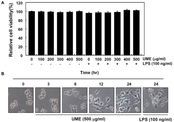
Fig. 1. Effects of root bark of U. macrocarpa water extracts (UME) on cell viability and morphology in RAW 264.7 macrophages.
(A) After RAW 264.7 cells were seeded, the cells were treated with the indicated concentrations of UME alone or pretreated with UME for 1 hr before 100 ng/ml of LPS treatment. After 24 hr, the cell viability was assessed using an MTT reduction assay. Data are expressed as the mean ± SD of three independent experiments. (B) RAW 264.7 cells were treated with 500 μg/ml of UME for the indicated times or with 100 μg/ml of LPS for 24 hr. The morphological changes of RAW 264.7 macrophage cell surfaces were observed by taking photographs using an inverted microscope (original magnification, ×200).
각의 membrane을 5% skim milk를 1 시간 처리하여 비특이적 인 단백질들에 대한 blocking을 실시한 후, 적정 항체 및 en- hanced chemiluminescence (ECL, Amersham Corp. Arling- ton Heights, IL, USA) 용액을 적용시킨 다음 X-ray film에 감광시켜 특정 단백질의 발현 양을 분석하였다. 본 연구에 사 용한 1 차 항체는 Santa Cruz Biotechnology Inc. (Santa Cruz, CA, USA) 및 Cell Signaling (Beverly, MA, USA)에서 구입하 였으며, 2 차 항체는 Amersham Life Science Co.에서 구입하 였다.
Immunofluorescence staining
유근피 추출물 처리에 따른 NF-κB의 세포 내 분포 변화를 위한 형광현미경학적 관찰을 위하여 6 well plate 내 coverslip 위에 부착시킨 RAW 264.7 세포에 유근피 추출물을 처리하였 다. 적정 시간 처리 후, 4% paraformaldehyde (Sigma-Aldrich Co.)가 함유된 phosphate buffer saline (PBS) 용액으로 cover- slip에 부착된 세포를 10 분간 고정하고, 100% 메탄올을 처리 하여 상온에 10 분간 반응시켰다. PBS로 세포를 수세 후, NF-κ B p65 항체(Santa Cruz Biotechnology Inc.)로 1 시간 염색하 고, fluorescein isothiocyanate (FITC) conjugated donkey an- ti-rabbit immunoglobulin G (IgG, Jackson Immuno Research
Laboratories, Inc., West Grove, PA, USA)로 1 시간 더 반응을 시켰다. 다시 PBS로 수세 후, 4,6-diamidino-2-phenylindole (DAPI, Sigma-Aldrich Co.) 용액으로 10 분간 핵을 염색한 후 형광현미경(Carl Zeiss) 하에서 NF-κB p65의 발현 정도를 관 찰하였다.
통계처리
모든 실험 결과는 평균(mean) ± 표준편차(SD)로 표시하였 고 Sigmaplot (Systat Software Inc., San Jose, CA, USA)을 이 용하여 Student t-test로 계적 유의성을 얻었다.
결과 및 고찰
유근피 추출물 처리에 의한 RAW 264.7 대식세포의 활성화 유근피 추출물이 RAW 264.7 대식세포의 면역 반응에 미치 는 영향을 조사하고자 다음과 같이 실험 조건을 설정하였다.
유근피 열수 추출물(UME)을 적정 농도로 RAW 264.7 대식세 포에 처리하거나, LPS (100 ng/ml) 단독 및 LPS 1 시간 선처리 후 유근피 추출물을 24 시간 동안 처리한 후 MTT assay를 실시하여 세포 생존도 변화를 측정하였다. Fig. 1A의 결과에서 알 수 있듯이, 본 연구에서 조사된 유근피 추출물 처리 농도
A B
C D
Fig. 2. Increased release of NO and cytokines by UME treatment in RAW 264.7 macrophages. (A) Cells were treated with the indicated concentrations of UME or 5 ng/ml of LPS for 24 hr, and the supernatants were collected, and then the NO concen- tration from supernatants was determined using the Griess reagent. (B-D) To quantify the amounts of cytokines produced by UME, the supernatants from cells grown under the same conditions as (A) were collected and the concentrations of TNF-α, IL-1β and IL-10 were measured using commercial ELISA kits. The data are shown as means ± SD of three independent experiments (*, p<0.05 vs. untreated control).
(100~500 mg/ml) 범위 내에서 RAW 264.7 대식세포의 생존율 에는 유의적인 변화가 관찰되지 않았으며, LPS 단독 및 유근 피 추출물과의 동시 처리군에서도 모두 유의적인 세포 독성을 보이지 않았다. 이 결과를 바탕으로 대식세포 면역 조절능 평 가를 위한 유근피 추출물의 최고 처리 농도를 500 mg/ml로 설정하였으며, 동일 조건에서 대식세포 활성화와 연관성이 있 는지를 조사하기 위해 형태적 변화를 관찰하였다. Fig. 1B에 나타낸 바와 같이, 유근피 추출물 처리 시간의 증가에 따라 LPS 처리군에서 관찰된 전형적인 대식세포 분화를 의미하는 형태적 변화[3]를 관찰할 수 있었으며, 이는 유근피 추출물이 RAW 264.7 대식세포의 활성을 촉진시킬 수 있었다는 간접적 인 증가가 될 수 있을 것으로 생각된다.
RAW 264.7 대식세포의 NO 생성에 미치는 유근피 추출 물의 영향
대식세포는 다양한 종류의 cytokine과 NO 등의 생성을 통 하여 감염성 인자나 종양세포에 대한 저항에 주요 작동 세포 (effector cell)로 작용한다. 또한 대식세포가 다양한 조직에 존 재하고 염증성 자극을 포함한 인자들에 대한 생체방어 기능을 한다는 점에서 이러한 인자들에 의하여 자가 활성화될 수 있 다[15, 25]. 따라서 유근피 열수 추출물 처리에 따른 RAW
264.7 세포의 분화가 대식세포 활성화와 직접 연관성이 있는 지를 조사하기 위하여 유근피 추출물이 처리된 배지에서 배양 된 세포 상등액에 존재하는 작동 분자(effector molecule) 중의 하나인 NO의 생성[28, 30] 변화를 먼저 조사하였다.
Fig. 2A의 결과에서 알 수 있듯이, 유근피 추출물 처리에 의하여 세포독성이 없는 범위에서 처리 농도 의존적으로 NO 의 생성이 증가되었으며, 대조군으로 사용한 5 ng/ml의 LPS 처리군에서도 유의적인 증가가 관찰되었다. 대식세포 활성화 지표 중의 하나인 NO는 대식세포에서 생성되는 반응질소 중 간체로서 NO synthase (NOS)에 의하여 L-arginine에서 만들 어지는 기체 분자이다[19]. 비록 과다한 NO의 생성은 혈관확 장이나 염증반응에 의한 조직 손상을 일으켜 생체 내 유해한 작용을 나타내는 것으로도 알려져 있으나, 적정량의 NO는 세 포 내 감염을 일으키는 미생물과 암세포를 제어하는 능력을 보유하고 있다. 따라서 세포독성을 유발하지 않는 적정 조건 에서 NO 생성의 촉진은 면역 기능을 증가시킬 수 있는 지표로 사용될 수 있다[7, 18]. 즉, 유근피 추출물 처리에 의한 NO 분비능의 증가는 유근피 추출물이 면역 기능 활성 증가 가능 성을 가질 수 있음을 보여주는 한 예로서 사용될 수 있을 것으 로 생각된다. 또한 유근피 추출물 처리에 의한 대식세포에서 NO의 생성 증가는 inducible NOS (iNOS)의 발현 증가에 의
Fig. 3. Effects of UME treatment on the levels of iNOS and cyto- kines proteins in RAW 264.7 macrophages. Total cellular proteins (30~50 μg) isolated from cells grown under the same conditions as Fig. 2 were resolved on 10% SDS-pol- yacrylamide gels, followed by Western blotting using the specific antibodies for iNOS, TNF-α, IL-1β and IL-10. β -actin was used as an internal control.
A
B
Fig. 4. Induction of nuclear translocation of NF-κB by UME treatment in RAW 264.7 macrophages. (A) Cells were treated with UME (500 μg/ml) for the indicated times.
Nuclear and cytosolic proteins were resolved on 10%
SDS-polyacrylamide gels followed by Western blotting using anti-NFκB p65 and anti-IκB-α antibodies. Lamin B and β-actin were used as internal controls for the nu- clear and cytosolic fractions, respectively. (B) Cells were stimulated with UME (500 μg/ml) for 30 and 60 min.
Localization of NF-κB p65 was visualized with a fluo- rescence microscope following immunofluorescence staining with anti-NFκB p65 and FITC-labeled IgG anti- bodies (red). Nuclei of the corresponding cells were vi- sualized with DAPI (blue). The cells were visualized us- ing a fluorescence microscope.
한 것임을 확인하였다(Fig. 3).
RAW 264.7 대식세포의 cytokine 생성에 미치는 유근피 추출물의 영향
다양한 cytokine 중, 면역 세포 조절에 중요한 인자인 TNF- α는 암세포에 자살 신호를 유도하거나 바이러스의 세포 내 복제를 저해하는 내재면역 반응(innate immune response)에 폭넓게 관여하는 신호분자의 일종이다. TNF-α는 주로 활성화 된 대식세포에 의해 분비되는데, 보조 T 세포, 자연살해세포 등과 같은 다양한 세포에서도 분비된다[17]. 또한 내인성 발열 cytokine으로 처음 알려진 IL-1은 서로 다른 유전자에 의해 발현되는 IL-1α와 IL-1β로 구성되어 있는데, 특히 IL-1β는 감 염에 대한 염증과 면역 반응들을 조절하는 cytokine으로 염증 과 숙주 방어에서 핵심적인 역할을 한다[13, 31]. 그리고 대표 적인 anti-inflammatory cytokine으로 알려진 IL-10은 면역조 절과 염증반응에서 다면 발현성의 효과를 가진다. IL-10은 B 세포의 생존과 분열을 촉진시키며, 염증성 cytokine의 분비를 억제하는 기능을 가진 것으로도 잘 알려져 있다[27]. 비록 이들 세 가지 cytokine이 생물활성 측면에서 면역 억제와 면역촉진 의 양면성을 가지지만, 과도하지 않은 적정량의 생성은 대식 세포의 활성화를 일으키는 신호전달을 매개 cytokine으로 작 용하며, 특정 자극에 의한 대식세포의 활성화 정도를 예측할 수 있는 지표로 사용될 수 있다.
본 연구의 결과에 의하면 유근피 추출물 처리에 따라 RAW 264.7 대식세포에서 세 가지의 cytokine 중, IL-1β의 생성에는 유의적인 증가 현상이 관찰되지 않았으며, 단백질 수준에서도 발현의 증가가 나타나지 않았다. 그러나 TNF-α와 IL-10의 생 성이 NO의 생성 증가와 동반되어 모두 증가되었으며(Fig.
2B~D), 이는 단백질 수준에서 각 cytokine의 발현 증가에 의한 것임을 알 수 있었다(Fig. 3). 이는 유근피 추출물에 의한 대식 세포 활성화에 NO, TNF-α와 IL-10의 생성 증가가 동반되어 나타났음을 의미하는 것으로 사료된다.
유근피 추출물이 RAW 264.7 대식세포의 세포 내 신호 전달계에 미치는 영향
세포 내 신호조절과 연관된 특정 단백질들의 활성 여부는 대식세포의 활성에 매우 중요한 조절자로서 역할을 한다[21,
Fig. 5. Effects of UME treatment on PI3K/Akt and MAPKs sig- naling pathways in RAW 264.7 macrophages. Cells were treated with 500 μg/ml of UME for the indicated times, and total cellular proteins (30~50 μg) were resolved on SDS-polyacrylamide gels, followed by Western blotting using the specific antibodies, as indicated.
A B
C D
Fig. 6. Effects of UME treatment on the production of NO and cytokines in LPS-stimulated RAW 264.7 macrophages. The cells were pretreated with various concentrations of UME for 1 hr and then incubated with or without LPS (100 ng/ml) for 24 hr. The amounts of NO (A) and cytokines (B-D) were measured in culture media using Griess reagent or commercial ELISA kits. The data are shown as means ± SD of three independent experiments (*,p<0.05 vs. untreated control; #, p<0.05 vs. LPS-treated group; n.s., non significant).
34]. 그 중, NF-κB는 iNOS와 cytokine을 포함한 면역 조절 유 전자들의 전사활성에 핵심 전사조절인자로서 작용하는데,
NF-κB는 일반적으로 inhibitor of NF-κB (IκB) 단백질과 복합 체를 형성하여 세포질에 존재 한다. 그러나 면역 활성 자극에 반응하여, IκB가 인산화 후 ubiqutine-proteasome 경로를 통 하여 분해되면, NF-κB가 유리되어 핵 내로 이동하여 면역 조 절 유전자 특정 promoter 영역을 활성화시킨다[4, 6]. 따라서 유근피 추출물에 의한 면역 조절 반응에서 NF-κB 활성 증가에 미치는 영향을 조사하기 위하여 핵과 세포질의 단백질을 분리 하여 NF-κB의 subunit의 하나인 p65 단백질의 발현 변화를 조사하였다. Fig. 4A의 결과에서 알 수 있듯이, 비록 세포질 내 IκB-α의 발현에는 큰 변화가 없었지만, 유근피 추출물 처리 15 분 후 부터 핵 내에서의 NF-κB p65의 발현이 관찰되었으 며, 60 분 경과 후 최고치를 보인 후 점차 발현이 감소되었다.
뿐만 아니라 면역형광염색법을 이용하여 NF-κB p65 단백질 의 세포 내 위치 변화를 확인 본 결과, 유근피 추출물 처리에 의해 핵 내로 NF-κB p65가 이동되었음을 관찰할 수 있었다 (Fig. 4B).
또한 세포의 증식 및 성장에 관여한다고 알려진 PI3K/Akt 신호 경로는 NF-κB와 함께 대식세포의 면역 조절 유전자들의 발현과 대식세포의 활성에 핵심적인 역할을 하는 것으로 알려 져 있다[20, 26]. 따라서 NF-κB와 PI3K/AKT 경로를 조절하는 것은 많은 면역 퇴행성질환 치료를 위한 유망한 신호전달경로 로 인식되고 있다. 아울러 extracellular signal-regulated kin- ase (ERK), c-Jun amino terminal kinase (JNK) 및 p38을 포함 하는 MAPKs 신호 경로 역시 면역 및 염증성 시스템의 중요한
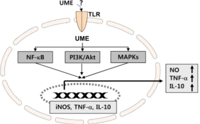
UME
Fig. 7. Schematic diagram of UME-induced immune responses through the activation of NF-κB, PI3K/Akt and MAPKs signaling pathways.
신호전달 반응을 매개하고, 대식세포 활성을 포함한 세포 활 성물질의 생성 및 다양한 생물학적 기능 조절의 주요 인자로 서 작용한다[1, 8]. 본 연구에서는 유근피 추출물에 의한 면역 조절 반응에 관여하는 세포 내 신호 전달계 역할의 이해를 위하여 PI3K의 하위 조절인자에 해당되는 Akt와, MAPKs에 속한 3가지 kinase (ERK, JNK 및 p38 MAPK)의 활성화 여부 를 부가적으로 조사하였다. Fig. 5의 결과에서 알 수 있듯이, Akt 단백질의 인산화는 유근피 추출물 처리 15 분부터 증가되 어 3 시간 후 최고치를 보였다. MAPKs의 경우 종류에 따라 다소 시간적 차이는 있으나, 유근피 추출물 처리에 의하여 인 산화형의 발현이 1 시간 이내에 모두 증가되었다. 이상의 결과 는 유근피 추출물에 의한 RAW 264.7 대식세포의 활성화에 NF-κB 신호계를 포함한 PI3K/Akt 및 MAPKs 신호전달계의 활성이 동시에 관여하고 있음을 의미하는 것이다.
RAW 264.7 대식세포에서 LPS에 의한 NO 및 cytokine의 과다 생성에 미치는 유근피 추출물의 영향
한편 염증 작용은 외부 감염에 대한 면역 방어기전과 같은 생리적 작용에 중요하게 작용하지만, 류마티스 관절염, 알츠 하이머병 등과 같은 자가면역 질환 및 여러 가지 만성적 질환 의 중요한 발병 및 진행 요인으로 작용 한다[5, 23, 29]. 따라서 비록 NO나 다양한 cytokine이 면역 활성 증가능을 가지고 있 으나, 이들의 과도한 생성 증가는 오히려 염증 반응을 야기할 수 있다. 따라서 유근피 추출물이 항염증 인자로서 작용할 수 있는지의 여부를 조사하기 위하여, LPS 처리 농도를 높여 염 증 유발 조건을 설정하였다. Fig. 6에 나타낸 결과에서 알 수 있듯이 LPS 처리 농도를 100 ng/ml로 높였을 경우, 5 ng/ml 처리군에 비하여 NO의 생성량이 6 배 이상 증가하였으며, 세 가지 종류의 cytokine 생성량도 모두 증가되었다. 비록 본 실 험 조건에서 처리된 유근피 추출물의 농도 조건에서 cytokine 의 생성을 억제하지는 못하였으나, NO의 생성은 유근피 추출 물의 처리 농도 증가에 따라 다소 유의적으로 억제되었음을
알 수 있었다(Fig. 6A). 이러한 결과들로 볼 때 유근피 추출물 이 항염증 효력이 없다고 결론 내릴 수는 없지만, 과도한 NO 의 생성 억제능을 보이는 것은 매우 의미 있는 결과로서, 이에 대한 추가적인 연구가 필요할 것으로 생각된다.
이상의 결과에서 유근피 추출물에 의한 대식세포의 활성화 과정은 NF-κB의 활성과 PI3K/Akt 및 MAPKs 신호 경로의 활성화를 통하여 iNOS 및 다양한 면역 조절 관련 cytokine의 전사 활성 촉진을 통하여 이루어 질 것으로 추정된다(Fig. 7).
따라서 이러한 면역 활성 증대에 관여하는 유근피 추출물의 주요 생리활성 물질의 탐색과 동물실험을 통한 추기적인 연구 가 수행되어야 할 것이다.
감사의 글
이 논문은 2014년 동의대학교 연구년제 지원과 산업통상자 원부ㆍ부산광역시 지원 지역혁신센터사업(RIC 08-06-07) 동 의대학교 블루바이오 소재 개발 및 실용화 지원센터의 지원으 로 이루어졌음.
References
1. Arthur, J. S. and Ley, S. C. 2013. Mitogen-activated protein kinases in innate immunity. Nat. Rev. Immunol. 13, 679-692.
2. Beutler, B. 2004. Innate immunity: an overview. Mol.
Immunol. 40, 845-859.
3. Choi, H. S., Kim, S. R., Hong, S. H., Ku, J. M., Kim, M.
K., Seo, H. S., Cho, S. G., Shin, S., Shin, Y. C. and Ko, S.
G. 2013. Water extract of deer bones activates macrophages and alleviates neutropenia. Evid. Based Complement. Alternat.
Med. 2013, 617302.
4. Gasparini, C. and Feldmann, M. 2012. NF-κB as a target for modulating inflammatory responses. Curr. Pharm. Des.
18, 5735-5745.
5. Guzik, T. J., Korbut, R. and Adamek-Guzik, T. 2003. Nitric oxide and superoxide in inflammation and immune regu- lation. J. Physiol. Pharmacol. 54, 469-487.
6. Hayden, M. S. and Ghosh, S. 2014. Regulation of NF-κB by TNF family cytokines. Semin. Immunol. 26, 253-266.
7. Hibbs, J. B., Taintor, R. R. and Vavrin, Z. 1987. Macrophage cytotoxicity: role for L-arginine deiminase and imino nitro- gen oxidation to nitrite. Science 235, 473-479.
8. Ivashkiv, L. B. 2011. Inflammatory signaling in macro- phages: transitions from acute to tolerant and alternative ac- tivation states. Eur. J. Immunol. 41, 2477-2481.
9. Kayser, O., Masihi, K. N. and Kiderlen, A. F. 2003. Natural products and synthetic compounds as immunomodulators.
Expert. Rev. Anti-Infect. Ther. 1, 319-335.
10. Klimp, A. H., de Vries, E. G., Scherphof, G. L. and Daemen, T. 2002. A potential role of macrophage activation in the treatment of cancer. Crit. Rev. Oncol. Hematol. 44, 143-161.
11. Kwon, J. H., Kim, S. B., Park, K. H. and Lee M. W. 2011.
Antioxidative and anti-inflammatory effects of phenolic
compounds from the roots of Ulmus macrocarpa. Arch.
Pharm. Res. 34, 1459-1466.
12. Kwon, Y. M., Lee, J. H. and Lee, M. W. 2002. Phenolic com- pounds from barks of Ulmus macrocarpa and its antioxida- tive activities. Kor. J. Pharmacogn. 33, 404-410.
13. Lane, T. and Lachmann, H. J. 2011. The emerging role of interleukin-1β in autoinflammatory diseases. Curr. Allergy Asthma Rep. 11, 361-368.
14. Lee, J. S. and Hong, E. K. 2011. Immunostimulating activity of the polysaccharides isolated from Cordyceps militaris. Int.
Immunopharmacol. 11, 1226-1233.
15. Liew, F. Y., Wei, X. Q. and Proudfoot, L. 1997. Cytokines and nitric oxide as effector molecules against parasitic infections. Philos. Trans. R. Soc. Lond. B Biol. Sci. 352, 1311- 1315.
16. Liles, W. C. 2001. Immunomodulatory approaches to aug- ment phagocyte-mediated host defense for treatment of in- fectious diseases. Semin. Respir. Infect. 16, 11-17.
17. Locksley, R. M., Killeen, N. and Lenardo, M. J. 2001. The TNF and TNF receptor superfamilies: integrating mamma- lian biology. Cell 104, 487-501.
18. Lorsbach, R. B., Murphy, W. J., Lowenstein, C. J., Snyder, S. H. and Russell, S. W. 1993. Expression of the nitric oxide synthase gene in mouse macrophages activated for tumor cell killing. Mmolecular basis for the synergy between inter- feron-gamma and lipopolysaccharide. J. Biol. Chem. 268, 1908-1913.
19. MacMicking, J., Xie, Q. W. and Nathan, C. 1997. Nitric oxide and macrophage function. Annu. Rev. Immunol. 15, 323-550.
20. Madrid, L. V., Wang, C. Y., Guttridge, D. C., Schottelius, A. J., Baldwin, A. S. Jr. and Mayo, M. W. 2000. Akt sup- presses apoptosis by stimulating the transactivation poten- tial of the RelA/p65 subunit of NF-kappaB. Mol. Cell. Biol.
20, 1626-1638.
21. Medzhitov, R. and Janeway, C. 2000. Innate immune recog- nition: mechanisms and pathways. Immunol. Rev. 173, 89-97.
22. Murray, P. J. and Wynn, T. A. 2011. Protective and patho- genic functions of macrophage subsets. Nat. Rev. Immunol.
11, 723-737.
23. Patel, T. N., Shishehbor, M. H. and Bhatt, D. L. 2007. A
review of high-dose statin therapy: targeting cholesterol and inflammation in atherosclerosis. Eur. Heart J. 28, 664-672.
24. Pinto, M. R., Melillo, D., Giacomelli, S., Sfyroera, G. and Lambris, J. D. 2007. Ancient origin of the complement sys- tem: emerging invertebrate models. Adv. Exp. Med. Biol. 598, 372-388.
25. Ritchlin, C. T., Haas-Smith, S. A., Li, P., Hicks, D. G. and Schwarz, E. M. 2003. Mechanisms of TNF-alpha- and RANKL-mediated osteoclastogenesis and bone resorption in psoriatic arthritis. J. Clin. Invest. 111, 821-831.
26. Saponaro, C., Cianciulli, A., Calvello, R., Dragone, T., Iacobazzi, F. and Panaro, M. A. 2012. The PI3K/Akt path- way is required for LPS activation of microglial cells.
Immunopharmacol. Immunotoxicol. 34, 858-865.
27. Schreiber, S., Heinig, T., Thiele, H. G. and Raedler, A. 1995.
Immunoregulatory role of interleukin 10 in patients with inflammatory bowel disease. Gastroenterology 108, 1434-1444.
28. Singh, V. K., Mehrotra, S., Narayan, P., Pandey, C. M. and Agarwal, S. S. 2000. Modulation of autoimmune diseases by nitric oxide. Immunol. Res. 22, 1-19.
29. Storck, M., Schilling, M., Burkhardt, K., Prestel, R., Abend- roth, D. and Hammer, C. 1994. Production of proinflamma- tory cytokines and adhesion molecules in ex-vivo xenogeneic kidney perfusion. Transpl. Int. 7, 647-649.
30. Tripathi, P. 2007. Nitric oxide and immune response. Indian J. Biochem. Biophys. 44, 310-319.
31. Wang, D., Zhang, S., Li, L., Liu, X., Mei, K. and Wang, X.
2010. Structural insights into the assembly and activation of IL-1β with its receptors. Nat. Immunol. 11, 905-911.
32. Wolf, S. F., Sieburth., D. and Sypek, J. 1994. Interleukin 12:
a key modulator of immune function. Stem Cell 12, 154-168.
33. Yang, W. K., Lee, J. J., Sung, Y. Y., Kim, D. S., Myung, C.
S. and Kim, H. K. 2013. Extract of Ulmus macrocarpa Hance prevents thrombus formation through antiplatelet activity.
Mol. Med. Rep. 8, 726-730.
34. Zhou, D., Huang, C., Lin, Z., Zhan, S., Kong, L., Fang, C.
and Li, J. 2014. Macrophage polarization and function with emphasis on the evolving roles of coordinated regulation of cellular signaling pathways. Cell Signal. 26, 192-197.
초록:유근피 추출물이 대식세포 면역조절에 미치는 영향
권다혜1․강혜주2․최영현2,3,4․정경태4,5․이종환6․강경화3․현숙경1,4․김병우4,7․황혜진1,4*
(1동의대학교식품영양학과, 2동의대학교 항노화연구소, 3동의대학교 한의학과, 4동의대학교 블루바이오소재개발
센터, 5동의대학교 임상병리학과, 6동의대학교 생명공학과, 7동의대학교 생명응용학과)
왕느릅나무(Ulmus macrocarpa)의 껍질을 말린 유근피는 오랫동안 부종, 감염 및 염증 제어의 목적으로 사용되어 져 왔음에도 불구하고 잠재적 면역조절 효과에 관해서는 연구가 이루어진 바 없다. 본 연구에서는 전통 약용자원 에서 새로운 면역기능 증가 신소재 발굴의 일환으로 유근피 열수 추출물의 면역 조절 효능을 RAW 264.7 대식세 포 모델을 이용하여 조사하였다. 이를 위한 대식세포의 활성화 관련 지표로서 NO, TNF-α, IL-1β 및 IL-10의 생성 량 변화를 조사하였다. 비록 유근피 추출물이 처리된 RAW 264.7 대식세포에서 IL-1β의 유의적인 유리는 관찰할 수 없었으나, NO, TNF-α 및 IL-10의 생성은 세포독성을 나타내지 않는 범위에서 유근피 추출물 처리 농도 의존적 으로 증가되었으며, 이는 또한 iNOS, TNF-α 및 IL-10의 단백질 발현 증가와 연관되어 있었다. 아울러 유근피 추출 물은 LPS에 의한 과도한 NO의 생성 억제능도 함유하고 있었으며, 유근피 추출물에 의한 대식세포의 활성화에는 NF-κB와 PI3K/Akt 및 MAPKs 등과 같은 면역 활성을 유도하는 신호전달계의 활성화가 연관되어 있음을 알 수 있었다. 따라서 본 연구의 결과는 유근피 추출물이 대식세포 활성화를 통한 면역 증강제로서의 개발 가능성이 매우 높음을 시사한다.