전이금속 갈륨(Ga(NO 3 ) 3 ) 을 이용한 biofilm을 형성하는 어류질병세균의 억제
김동휘1, 수브라마니안 다라니드다란1, 장영환2, 허문수1*
1제주대학교해양과학대학수산생명의학과
2제주도특별자치도해양수산자원연구소
Received: July 28, 2016 / Revised: September 23, 2016 / Accepted: October 4, 2016
서 론
우리나라는 1980년대부터어류양식기술을도입하여육성 하였다. 그중우리나라의대표적인해상양식어류인넙치는 1989년부터종묘생산기술이확립되어경제성증대를위한 고밀도사육방법으로양식기술이보급되어왔다. 양식넙치 의생산력증대는다각적인연구노력으로 2004년 32,141톤, 2009년 56,674톤, 2011년 40,805톤, 2013년 36,944톤, 2014년 42,137톤으로꾸준히성장해왔다[14].
국내의어류양식산업은크게성장하고있지만, 양식도 중발생하는어류질병이양식산업발전에심각한악영향 을미치는것으로알려져있다. 일반적으로육상수조양식의
경우단위면적당생산량을극대화시키기위해고밀도사육 을하고있는데, 이러한고밀도사육은증체율저하및각종 질병발생의직접적인원인을제공하여폐사에따른경제적 손실을일으킨다[13].
제주양식넙치의경우폐사현황을보면 2010년 4,519톤 (생산량 21,370톤), 2011년 4,427톤(생산량 22,823톤), 2012년 5,601톤(생산량 24,575톤), 2013년 5,760(생산량 23,002톤), 2014년 6,710톤(생산량 26,283톤), 2015년 6,928톤(생산량 27,142톤)으로해마다증가하고있다. 또한폐사로인한피 해액은 2010년 294억원, 2011년 376억원, 2012년 513억원, 2013년 403억원, 2014년 485억원, 2015년 529억원으로조 사되었다[18].
넙치의양식과정에서발생하는주요세균성질병의원인균 으로에드워드증(Edwardsiella sp.), 연쇄구균증(Streptococcus sp.), 비브리오증(Vibrio sp.), 활주세균증(Flexibacter maritimus)
등이보고되고있다[10, 15]. 하지만우리나라의어류양식
Inhibitory Effect of Transition Metal Gallium [Ga(NO3)3] on Biofilm Formation by Fish Pathogens Dong-Hwi Kim1, Subramanian Dharaneedharan1, Young-Hwan Jang2, and Moon-Soo Heo1*
1Marine Applied Microbes and Aquatic Organism Disease Control Lab, Department of Aquatic Biomedical Sciences, School of Marine Bio- medical Sciences & Marine and Environmental Research Institute, Jeju National University, Jeju 63243, Republic of Korea
2Jeju Special Self-Governing Province Ocean and Fisheries Research Institute, Jeju 63629, Republic of Korea
The prevalence of pathogenic bacteria such as Streptococcus parauberis (Sp), Streptococcus iniae (Si), and Edwardsiella tarda (Et) in flounder fish farms in Jeju Island and their management by gallium treatment was studied. Sp, Si, and Et were found to exhibit a low rate of cell growth and high biofilm formation. Hence, in the present study, cell growth and biofilm formation were measured spectrophotometrically 72 h after the addition of different concentrations of gallium (2, 4, or 8 mg/ml). In addition, cell death was measured by resazurin and propidium iodide staining assays. The results showed that bacterial cell death increased and biofilm formation decreased with an increasing concentration of gallium. Hence, the present study signifies that the use of gallium against bacterial pathogens could be useful for disease management in flounder farms.
Keywords: Biofilm, fish pathogen, gallium, olive flounder, propidium iodine, resazurin
*Corresponding author
Tel: +82-64-754-3473, Fax: +82-64-756-3493 E-mail: [email protected]
© 2016, The Korean Society for Microbiology and Biotechnology
산업은어류질병의예방보다는치료위주의대책으로, 항생 제및극약으로지정된화학약품사용으로인해항생제잔 류독성과항생제에대한저항성세균출현과같은문제로 소비자들의불신이사회적으로팽배해있다.
Biofilm은세균이스스로분비한다량체기질(Polymeric matrix)을이용하여 다당류 중합체인 EPS (Extracellular Polymeric Substances)로둘러쌓여있다[6, 11]. Biofilm은 주로단일세균으로이루어지나, 다양한종이모여형성하고 그안에존재하기도하며, 그들이생존하기위한이온매개체 로써 3가이온인철이온이필수적이다[5, 11]. 특히병원균
에있어생체내에부착하고 biofilm과같이병원성에관련
되는작용을할때다량의철이온이필요하게된다[17, 19].
이러한미생물의 biofilm은항생제와숙주방어에대한저항
성이높기때문에억제하는데어려움이있다.
전이금속갈륨(Ga(NO3)3)은 3가이온형태로수용액상에 존재하며, 고칼슘혈증을치료하는용도로사용되고있으며
생체내에안전성을가지고있다고알려져있다[8]. 이러한
3가이온형태의갈륨이세균의 철흡수 기작을교란하여
biofilm 형성을억제하는것으로사료된다[3, 7].
따라서본연구에서는전이금속갈륨을이용하여어류질
병세균이생성하는 biofilm을억제하여세균을사멸시키고
자한다.
재료 및 방법
어류질병세균 확보
제주도내넙치양식장에서주로발생하는 Streptococcus parauberis KCTC 3651 (Sp), Streptococcus iniae KCTC 3657 (Si), Edwardsiella tarda KCTC 12267 (Et)을생물자 원센터(Korea Collection for Type Culture, KCTC)에서분 양받았다. 분양받은 균주를 Brain Heart Infusion Agar (BHIA, Difco., USA)에접종하여 25℃에서 48시간배양하였 다[3, 9]. 배양된균주를 NaCl이 1.5% 첨가된 Brain Herat Infusion Broth (BHIB, Difco., USA)에접종하여 25℃에서 48시간배양시킨후, −80℃에 20% (v/v) glycerol에보관하 였다[7, 19].
어류질병세균의 성장능 및 biofilm 형성능 확인
어류질병세균의성장능을측정하기위해전배양한균주를 멸균 Phosphate Buffered Saline (PBS)에 105 CFU/ml로희 석하여 96 well plate (Thermo Scientific Nunc., USA)에 200 µl씩접종하여 660 nm에서흡광도를측정하였다. 또한 어류질병세균의 biofilm 형성능을 알아보기 위해 96 well plate에 BHIB 배지 100 µl를분주한뒤, 105 CFU/ml로희석
된균주를분주한배지에 100 µl씩접종하여 25℃에서 48시 간동안정치배양시켰다. 부유세균이포함된배양액을제 거하고수세후 0.1% w/v crystal violet (CV)를부착세균에 30분간 염색하였다. 이후염색된 CV를 3회수세후, 95%
ethanol에다시용해시켜 595 nm에서흡광도를측정하였다 [2].
Biofilm 저해능
갈륨(Gallium nitrate, Sigma., USA)을이용하여어류질 병세균의 biofilm 저해능을확인하기위해 96 well plate에 Muller Hinton Broth (MHB, Difco., USA)를 100 µl를분 주한뒤, 어류질병세균을 PBS에 105 CFU/ml의농도로희 석하여에접종하여 100 µl 접종하였다. 갈륨을농도별(2.0, 4.0, 8.0 mg/ml)로처리하여각각 25℃에서 24시간, 48시간, 72시간동안배양하여 biofilm의저해능을측정하였다[2, 16].
Resazurin Reduction Test
Resazurin reduction test (RRT)는지시약인 resazurin이
세포에의해 resorufin으로환원되는양을측정하는것이다
[1, 6]. 증식중인세포는청색의산화형 resazurin을적색의 환원형 resorufin으로환원하며, 이양은세포의증식도를반 영한다[1, 4, 6]. RRT의실험법은 96 well plate에 MHB를 100 µl씩분주를한후, 전배양한균주를멸균 PBS에 105 CFU/ml로희석하여 MHB에 50 µl씩접종한다. 접종한후갈 륨을 2.0, 4.0, 8.0 mg/ml의농도로처리하여 25℃에서 48시 간정치배양한다. 차광된플라스크에 멸균 증류수 1 L와 Resazurin sodium salt (Resazurin, Sigma Aldrich., USA) 11 mg을섞어용해시킨후, pH 7.0으로보정하여최종농도
0.11%의농도로사용하였다. 48시간배양된균주에따라각
각 resazurin을 20 µl씩접종하여 6시간동안반응대기후색 의변화를관찰하였다[12].
Vitality staining
갈륨을이용하여어류질병세균에대한저해능을확인하기 위해 Chamber slide (NUNK, USA)에 MHB를 150 µl씩분 주를한후, 105 CFU로희석한어류질병균주를 100 µl씩접 종하였다. 접종후갈륨을 2.0, 4.0, 8.0 mg/ml의농도로처 리하여 25℃에서 48시간동안배양하였다. Propidium Iodide (PI, Sigma., USA)는세포가사멸할시세포의 DNA와결합 하여빨간색으로염색이되는시약이다. PI를 2 mg/ml의농 도로 chamber slide에 10 µl씩분주하여 25℃에서 30분반 응시켰다[3]. 반응후공촛점현미경(Confocal Laser Scanning Microscope, Olympus Optical Co., Japan)을 이용하여관 찰하였다.
결과 및 고찰
Biofilm 형성능 확인
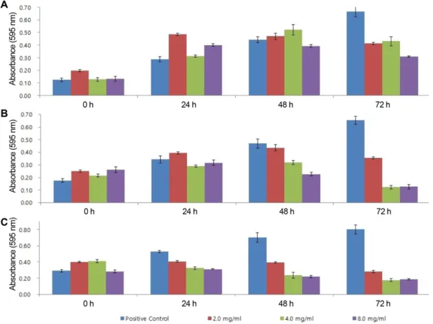
어류질병균주를이용하여 biofilm 형성능과생육도를측 정하였다(Fig. 1). 어류질병세균의생육도의경우 Et가 Sp, Si에비해높게나타났지만, biofilm 형성능의경우 Sp가 Si,
Et에비해높게나타났다. 이러한결과로미루어보아균주
별성장도와 biofilm 형성능간의특정한패턴이없이나타났
으며, 세균의성장도가높다하더라도꼭 biofilm을활발히
형성하지않음을알수있었다. Biofilm 저해능 확인
어류질병세균에대해갈륨이 biofilm을저해하는지알아
보기위해각각의균주를배양한후갈륨을농도별(2.0, 4.0,
8.0 mg/ml)로첨가하여 biofilm을측정하였다(Fig. 2). 3균주 모두 48시간째부터 biofilm 형성이저해되기시작하여, 72시
간째갈륨을처리하지않은컨트롤에비해 biofilm이크게저
해되는것을확인할수있었다.
Sp는 72시간 째 갈륨의 모든 농도에서 컨트롤에 비해
biofilm 형성능이저해되는것을확인하였으며, 이중갈륨농 도 8.0 mg/ml에서 가장 큰 biofilm 저해능을 보였다(Fig.
2A). Si, Et는 72시간째갈륨의모든농도에서컨트롤에비 해 biofilm 형성능이저해되는것을확인하였으며, 이중갈 륨농도 4.0 mg/ml에서 가장 큰 biofilm 저해능을 보였다 (Fig. 2B, 2C).
어류질병 3균주중 Et에서가장큰 biofilm 저해능을나타 났다. 이는갈륨이이미형성된 biofilm 자체를파괴하는효과 가있으며, biofilm의형성능이높고낮음에상관없이 biofilm
을억제하기때문에매우긍정적인결과가할수있다[2, 9].
Fig. 2. Biofilm inhibition activity of gallium for fish pathogen. (A) S. parauberis, (B) S. iniae, (C) E. tarda.
Fig. 1. Biofilm formation activity and growth of fish pathogen.
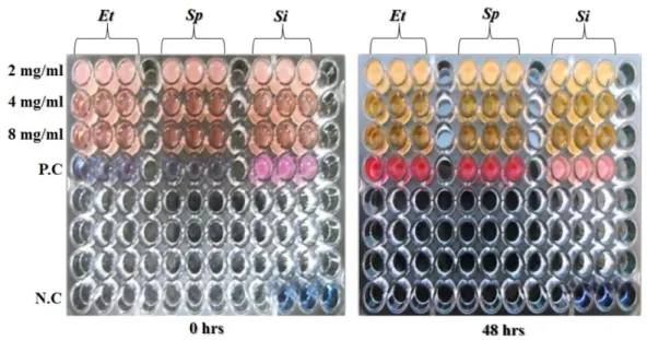
Resazurin reduction test
갈륨이 biofilm을억제하는동시에균주를직접적으로사
멸시키는지알아보기위해, RRT를이용하여어류질병세균 의사멸을확인하였다. 갈륨을처리하지않은어류질병세균 에 resazurin을각각처리하였을시, 청색에서진한분홍색 으로색이변하였다(Fig. 3).
균주를 105 CFU/ml로접종하여갈륨을 2.0, 4.0, 8.0 mg/
ml의농도로처리한그룹에서는연한분홍색에서진한노란
색으로색이변한것을확인할수있었다(Fig. 3). 레사주린
을첨가한직후의 pH가 7.0이고, 색의변화가노란색으로변 하였을때 pH는 4.8로나왔다.
이는갈륨을처리하지않은컨트롤그룹에서세균의증식 에따라청색에서진한분홍색으로의색의변화가일어난반 면, 갈륨을처리한그룹에선세포의사멸로인한세포호흡의 저해로인하여색의변화가연분홍에서노란색으로변한것 을확인하였다[12].
Vitality staining 결과
갈륨이병원균을직접적으로사멸시키는지를확인하기위 해균주를배양한후갈륨을처리하여 PI로염색을하여병 원균의사멸을확인하였다. 갈륨을처리하지않은그룹에서 세포의사멸이일어나지않았지만, 갈륨을처리한그룹에서
세포의사멸이눈에띄게증가한것을확인할수있었다(Fig.
4). 또한갈륨의농도가높아질수록사멸되는균주도같이증
가하는것을확인할수있었다[19].
이러한일련의실험을통하여갈륨이어류질병을유발시
키는 3가지균주에 biofilm을억제시키는동시에사멸까지
일으키는것을확인할수있었다. 본연구에서수행된갈륨 이어류질병세균을억제하고항생제내성균의요인이되는
biofilm을억제하는물질로서가치가있다고사료된다.
추후이러한 in vitro 상에서의실험결과를이용하여양식
넙치에적용하여독성및안전성에대한실험을진행하고자 한다.
Fig. 3. Effect of gallium on viable cell count of bacterial fish pathogen. (P.C) Positive Control (well contain untreated bacterial cells), (N.C) Negative Control (well contain only culture medium), (Et) E. tarda, (Sp) S. parauberis, (Si) S. iniae.
Fig. 4. Confocal microscopic image of gallium treated fish pathogenic bacterial cells after 48 h. (P.C) Positive Control (well contain untreated bacterial cells), (Et) E. tarda, (Sp) S. parauberis, (Si) S. iniae.
요 약
제주도넙치양식장에서주로발생하는어류질병세균인 S.
parauberis, S. iniae, E. tarda에대한피해를줄이고자갈륨 을이용하여어류질병세균을억제하고자한다. 본연구진은 Sp, Si, Et의생육도와 biofilm 형성능을확인하였으며, 세균
의성장도와 biofilm 형성능간의특정한패턴은없었으며세
균의성장도가높다하더라도 biofilm을활발히형성하지않
음을알수있었다. 어류질병세균이형성하는 biofilm을저 해하기위해갈륨을 2.0, 4.0, 8.0 mg/ml로첨가하여저해능 을확인한결과 72시간째 biofilm의형성이크게저해되는 것을확인하였다. 또한 biofilm을저해하는동시에균주의사 멸능을확인하기위해 resazurin assay와 propidium iodide 를이용하여염색한결과갈륨의농도에따라균주의사멸 이증가하는것을확인하였다. 이에따라 in vitro 상에서갈 륨이어류질병세균의 biofilm을억제하는동시에균주를사 멸시키는물질로서가치가있다고사료된다.
Acknowledgments
This research was supported by The Leading Human Resource Training Program of Regional Neo industry through the National Research Foundation of Korea (NRF) funded by the Ministry of Science, ICT and future Planning (2016H1D5A1911152 &
2013H1B8A2032163).
References
1. Ansar AS, Gogal RM, Walsh JE. 1994. A new rapid and simple non-radioactive assay to monitor and determine the prolifera- tion of lymphocytes: an alternative to [3H] thymidine incorpora- tion assay. J. Immunol. Method. 170: 211-224.
2. Banin E, Lozinski A, Brady KW, Berenshtein E, Butterfield PW, Moshe M, et al. 2008. The potential of desferrioxamine-gallium as an anti-Pseudomonas therapeutic agent. Proc. Natl. Acad. Sci.
USA 105: 16761-16766.
3. Chiang WC, Cilsson M, Jensen PØ, Høiby N, Nielsen TE, Givskov M, et al. 2013. Extracellular DNA Shields against Aminoglyco- sides in Pseudomonas aeruginosa Biofilms. Antimicrob. Agents Chemother. 57: 2352-2361.
4. De Fries R, Mitsuhashi M. 1995. Quantification of mitogen induced human lymphocyte proliferation: Comparison of ala- marbluetm assay to 3h‐thymidine incorporation assay. J. Clin.
Lab. Anal. 9: 89-95.
5. Deleon K, Balldin F, Watters C, Hamood A, Griswold J, Sreedha- ran S, et al. 2009. Gallium maltolate treatment eradicates Pseu- domonas aeruginosa infection in thermally injured mice.
Antimicrob. Agents Chemother. 53: 1331-1337.
6. Im JT, Park IK, Koh TS. 2007. Proliferation assay of splenocyte and PBMC by the evaluation of alamar blue dye reduction value in broiler chicks. J. Anim. Sci. Technol. 49: 213-224.
7. Jung SH, Choi DL, Kim JW, Jo MR, Jee BY, Seo JS. 2009. Pharma- cokinetics of oxolinic acid in cultured olive flounder Paralichthys olivaceus by oral administration, injection and dipping. J. Fish.
Pathol. 22: 125-135.
8. Kaneko Y, Thoendel M, Olakanmi O, Britigan BE, Singh PK. 2007.
The transition metal gallium disrupts Pseudomonas aeruginosa iron metabolism and has antimicrobial and antibiofilm activity.
J. Clin. Invest. 117: 877-888.
9. Kim JH, Kim CH, Hacker J, Ziebuhr W, Lee BK, Cho SH. 2008.
Molecular characterization of regulatory genes associated with biofilm variation in a Staphylococcus aureus strain. J. Microbiol.
Biotechnol. 18: 28-34.
10. Lee CH, Kim PY, Ko CS, Oh DC, Kang BJ. 2007. Biological charac- teristics of Streptococcus iniae and Streptococcus parauberis iso- lated from cultured flounder, Paralichthys olivaceus, in Jeju. J.
Fish. Pathol. 20: 33-40.
11. Lee JH. 2015. A review on microbialites: a Korean perspective. J.
Petrol. Soc. Korea 24: 291-305.
12. Martin A, Camacho M, Portaels F, Palomino JC. 2003. Resazurin microtiter assay plate testing of Mycobacterium tuberculosis sus- ceptibilities to second-line drugs: Rapid, simple, and inexpen- sive methods. Antimicrob. Agents Chemother. 47: 3616-3619.
13. Ministry for Food Agriculture Forestry and Fisheries. 2014. Statis- tical year book of maritime affairs and fisheries.
14. National Fisheries Research & Development Institute. 2015. Pre- vention of bacterial fish disease and medical treatment for pro- duce health fish.
15. Nguyen HT, Kanai K. 1999. Selective agars for the isolation of Streptococcus iniae from Japanese flounder, Paralichthys oliva- ceus, and its cultural environment. J. Appl. Microbiol. 86: 769-776.
16. Oh KO, Kim KK. 2009. Prevention of Biofilm Infections. J. Bacte- riol. Virol. 39: 237-246.
17. Park JS, Ju SA, Heo MS, Jung CR, Ju JW. 1999. Purification sidero- phore from Vibrio mimicus ATCC 33653 and its effect to bacterial pathogenicity. J. Korean Soc. Microbiol. 34: 461-470.
18. Statistics Korea. 2015. Aquaculture Status Survey.
19. Woo SH, Lee JH, Kim YK, Cho MY, Jung SH, Kim JW, et al. 2010.
Effects of garlic Allium sativum extract immersion on the immune responses of olive flounder Paralichthys olivaceus pre- challenged with pathogenic bacteria. J. Fish. Pathol. 23: 199-209.