"J. Korean Soc. Radiol., Vol. 12, No. 2, April 2018"
Evaluation of Absorbed Dose According to the Nanoparticle in Prostate Cancer Brachytherapy
Eun-tae Park,1 Deuk-hee Lee,1 In-chul Im2,*
1Department of Radiation Oncology, Busan Paik Hospital, Inje University, Korea
2Department of Radiological Science, Dongeui University, Korea
Received: January 30, 2018. Revised: April 15, 2018. Accepted: April 30, 2018
ABSTRACT
This study evaluated absorbed dose of brachytherapy according to the nanoparticle in prostate cancer which many occurred in Korean man and provided basic data. Absorbed dose evaluation was using MCNPX program which was applied Monte Carlo simulation. Source was applied 192Ir which was many using in Korean HDR machine and gold, ferric oxide, gadolinium and iodine nanoparticle were applied. Prostate absorbed dose result was increased when using nanoparticle, in particular gold nanoparticle was the highest result as 3.13E-13 J/kg·e.
Absorbed dose of surrounding organs and distance was similar between using nanoparticle and non-using nanoparticle. Therefore, brachytherapy was used nanoparticle was increased therapeutic ratio and efficiency of radiation therapy.
Keywords: Prostate cancer, Brachytherapy, Nanoparticle, Monte Carlo
I. INTRODUCTION
우리나라 남성에게서 발생하는 전립선암은 식습 관 및 생활습관의 서구화로 변함에 따라 증가하고 있는 추세이며[1], 2017년 국가 암 통계에 따르면 현 재까지도 남성 암 발생률 1위를 기록하고 있다[2].
이러한 전립선암을 치료하는 방법에는 남성호르 몬 차단요법이나 화학요법이 시행되고 있으나, 완 치를 목적으로 하는 치료법으로는 외과적 절제, 체 외방사선치료(external radiation therapy) 및 근접방 사선치료(brachytherapy) 등이 있다[3,4]. 특히 방사선 치료는 종양조직의 제어율을 높임과 동시에 주변 정상조직의 장애 발생률을 충분히 낮게 유지함으 로서 치료가능비(therapeutic ratio)를 높이는 것이 중 요한 요소이다[5,6]. 이러한 치료가능비를 향상시키 는 방법으로 방사선과 종양조직 내 물질간의 반응 단면적을 높여 종양 부위에 대한 흡수에너지를 높 이는 것이 제안되고 있다[7].
나노입자를 이용한 나노의학(nanomedicine)과 방
사선을 결합한 하이브리드 치료에 대한 연구가 활 발히 진행되고 있으나, 국외의 경우 근접방사선치 료의 표적장기에 대한 연구가 주를 이루고 있다[7-9]. 국내의 경우 과거 근접방사선치료 시 인접장기와 주변인에 대한 피폭연구가 진행되었으나 나노의학 을 결합한 연구는 미비한 실정이다[10,11].
이에 본 연구는 전립선암 환자를 대상으로 한 근 접방사선치료 시 나노입자 사용여부에 따른 표적 장기의 선량변화와 주변장기 그리고 환자 주위에 대한 선량평가를 모의피폭체를 이용하여 Monte Carlo 시뮬레이션 기법으로 평가하고자 한다.
II. MATERIAL AND METHODS
1. Monte Carlo 시뮬레이션
모의모사는 MCNPX(Monte Carlo N-Particle Extended) 프로그램(ver.2.6.0, USA)을 이용하여 구 현하였다. 모의피폭체는 플로리다 대학교에서 수정 한 MIRD(medical internal radiation dose)형 팬텀을 https://doi.org/10.7742/jksr.2018.12.2.167
대상으로 하였으며, 근접치료를 위한 선원은 국내 HDR(high dose rate)장비에서 일반적으로 이용되는
192Ir선원을 이용하였다[12]. 선량증가 물질은 Zabihzadah 등과 Moghaddas 등, Hwang 등의 선행연 구를 바탕으로 금(aurum, Au), 가돌리늄(gadolinium, Gd), 요오드(iodine, I), 산화철(ferric oxide, Fe2O3)을 사용하였으며, 50 nm의 크기로 설정하였다[7-9].
선원의 배치는 근접방사선치료 시 사용하는 grid 의 간격인 5 mm를 기준으로 균일하게 배치를 하였 으며, 나노입자의 밀도는 선행연구 중 가장 낮은 밀도인 7 mg/g을 기준으로 하였다[7].
2. 선량측정
선량의 측정은 *F8 tally를 지정하여 에너지 (MeV)를 계측하여 각 장기의 단위질량으로 환산하 여 측정하였다. 표적장기인 전립선의 선량을 나노 입자 종류에 따라 평가하였으며, 동일한 방식으로 인접장기에 대한 선량의 변화를 측정하였다. 주변 인에 대한 선량평가는 인체를 기준으로 하여 거리 30, 50, 100, 200 cm에 공기벽을 설치하여 흡수선량 을 측정하였으며, Fig. 1에 나타내었다.
Fig. 1. Absorbed dose evaluation point in MCNPX.
III. RESULT
1. 전립선의 선량평가
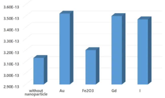
전립선의 선량은 Fig. 2와 같이 나타났다. 나노입자 를 사용하지 않은 선량이 가장 낮은 것으로 나타났으 며, 나노입자 종류에 따라서 금, 가돌리늄, 요오드, 산
화철 순으로 선량이 증가하는 경향성을 보였다.
(unit : J/kg·e) Fig. 2. Absorbed dose of prostate according to the
nanoparticle.
2. 인접장기 선량평가
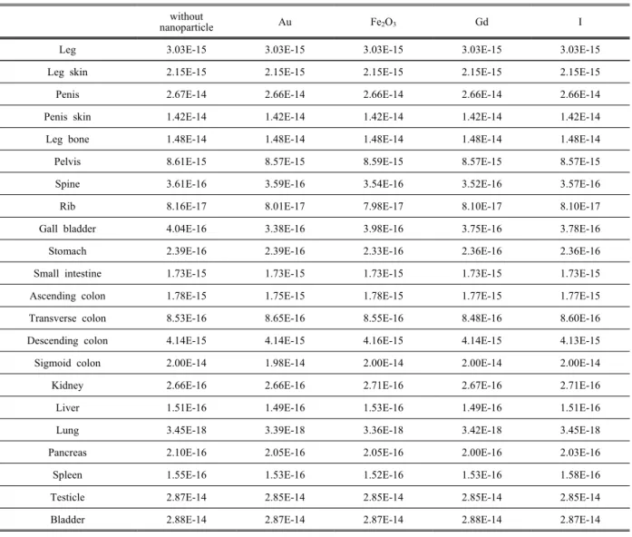
인접장기의 선량 결과는 Table 1에 나타내었다.
전립선을 기준으로 하여 인접장기인 방광에서 2.88E-14 J/kg·e로 가장 높게 나타났으며 다음으로 생식기와 S자 결장 순으로 선량이 가장 높은 것으 로 나타났다. 나노입자 종류에 따라서는 큰 차이를 나타내지는 않았지만 나노입자를 사용하지 않은 경우보다 선량이 소폭 감소하는 결과를 보였다.
3. 거리에 따른 선량결과
거리에 따른 결과는 Fig. 3과 같은 결과를 나타내 었다. 거리가 멀어질수록 선량이 감소하는 결과를 나타내었으나, 나노입자 사용에 따른 차이는 크게 나타나지 않는 것으로 나타났다.
(unit : J/kg·e) Fig. 3. Absorbed dose according to the nanoparitcle
and distance.
"J. Korean Soc. Radiol., Vol. 12, No. 2, April 2018"
Table 1. Absorbed dose of surrounding organs according to the nanoparticle (unit : J/kg·e)
without
nanoparticle Au Fe2O3 Gd I
Leg 3.03E-15 3.03E-15 3.03E-15 3.03E-15 3.03E-15
Leg skin 2.15E-15 2.15E-15 2.15E-15 2.15E-15 2.15E-15
Penis 2.67E-14 2.66E-14 2.66E-14 2.66E-14 2.66E-14
Penis skin 1.42E-14 1.42E-14 1.42E-14 1.42E-14 1.42E-14
Leg bone 1.48E-14 1.48E-14 1.48E-14 1.48E-14 1.48E-14
Pelvis 8.61E-15 8.57E-15 8.59E-15 8.57E-15 8.57E-15
Spine 3.61E-16 3.59E-16 3.54E-16 3.52E-16 3.57E-16
Rib 8.16E-17 8.01E-17 7.98E-17 8.10E-17 8.10E-17
Gall bladder 4.04E-16 3.38E-16 3.98E-16 3.75E-16 3.78E-16
Stomach 2.39E-16 2.39E-16 2.33E-16 2.36E-16 2.36E-16
Small intestine 1.73E-15 1.73E-15 1.73E-15 1.73E-15 1.73E-15
Ascending colon 1.78E-15 1.75E-15 1.78E-15 1.77E-15 1.77E-15
Transverse colon 8.53E-16 8.65E-16 8.55E-16 8.48E-16 8.60E-16
Descending colon 4.14E-15 4.14E-15 4.16E-15 4.14E-15 4.13E-15
Sigmoid colon 2.00E-14 1.98E-14 2.00E-14 2.00E-14 2.00E-14
Kidney 2.66E-16 2.66E-16 2.71E-16 2.67E-16 2.71E-16
Liver 1.51E-16 1.49E-16 1.53E-16 1.49E-16 1.51E-16
Lung 3.45E-18 3.39E-18 3.36E-18 3.42E-18 3.45E-18
Pancreas 2.10E-16 2.05E-16 2.05E-16 2.00E-16 2.03E-16
Spleen 1.55E-16 1.53E-16 1.52E-16 1.53E-16 1.58E-16
Testicle 2.87E-14 2.85E-14 2.85E-14 2.85E-14 2.85E-14
Bladder 2.88E-14 2.87E-14 2.87E-14 2.88E-14 2.87E-14
IV. DISCUSSION
방사선치료 시 높은 원자번호의 나노입자를 사용 하는 것은 광전효과(photoelectric effect)와 콤프턴산 란(compton scattering)으로 인 방사선의 반응단면적 (cross-section)을 증가시켜 치료 효율을 좋게 한다[13].
본 연구에서 나노입자에 따른 표적장기의 선량 을 비교한 결과 원자번호가 가장 높은 금 나노입자 에서 가장 큰 선량증가를 보였다. 이러한 결과는 Zabihzadeh 등의 선행연구에서도 금 나노입자의 선 량증가효과(dose enhancement effect, DEF)가 가장 크다고 보고하였다[7].
다음으로 인접장기에 대한 피폭선량은 전립선에
인접한 비뇨기 계통에서 피폭이 가장 높은 것으로 나타났다. Kim 등은 전립선의 근접치료 시 음경 및 음낭에서 가장 높은 피폭이 나타나는 것으로 보고하 였다[9]. 이러한 차이는 본 연구에서 사용한 팬텀은 서양인을 기준으로 하여 제작 된 MIRD형 팬텀으로 선량평가를 하였으나, Kim 등은 아시아인을 기준으 로 한 팬텀을 이용한 차이인 것으로 사료된다. 반면 전립선 인근 비뇨기 및 생식기 장기에서 높은 선량 이 나타나며 거리가 먼 장기일수록 피폭이 감소하는 것은 같은 경향성을 보였다. 나노입자에 따른 선량 의 차이는 크지 않았으나, 나노입자를 미사용한 경 우보다 소량 감소하는 결과로 나타났다.
거리에 따른 선량은 거리에 비례하여 감소하는 것으로 나타났다. 이는 거리에 따라 방사선이 감소
하는 특징에 따른 것으로 판단되며 , Park 등의 선 행연구와 같은 경향성을 보였다[11]. 반면 나노입자 사용 여부에 따른 차이는 없는 것으로 나타났다.
본 연구를 통하여 저 밀도의 나노입자 사용 시 표 적장기의 선량은 증가하나, 인접장기 및 주위 선량 은 큰 차이를 나타내지 않는 것으로 나타났다. 이러 한 현상은 방사선치료 중 중요한 요소인 치료가능 비를 증가시키는 것으로서, 정상조직의 피폭을 감 소하는 것과 같은 결과를 나타낼 것으로 생각된다.
Duc 등은 나노입자 밀도가 5 mg/g 이상일 경우 유효한 차이를 보이며 밀도가 높아질수록 흡수선 량이 증가되는 것으로 보고하였다[14]. 또한 Hwang 등과 Unezaki 등은 나노입자의 크기가 커질수록 흡 수선량이 증가하는 연구결과를 보였다[9,15]. 따라서 차후 나노입자의 밀도와 크기를 변화시킨 표적장 기의 선량과 인접장기 및 주위 선량에 대한 연구도 필요할 것으로 사료된다.
V. CONCLUSION
국내 남성에게서 발병률이 높은 전립선암을 대 상으로 근접치료 시 나노입자 사용에 따른 선량평 가를 하였다. 표적장기인 전립선에 나노입자를 주 입 시 표적장기의 흡수선량이 증가하였으며, 이중 금 나노입자가 가장 높게 나타났다. 반면 정상조직 및 주위선량에 대해서는 큰 차이를 보이지 않았다.
그러므로 근접치료 시 나노입자의 활용은 치료가 능비를 상승시켜 치료효율을 상승시킬 수 있을 것 으로 판단된다.
Reference
[1] S. R. Im, “Analysis of Relative Factor and Estimate of Incidence Rate in Prostate Cancer: The Korean Ca ncer Prevention Study-II (KCPS-II),” Graduate School of Public Health Yonsei University Master`s Thesis, pp. 71-72, 2015.
[2] National Cancer Information Center(https://www.cance r.go.kr), 2017
[3] W. Park, S. J. Huh, H. H. Choi, H. M. Lee, S. E.
Chai, Y. C. Ahn, D. H. Lim, “Results of Definitive Radiotherapy in the Treatment of Prostate Cancer,” T
he Korean Urological Association, Vol. 46, No. 3, p p. 201-228, 2005.
[4] H. S. Chu, “Factors Affecting the Biochemical Recurr ence after Radical Prostatectomy or Radiotherapy in I ntermediate and High-risk Patients with Prostate Canc er,” University of Ulsan Master`s Thesis, pp. 1, 200 9.
[5] S. S. Kang, I. H. Go, G. J. Kim, S. H. Kim, Y. S.
Kim, Y. J. Kim, Radiation Therapeutics, third edition, Chung-ku munhwasa, Korea, 2014.
[6] D. H. Lee, E. T. Park, J. H. Kim, I. C. Im, “Evaluat ed Absorbed Dose According to Prescribed Dose and Therapeutic Technique in Radiation Therapy,” Journal of the Korean Society of Radiology, Vol. 10, No. 6, pp. 469-476, 2016.
[7] M. Zabihzadah, S. Arefian, “Tumor dose enhancement by nanoparticles during high dose rate 192Ir brachyth erapy,” Journal of Cancer Research and Therapeutics, Vol. 11, No. 4, pp. 752-759, 2015.
[8] T. A. Moghaddas, M. Ghorbani, A. Haghparast, R.
T. Fylnn, M. T. Eivazi, “Monte Carlo Study on Dose Enhancement Effect of Various Paramagnetic Nanoshe lls in Brachytherapy,” Journal of Medical and Biologi cal Engineering, Vol. 34, No. 6, pp. 559-567, 2014.
[9] C. H. Hwang, S. S. Kang, J. H. Kim, “A Monte Car lo Study of Secondary Electron Production from Gold Nanoparticle in Kilovoltage and Megavoltage X-rays,”
Journal of the Korean Society of Radiology, Vol. 10, No. 3, pp. 153-159, 2016.
[10] J. H. Kim, C. S. Im, J. H. Hwang, “Radiation Dose Calculation in the Surrounding Organs during Brachy therapy of Prostate Cancer,” Korean Journal of Medi cal physics, Vol. 19, No. 13, pp. 172-177, 2008.
[11] E. T. Park, J. H. Kim, “Dose Evaluation of the Ma n Adjacent to an Implanted Patient During the Prost ate Cancer Brachytherapy,” Journal of the Korean S ociety of Radiology, Vol. 10, No. 1, pp. 39-44, 201 6.
[12] O. N. Yang, S. S. Shin, W. S. Ahn, D. Y. Kim, W. S. Choi, K. T. Kwon, C. H. Lim, S. H. Lee,
“Comparison of Treatment Planning on Dosimetric D ifferences Between 192Ir Sources for High-Dose Rat e Brachytherapy,” Radiology and Nuclear Medicine, Vol. 39, No. 2, pp. 163-170, 2016.
"J. Korean Soc. Radiol., Vol. 12, No. 2, April 2018"
[13] P. Retif, S. Pinel, M. Toussaint, C. Frochot, R. Cho uikrat, T. Bastogne, M. B. Heyob, “Nanoparticles fo r Radiation Therapy Enhancement: the Key Paramete rs,” Theranostics, Vol. 5, No. 9, pp. 1030-1044, 201 5.
[14] G. L. Duc, I. Miladi, C. Alric, P. Mowat, E. B. K risch, A. Bouchet, E. Khalil, C. Billotey, M. Janier, F. Lux, T. Epicier, P. Perriat, S. Roux, O. Tillemen t, “Toward an Image-Guided Microbeam Radiation T herapy Using Gadolinium-Based Nanoparicles,” Amer ican Chemical Society Nano, Vol. 5, No. 12, pp. 95 66-9574, 2011.
[15] S. Unezaki, K. Maruyama, J. I. Hosoda, I. Nagae, Y. Koyanagi, M. Nakata, O. Ishida, M. Iwatsuru, S.
Tsuchiya, “Direct measurement of the extravasation of polyethy- leneglycol coated liposomes into solid t umor tissue by in vivo fluorescence microscopy,” Int ernational Journal of Pharmaceutics, Vol. 144, No.
1, pp. 11-17, 1996.
전립선암의 근접치료 시 나노입자에 따른 흡수선량평가
박은태,1 이득희,1 임인철2,*
1인제대학교 부산백병원 방사선종양학과
2동의대학교 방사선학과
요 약
국내 남성에게서 많이 발생하는 전립선암을 대상으로, 근접치료 시 나노입자 사용에 따른 선량을 평가하 여 기초자료를 제시하고자 하였다. 선량평가는 몬테카를로 시뮬레이션 기법인 MCNPX 프로그램을 이용하 였다. 선원은 국내 HDR장비에 다용하는 192Ir으로 선정하고 나노입자는 금, 가돌리늄, 산화철, 요오드를 사 용하였다. 그 결과 표적장기인 전립선은 나노입자를 사용 시, 사용하지 않은 경우에 비해 모두 흡수선량이 높게 나타났다. 특히 금 나노입자가 3.13E-03 J/kg·e의 값으로 가장 높았다. 주변장기 및 주변인에 대한 선 량은 나노입자 사용에 따른 차이가 크지 않은 것으로 나타났다. 나노입자 사용은 치료가능비를 상승시켜 치료효율을 증가시킬 수 있을 것으로 판단된다.
중심단어: 전립선암, 근접치료, 나노입자, 몬테카를로