Effect of Posterior-Anterior Mobilization of the Thoracic Spine on Pain, Respiratory Function, and Thoracic Circumference in
Patients With Chronic Low Back Pain
Ju-jung Park1, MSc, PT, Seung-chul Chon2, PhD, PT
1Dept. of Physical Therapy, Jaseng Korean Medicine Hospital
2Dept. of Physical Therapy, College of Medical Science, Konyang University
Abstract
1)Background: Posterior-anterior (PA) vertebral mobilization, a manual therapy technique has been used for relieving pain or stiffness treating in spinal segment for in clinical practice, however evidence to gauge efficacy is yet to be synthesised.
Objects: This study aimed to investigate the effect of PA mobilization of the thoracic spine on the respiratory function in patients with low back pain (LBP).
Methods: The study participants included 30 patients with chronic LBP. They were randomly allocated to the experimental and control groups. The experimental and control groups received PA mobilization of the T1-T8 level of the thoracic spine and placebo mobilization, respectively. All patients received interventions for 35 minutes a day, five times a week, over 2-week period, respectively. Forced vital capacity (FVC), forced expiratory volume in 1 second (FEV1), peak expiratory flow (PEF), forced expiratory flow 25∼75% (FEF25∼75%), and chest wall expansion were measured before and after the intervention. Statistical analysis was performed using independent t-test and two-way analysis of variance, and Pearson’s correlation analysis was used to compare the correlation between respiratory function and chest measurement.
Results: The experimental group showed significant improvements in FVC, FEV1, PEF, FEF25∼75%
(p<.05), and chest wall expansion (p<.05) compared with the control group.
Conclusion: PA mobilization of the upper thoracic spine may be beneficial for improving respiratory function parameters including FVC, FEV1, PEF, FEF25∼75%, and chest wall expansion in patients with chronic LBP.
Key Words: Low back pain; Respiratory function; Thoracic mobilization; Vital capacity.
Introduction
Low back pain (LBP) is one of the most common neuromusculoskeletal disorders, with a lifetime preva- lence rate of >75% (Hanney et al, 2016). Research into the pathogenesis of chronic LBP has identified many contributing factors, including socioeconomic and psychological influences, genetic predisposition, degenerative changes, and muscle imbalance (Verkerk et al, 2012). Moreover, the medical cost of these
problems is causing yearly economic losses. Patrick et al (2014) classified cases of persistent symptoms lasting longer than 3 months as chronic, and re- ported the importance of early treatment because of the slow recovery of chronic LBP over time.
LBP has a high correlation with respiratory func- tion (Grimstone and Hodges, 2003; Smith et al, 2006). Respiration regulates the movement of the di- aphragm through the up-and-down movement and contraction and relaxation of the muscles between
Corresponding author: Seung-chul Chon [email protected]
This research was supported by Basic Science Research Program through the National Research Foundation of Korea (NRF) funded by the Ministry of Education (2017R1D1A3B03031876).
the ribs and problems with the movement of the rib cage can cause dysfunction of these respiratory muscles (Cahalin et al, 2002). Patients with various pulmonary diseases have problems not only with the lungs but also with the respiratory muscles, and they also experience musculoskeletal disorders (Wagner, 2006). In other words, patients with LBP commonly have musculoskeletal disorders with dys- function of the respiratory muscles, and abnormal symptoms and rapid fatigue of the respiratory mus- cles are also observed during low-intensity exercise (MacIntyre, 2006).
The spine movement of patients with back pain is partially restricted and limited, particularly in terms of extension mobility of the upper thoracic spine (Mohanty and Pattnaik, 2016). Specifically, unstable lumbar spine with pain and abnormal functional movement reduces the mobility of adjacent spinal joints such as the thoracic region. And also, the thoracic spine is directly connected to the thorax and joints, the movement of the thoracic spine is inter- dependent, and the thoracic mobility is an important factor in the respiratory system. Increased chest wall mobility positively influences respiratory control, coughing ability, lung capacity, and spinal motion by inducing smooth movement and contraction of the respiratory muscle (Hodges and Gandevia, 2000).
Hypomobility of the thoracic spine causes the chest to sink and limits the expansion of the circumference of the thorax during inspiration.
Decreased respiratory function is also associated with back pain because it affect the posture control ability (Ruhe et al, 2011). In particular, the dia- phragm is an important muscle in regulating the spine during postural control on inspiration (Hodges and Gandevia, 2000). In previous studies in patients with chronic LBP and chronic obstructive pulmonary disease, the proprioceptive sensations needed for pos- tural control were reduced when the inspiratory muscle was used (Janssens et al, 2010; Janssens et al, 2013). In addition, patients with chronic LBP ex- perienced greater diaphragmatic fatigue than healthy
controls, and training of the inspiratory muscle in these patients improved postural control and reduced pain intensity (Janssens et al, 2010).
Spinal joint mobilization in patients with LBP has been reported to help relieve pain (Savigny et al, 2009). Maitland has categorized and applied joint mobilization from grade 1 to grade 5 for the treat- ment of joint dysfunction (Banks, 2010). Cervical and thoracic joint operations can control the autonomic nervous system to regulate respiratory muscles and pulmonary function by promoting the activity of the sympathetic nerves emerging between the vagus nerves and thoracic vertebrae 1 and 5, which are the 11th cranial nerves under the control of para- sympathetic nerves (Engel and Vemulpad, 2007).
Yang and Kim (2015) reported that the application of thoracic spine mobilization to patients with chronic LBP had a positive effect on pain and proprioceptive sensation. Babina et al (2016) reported improved pul- monary function in patients with chronic LBP when thoracic spine mobilization was applied, and Ito et al (1999) reported that reduction of the range of motion of the thoracic spine reduced pulmonary function, in- creased chest circumference (CC), and has been as- sociated with improved pulmonary function.
Although several reports are available on pain re- duction and motor function improvement in patients with LBP, data on respiratory ability to evaluate the effects of joint mobilization and pulmonary function are still lacking. This study aimed to investigate the effect of joint mobilization of the thoracic spine on pain and respiratory function and CC length in pa- tients with chronic LBP.
Methods
Subjects
The study subjects included 30 patients with chronic LBP. A physical therapist with 6 years of experience made the diagnosis of LBP according to the clinical assessment criteria. Medical diagnosis of
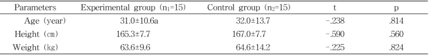
Parameters Experimental group (n1=15) Control group (n2=15) t p
Age (year) 31.0±10.6a 32.0±13.7 -.238 .814
Height (㎝) 165.3±7.7 167.0±7.7 -.590 .560
Weight (㎏) 63.6±9.6 64.6±14.2 -.225 .824
amean±standard deviation.
Table 1. General characteristics of subjects (N=30)
Figure 1. Thoracic mobility test.
LBP was made by an orthopaedist or a physician in hospital. The inclusion criteria were subjects who had >3 months of back pain, had visual analog scale (VAS) score of >4 points, and showed positive on thoracic motility test. The exclusion criteria were cardiopulmonary system and nervous system prob- lems, smoking, spinal fractures, and spinal joint surgery. Explanations about the procedure and sta- bility were given to all subjects before the experi- ment, and informed consent was obtained from all the subjects, and this study was approved by the university ethics and institutional review board (approval number: 2018-057-01). Table 1 shows the general characteristics of the study subjects.
Instrumentation and measurement
Visual analogue scale
The visual analogue scale (VAS) is considered to be one of the best methods available for the estima- tion of the intensity of pain. VAS is self-report measure consisting simply of a 10 centimeter line with a statement at each end representing one di- mension being measured. For pain intensity, the scale is most commonly anchored by “no pain” (score of 0) and “pain as bad as it could be” (score of 10).
Thoracic spine mobility test
Heiderscheit and Boissonnault (2008) examined the mobility of the thoracic spine using a tester’s hand in the prone position by pushing the spinous processes from the posterior to the anterior direction in the thoracic vertebrae 1∼8, and Gonnella et al (1982) graded the results by using a 7-point (0-6) scale: 0 point means the movement of the segment cannot be detected in a rigid state; 1 point and 2 points, the
low range of the resistance before the normal range;
3 points, the normal range; 4 points and 5 points, the normal range and a significantly reduced resistance to an increased range; and 6 points, an excessive range, meaning no ligament and capsular limitation.
Respiratory function test
Spirobank G (Spirobank G, MIR, Rome, Italy) was used to examine the respiratory function of the subjects. Forced vital capacity (FVC), forced ex- piratory volume in 1 second (FEV1), peak expiratory flow (PEF), and forced expiratory flow 25∼75%
(FEF25∼75%) were measured. Before starting the ex- periment, a full explanation about the method of the pulmonary function test was given to the subjects.
During the respiratory function test, the subjects were in the following normal posture: seating on a chair with the legs open and the back not leaning against the back of the chair. The nose was closed using a nasal plug; the measuring instrument placed inside the mouth was held in one hand; and maximum expiration was performed after maximum inspiration. At this time, the upper body was controlled so as not to bend in a compensating motion. Measurements were taken
A B C
Figure 2. Chest measurement.
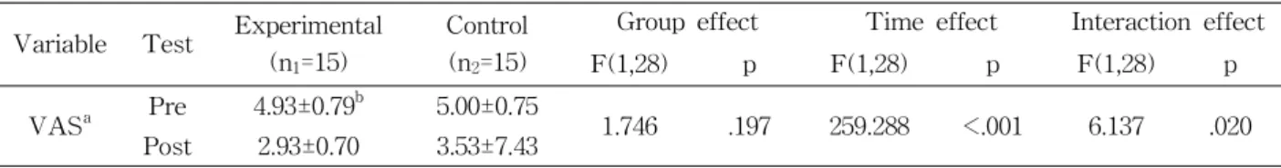
Variable Test Experimental (n1=15)
Control (n2=15)
Group effect Time effect Interaction effect
F(1,28) p F(1,28) p F(1,28) p
VASa Pre 4.93±0.79b 5.00±0.75
1.746 .197 259.288 <.001 6.137 .020 Post 2.93±0.70 3.53±7.43
avisual analog scale,bmean±standard deviation.
Table 2. Comparisons visual analog scale in both groups
A B C
Figure 3. Upper and middle thoracic spine mobilization (A: upper thoracic spine superior view, B: upper thoracic spine lateral view, C: middle thoracic spine superior view).
three times consecutively, and the mean value was used in the analysis.
Chest measurement
Before and after intervention, the CC was meas- ured at the time of maximum inspiration. According to the study by Bockenhauer et al (2007), the point in the 3rd intercostal region to the mid-clavicular line (Figure 2, A) and the point at the 5th spinous processes from the thoracic spine (Figure 2, B) were marked, and the circumference was measured where the two points meet (Figure 2, C).
Intervention
The experimental group was treated for chronic LBP by performing Maitland grade 3 on thoracic spine vertebrae 1∼8 (Figure 3). The control group was also treated for chronic LBP by applying Maitland grade 1 on thoracic spine vertebrae 1∼8. Both groups received interventions for 35 minutes a day, five times a week, over 2-week period, respectively. Grade 3 mobilization was applied to the experimental group to obtain the range of motion of the normal joint by imparting a large vibration to the hypomobility region and stretch- ing the connective tissue of the joint or the joint capsule. Grade 1 mobilization refers to a small amount of vibration at the beginning of the range of motion of the joint to control pain or muscle spasm.
Statistical analysis
The general characteristics of the subjects such as age, height, and weight, were used in the in- dependent t-test. Two-way analysis of variance with interindividual factors was used to compare respira- tory function and CC length before and after the ex- periment in the experimental and control groups. The Bonferroni post-hoc test was performed when the effect within the individual was significant. Pearson’s correlation analysis was used to compare the corre- lation of respiratory function and CC length. The collected data were analyzed using the statistical program SPSS ver. 20.0 (IBM Corp., Armonk, NY, USA), and the correlation between lung function and CC was set at p<.01. A p value of <.05 was con- sidered statistically significant.
Results
In the VAS, the group effect was not significant (F=1.746, p=.197), and the time effect was significant (F=259.288, p<.001). The interaction effect was sig- nificant (F=6.137 p=.020) (Table 2).
In the FVC, the group effect was not significant
Variable Test Experimental (n1=15)
Control (n2=15)
Group effect Time effect Interaction effect
F(1,28) p F(1,28) p F(1,28) p
FVCa Pre 3.42±.88b 3.50±.88
.002 .962 37.105 <.001 13.417 .001 Post 3.58±.93 3.54±.89
FEV1c Pre 2.80±.85 3.03±.80
.158 .694 25.212 <.001 12.406 .001 Post 3.06±.79 3.07±.81
PEFd Pre 5.76±2.28 6.41±1.90
.045 .834 18.322 <.001 13.938 .001 Post 6.81±2.06 6.48±1.91
FEF25∼75%e Pre 3.46±1.14 3.52±.81
.043 .837 15.385 .001 7.608 .010
Post 3.79±1.07 3.58±.84
aforced vital capacity, bmean±standard deviation, cforced expiratory volume 1 second, dpeak expiratory flow, eforced expiratory flow 25∼75%.
Table 3. Comparison of respiratory function between two groups
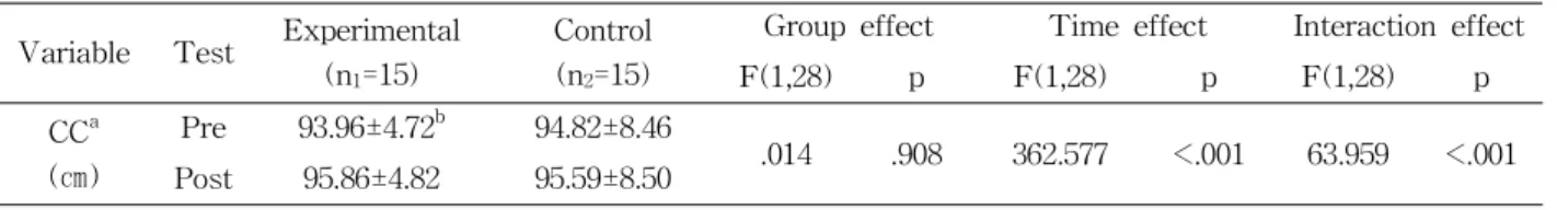
Variable Test Experimental (n1=15)
Control (n2=15)
Group effect Time effect Interaction effect
F(1,28) p F(1,28) p F(1,28) p
CCa (㎝)
Pre 93.96±4.72b 94.82±8.46
.014 .908 362.577 <.001 63.959 <.001 Post 95.86±4.82 95.59±8.50
achest circumference, bmean±standard deviation.
Table 4. Comparisons chest circumference between in both groups (F=.002, p=.962), while the time effect (F=37.105,
p<.001) and the interaction effect (F=13.417, p=.001) were significant, respectively. In the FEV1, the group effect was not significant (F=.158, p=.694), while the time effect (F=25.212, p<.001) and the interaction ef- fect (F=12.406, p=.001) were significant, respectively.
In the PEF, the group effect was not significant (F=.045, p=.834), while the time effect (F=18.322, p<.001) and the interaction effect (F=13.938, p=.001) were significant, respectively. In the FEF25∼75%, the group effect was not significant (F=.043, p=.837), while the time effect (F=15.385, p=.001) and the in- teraction effect (F=7.608, p=.010) were significant, re- spectively (Table 3).
In the CC, the group effect was not significant (F=.014, p=.908), while the time effect (F=362.577, p<.001) and the interaction effect (F=63.959, p<.001) were significant, respectively (Table 4).
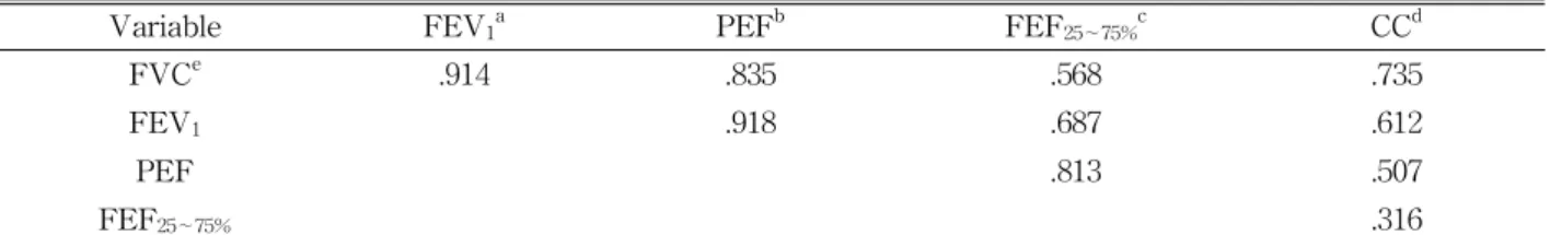
The correlation coefficients between respiratory function and CC were positively correlated with FVC=.735 (p<.01), FEV1=.612 (p<.01) and PEF=.507 (Table 5).
Discussion
Although improvement in pain and respiratory function in patients with back pain is important for the normal movement of the vertebrae through pos- tural control, studies applying appropriate intervention to the thoracic region directly associated with respira- tory function are limited. In this study, we evaluated pain, respiratory function, and CC length by applying Posterior-Anterior mobilization to the thoracic region in patients with LBP. We also investigated the corre- lation between respiratory function and CC length. As a result, a positive effect on pain, respiratory function, and CC length and a moderate correlation among res- piratory function parameters such as FVC, FEV1, and PEF, except for FEF25∼75%, were found.
The LBP group included people who had recurrent back pain in clinical field and had not improved with nonoperative treatments. In participants with LBP, we observed a reduction of pain specifically improve- ments in VAS (from 5 to 3) after the intervention.
Our findings are consistent with those of Ko et al
Variable FEV1a PEFb FEF25∼75%c CCd
FVCe .914 .835 .568 .735
FEV1 .918 .687 .612
PEF .813 .507
FEF25∼75% .316
aforced expiratory volume 1 second, bpeak expiratory flow, cforced expiratory flow 25∼75%, dchest circumference,
eforced vital capacity.
Table 5. Correlation between respiratory function and chest circumference
(2009), who showed that engaging in thoracic mobi- lization like PA mobilization with lumbar stabilization reduced the oswestry disability index cores of pa- tients with chronic LBP. Kaltenborn et al (1993) re- ported that unstable lumbar spine stability reduces the mobility of adjacent spinal joints such as the thoracic part. The results of this study suggest that the lumbar spine can be treated by increasing the mobility of the thoracic spine with reduced motility, and that the side effects are less severe than the di- rect treatment of the painful lumbar spine (Singer and Giles, 1990).
Although improvement in respiratory function should be included in the field of rehabilitation and physical therapy for improving the pain control and exercise capacity of patients with back pain, con- crete intervention methods are lacking. This study suggests that the use of joint mobilization indirectly applied to the thoracic region rather than to the lumbar region could solve the difficulties of patients with back pain, because direct intervention is diffi- cult owing to pain in the lumbar region and the ability to control the abnormal movement, and can be introduced as an intervention method. In addition, considering the anatomical structure of the thoracic region, mobilization in only the PA direction was applied to the thoracic vertebrae 1∼8 region.
Therefore, various intervention methods may be considered in combination with the functional move- ment of the thoracic region. In this study, the FVC, FEV1, PEF, and FEF25∼75% were measured using the most common and easily used method of spirometry, to evaluate respiratory function. For the CC meas- urement, the point in the 3rd intercostal region to
the mid-clavicular line and the point at the 5th spi- nous processes from the thoracic spine were marked, and the circumference was measured where the two points meet, by using a tapeline (Bockenhauer et al, 2007).
Compared with the control group, all respiratory function parameters were improved in the ex- perimental group. FVC was improved by about 5%
in the experimental group after the intervention, but by only about 1% in the control group. FVC is the total amount of air that can be blown out of the lungs during forced exhalation after maximum in- halation and is generally considered normal when it is >80% of the predicted value (Lima et al, 2011).
FEV1 increased by approximately 9% after the inter- vention in the experimental group, but increased by only approximately 1% in the control group. FEV1 is the maximum amount of air that can be released within 1 second and is an indicator of whether the large airway has been shut down. The PEF in- creased by approximately 5% after the intervention in the experimental group but increased by only ap- proximately 1% in the control group. PEF is the maximum flow rate generated during forceful ex- halation, and reflects the bronchial condition.
Moreover, the CC was further improved in the experimental group compared with the control group.
This may be due to the mechanical advantage of PA mobilization applied to the thoracic spine. In particular, passive external forces, such as joint mo- tion, expand the ribcage during breathing, and smooth movements of the associated connective tis- sues appear to enhance the mobility of the thoracic joints. The increase in the length of the thoracic
cavity seems to have a positive effect on the respi- ratory function, leading to minimization of car- diopulmonary pressures and improvement in function by expansion of the thoracic cavity (Hussain and Pardy, 1985). In a previous study supporting this finding, it was suggested that restricting the move- ment of the chest could decrease the values of res- piratory function parameters such as FVC and FEV1
(Gonzalez et al, 1999). Brenner et al (2007) reported that direct joint mobilization of the thoracic spine contributes to respiratory function by increasing the mobility of the muscles and joints between the ribs constituting the thorax. These results suggest that the joint mobilization applied to the thoracic spine relaxes the surrounding joints and soft tissues, and aids in thoracic expansion.
The correlation analysis of respiratory function and CC length showed the greatest correlation between FVC and CC (r=.735, p<.01). According to Cline et al (1999), the increase in thoracic mobility is asso- ciated with an increase in the length of the CC due to an increase in the optimal length of the in- spiratory muscle. The positive correlations between FEV1 and CC (r=.612, p<.01) were also similar, and Ozgocmen et al (2002) showed that the increase in CC length correlated with maximal exhalation pressure. The positive correlation between PEF and CC (r=.507, p<.01) also suggests that force gen- eration by the inspiratory muscle should precede the increase in the maximum expiratory flow (Tzelepis et al, 1997). Increased CC may be considered to im- prove aerobic flow by optimizing the inspiratory muscle function.
This study has some limitations, including the sample recruitment, because we only included pa- tients with LBP, and the use of a universal and easy-to-use measurement instrument, mainly focus- ing on expiratory function, to measure respiratory function. In future studies, a high-function breathing apparatus capable of measuring the inspiratory ca- pacity of a number of patients with LBP should be used to overcome these limitations.
Conclusion
The purpose of this study was to investigate the effects of PA joint mobilization on pain, respiratory function, and CC length in 30 patients with LBP, as well as to investigate the correlation between respi- ratory function and CC length. Joint mobilization ap- plied to the thoracic spine improved pain, respiratory function, and CC length. A moderate correlation among respiratory function parameters such as FVC, FEV1, and PEF, except for FEF25∼75%, was found.
Therefore, we would like to recommend indirect PA joint mobilization in the thoracic region to improve the pain and respiratory function of patients with back pain with difficulty in posture control.
References
Babina R, Mohanty PP, Pattnaik M. Effect of thoracic mobilization on respiratory parameters in chronic non-specific low back pain: A randomized con- trolled trial. J Back Musculoskelet Rehabil.
2016;29(3):587-595. https://doi.org/10.3233/BMR- 160679
Banks, K. Geoffrey D. Maitland, 1924-2010. Physical Therapy. 2010;90(3):326.
Brenner AK, Gill NW, Buscema CJ, et al. Improved activation of lumbar multifidus following spinal manipulation: a case report applying rehabilitative ultrasound imaging. J Orthop Sports Phys Ther.
2007;37(10):613-619. https://doi.org/10.2519/jospt.2 007.2470
Bockenhauer SE, Chen H, Julliard KN, et al.
Measuring thoracic excursion: Reliability of the cloth tape measure technique. J Am Osteopath Assoc. 2007;107(5):191-196.
Cahalin LP, Braga M, Matsuo Y, et al. Efficacy of diaphragmatic breathing in persons with chronic obstructive pulmonary disease: A review of the literature. J Cardiopulm Rehabil. 2002;22(1):7-21.
Cline CC, Coast JR, Arnall DA. A chest wall re-
strictor to study effects on pulmonary function and exercise. 1. Development and validation.
Respiration. 1999;66(2):182-187. https://doi.org/10.
1159/000029366
Engel RM, Vemulpad S. The effect of combining manual therapy with exercise on the respiratory function of normal individuals: A randomized control trial. J Manipulative Physiol Ther. 2007;
30(7):509-513. https://doi.org/10.1016/j.jmpt.2007.
07.006
Gonnella C, Paris SV, Kutner M. Reliability in eval- uating passive intervertebral motion. Phys Ther.
1982;62(4):436-444.
Gonzalez J, Coast JR, Lawler JM et al. A chest wall restrictor to study effects on pulmonary function and exercise. 2. The energetics of restrictive breathing. Respiration. 1999;66(2):188-194.
Grimstone SK, Hodges PW. Impaired postural compen- sation for respiration in people with recurrent low back pain. Exp Brain Res. 2003;151(2):218-224.
https://doi.org/10.1007/s00221-003-1433-5
Hanney WJ, Masaracchio M, Liu X, et al. The influ- ence of physical therapy guideline adherence on healthcare utilization and costs among patients with low back pain: A systematic review of the literature. PLoS One. 2016;11(6):e0156799. https://
doi.org/10.1371/journal.pone.0156799
Heiderscheit B, Boissonnault W. Reliability of joint mobility and pain assessment of the thoracic spine and rib cage in asymptomatic individuals.
J Man Manip Ther. 2008;16(4):210-216.
Hodges PW, Gandevia SC. Changes in intra-abdomi- nal pressure during postural and respiratory ac- tivation of the human diaphragm. J Appl Physiol.
2000;89(3):967-976. https://doi.org/10.1152/jappl.2000.
89.3.967
Hussain SN, Pardy RL. Inspiratory muscle function with restrictive chest wall loading during ex- ercise in normal humans. J Appl Physiol. 1985;
58(6):2027-2032. https://doi.org/10.1152/jappl.1985.
58.6.2027
Ito M, Kakizaki F, Tsuzura Y, et al. Immediate effect
of respiratory muscle stretch gymnastics and di- aphragmatic breathing on respiratory pattern.
Respiratory muscle conditioning group. Intern Med. 1999;38(2):126-132.
Janssens L, Brumagne S, McConnell AK, et al.
Proprioceptive changes impair balance control in individuals with chronic obstructive pulmonary disease. PLoS One. 2013;8(3):e57949. https://do- i.org/10.1371/journal.pone.0057949
Janssens L, Brumagne S, Polspoel K, et al. The effect of inspiratory muscles fatigue on postural control in people with and without recurrent low back pain. Spine. 2010;35(10):1088-1094. https://doi.org/
10.1097/BRS.0b013e3181bee5c3
Kaltenborn FM, Evjenth O, Kaltenborn TB, et al.
The Spine: Basic evaluation and mobilization techniques. 3rd ed. Oslo, Olaf Norli Bokhandel, 1993:11-87, 163-216.
Ko TS, Jung HB, Kim JA. The effects of thoracic mobilization on pain, disability index and spinal mobility in chronic low back pain. J Special Edu Rehabil Sci. 2009;48(2):115-137.
Lima IS, Florêncio de Moura Filho O, Cunha FV, et al. Chest and neck mobilization effects on spiro- metric responses in healthy subjects. J Mani- pulative Physiol Ther. 2011;34(9):622-626. https://
doi.org/10.1016/j.jmpt.2011.08.004
MacIntyre NR. Muscle dysfunction associated with chronic obstructive pulmonary disease. Respir Care. 2006;51(8):840-847.
Mohanty P, Pattnaik M. Mobilisation of the thoracic spine in the management of spondylolisthesis. J Bodyw Mov Ther. 2016;20(3):598-603. https://do- i.org/10.1016/j.jbmt.2016.02.006
Ozgocmen S, Cimen OB, Ardicoglu O. Relationship between chest expansion and respiratory muscle strength in patients with primary fibromyalgia.
Clin Rheumatol. 2002;21(1):19-22.
Patrick N, Emanski E, Knaub MA. Acute and chron- ic low back pain. Med Clin North Am.
2014;98(4):777-789. https://doi.org/10.1016/j.mcna.
2014.03.005
This article was received September 21, 2018, was reviewed September 21, 2018, and was accepted November 5, 2018.
Ruhe A, Fejer R, Walker B. Center of pressure ex- cursion as a measure of balance performance in patients with non-specific low back pain com- pared to healthy controls: A systematic review of the literature. Eur Spine J. 2011;20(3):358-368.
https://doi.org/10.1007/s00586-010-1543-2
Savigny P, Watson P, Underwood M, et al. Early management of persistent non-specific low back pain: Summary of NICE guidance. BMJ. 2009;
338:b1805. https://doi.org/10.1136/bmj.b1805 Singer KP, Giles LG. Manual therapy considerations
at the thoracolumbar junction: An anatomical and functional perspective. J Manipulative Physiol Ther. 1990;13(2):83-88.
Smith MD, Russell A, Hodges PW. Disorders of breathing and continence have a stronger asso- ciation with back pain than obesity and physical activity. Aust J Physiother. 2006;52(1):11-16.
Tzelepis GE, Zakynthinos S, Vassilakopoulos T, et al.
Inspiratory maneuver effects on peak expiratory flow. Role of lung elastic recoil and expiratory pressure. Am J Respir Crit Care Med. 1997;
156(5):1399-1404. https://doi.org/10.1164/ajrccm.156.
5.9702009
Verkerk K, Luijsterburg PA, Miedema HS, et al.
Prognostic factors for recovery in chronic non- specific low back pain: A systematic review.
Phys Ther. 2012;92(9):1093-1108. https://doi.org/
10.2522/ptj.20110388
Wagner PD. Skeletal muscles in chronic obstructive pulmonary disease: Deconditioning, or myopathy?
Respirology. 2006;11(6):681-686. https://doi.org/10.
1111/j.1440-1843.2006.00939.x
Yang JM, Kim SY. The effect of thoracic joint mo- bilization on pain, proprioception and static bal- ance in patients with chronic low back pan.
Phys Ther Korea. 2015;22(3):1-11.