Ⅰ. 서 론
Plasma cell myeloma 혹은 다발성 골수종은 bone mar- row 내에 비정형적인 plasma cell이 증식한 것으로 이러한 암종은 단일 세포종에서 유래되어 단일크론성을 보인다
1).
Plasma cell neoplasm은 다음과 같이 4가지로 분류할 수 있다: multiple myeloma, plasma cell leukemia, solitary plasmacytomas of the bone(SPB), extra-
medullary plasmacytomas(EMP). 또한 국소화 되었느 냐 파종성으로 나타나느냐에 따라서도 다른 방식으로 세분 할 수 있다
2,3). 국소화되어 나타나는 plasma cell neo- plasm은 드물며 모든 plasma cell neoplasm의 2-5%를 차지하는 solitary plasmacytoma, 3%를 차지하는 EMP of the soft tissue가 있으며 파종성으로 나타나는 경우는 multiple myeloma, plasma cell leukemia가 있다. 이러 한 다양한 형태의 plasma cell neoplasm은 임상 증상, 방 이정아∙박지영∙윤성훈∙박민규∙김창현∙박재억
가톨릭대학교 강남성모병원 구강악안면외과
악골에서 발견된 multiple myeloma의 구강내 plasmacytoma 병소: 증례보고
MULTIPLE MYELOMA IN THE MANDIBLE MANIFESTED AS ORAL LESION OF PLASMACYTOMA: A CASE REPORT
Jeong-A Lee, Ji-Young Park, Sung-Hun Yun, Min-Kyu Park, Chang-Hyun Kim, Je-Uk Park Department of Oral and Maxillofacial Surgery, Kang-nam St. Mary’s Hospital,
College of Medicine, The Catholic University of Korea
Plasma cell neoplasms are generally categorized into four groups; multiple myeloma(MM), solitary plasmacytoma of the bone(SPB), plasma cell leukemias, and extramedullary plasmacytomas(EMP).
These tumors may be further described as localized or diffuse in presentation. Localized plasma cell neoplasms are rare occurrences and include solitary plasmacytomas of the skeletal system, which account for 2-5% of all plasma cell neoplasms and extramedullary plasmacytomas of the soft tissue, which account for approximately 3% of all such neoplasms. A plasmacytoma is defined as any dis- crete, most likely solitary mass of neoplastic plasma cells either in the bone marrow or in various soft tissue sites. Diffuse lesions include the other two groups, multiple myeloma and plasma cell leukemia. The relationship between these processes has not yet been definitively characterized, but there appears to be a continuum in which both SPB and EMP often progress to MM.
The patient was referred who had continuous deep throbbing bone pain and swelling on the left posterior gingival area of the mandible after extraction of the first and second molar. The result of intraoperative excisional biopsy of the lesion was confirmed as a plasmacytoma. And it revealed sys- temic multiple myeloma through the further diagnostic work-up.
It is worth to report because of a rare case of multiple myeloma found in oral cavity as a form of plasmacytoma.
Key words: Multiple myeloma, Plasmacytoma
Abstract
사선 특징에 의해 근거하며 진단은 치료 방법 및 예후 결정 에 중요하다. 다발성 골수종은 전신 파종성 질환으로 그 예 후가 좋지 않은 것으로 알려져 있다
2). 이것들 간의 상관관 계 및 진행여부는 확실하게 알려지지 않았으나 SPB와 EMP가 종종 multiple myeloma로 진행되는 것으로 보이 며 비록 초기 진단시 전신적인 파급이 없었다 하더라도 후 에 파종성으로 발전할 수 있다
2,3).
본 증례는 구강내 연조직 부종 및 하악골 심부 동통을 주 소로 내원하여 조직검사를 시행한 결과 plasmacytoma로 진단된 환자로 추가적인 검사를 통하여 전신적인 multiple myeloma가 plasmacytoma의 형태로 구강내에서 먼저 발 현된 것으로 파악되어 그 증례를 보고하고자 함이다.
Ⅱ. 증례보고
65세 여자환자로 좌측 하악 대구치 발치 부위 상방의 연 조직 부종 및 동측 하악골 심부 동통, 좌측 하순부위의 찌릿 찌릿한 감각이상을 주소로 본과에 내원 하였다.
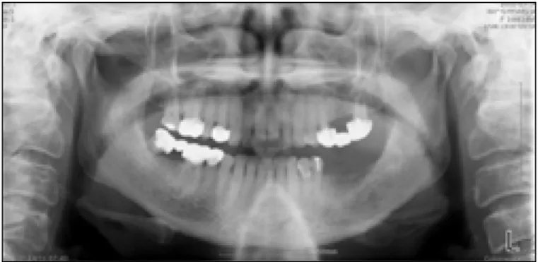
환자는 약 3개월 전 타병원에서 좌측 하악 대구치 동요 및 동통을 주소로 발거한 후 상기 증상이 발생하여 본원에 의 뢰되었다. 본원 초진시 임상검사상 좌측 하악 대구치 발치 와는 새로운 연조직으로 완전히 치유되었고, 상부 치은이 상방 및 협부로 증식되었으나 염증소견은 없었다. 이 부위 의 촉진시 치조골이 팽창된 듯 단단하였고 당일 촬영한 파 노라마 상에서는 좌측 대구치 발치와 부위에 약간의 방사선 불투과상이 관찰되었다(Fig. 1). 좌측 구치부 발치와 상방 연조직의 조직검사를 시행하였고 결과 만성염증으로 진단 되어 전신마취하에 단순 절제 및 생검을 계획하였고 전신마 취를 위한 술전검사를 시행하였다. 심전도 검사, 흉부 방사 선 사진 촬영, 혈액검사, 뇨검사를 시행하였고 검사결과 특
이소견은 없었다. 또한 환자는 고막 천공 수술, 자궁외 임신 수술의 경력 이외에 전신질환은 없었고 복용중인 약물도 없 었다.
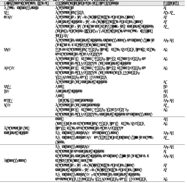
비기도 삽관 후 좌측 하악 구치 발치와 상방의 연조직에 절개선을 가하고 연조직 박리를 시작하였다. 병변은 주변 하악골과 잘 경계지어졌으며 좌측 하치조신경 및 혈관을 둘 러싼 연조직 덩어리 형태로 발견되었다(Fig. 2). 하치조신 경 및 혈관을 포함하여 덩어리를 전체로 절제해 낸 후 병소 는 임상병리과에 보냈고 수술부위는 골이식 없이 봉합하였 다. 수술 후 환자는 좌측 하치조신경의 신경지배를 받는 좌 측 하순과 턱부위의 감각 소실과 약간의 통증을 호소하였으 나 그 외 특이소견 없이 치유되었고 조직검사 결과가 나오 기 전에 퇴원하였다. 수술 중 시행한 조직검사상 병변은 plasmacytoma로 진단되었다(Fig. 3). 이후 plasmacy- toma는 radiosensitive한 특성을 가지므로 방사선 치료를 계획하였으며 전신적인 multiple myeloma를 배제하기 위 한 검사를 시행하였다. general lab, plain X-ray(skull series, both humerus AP, both clavicle AP, both femur AP & lateral) 촬영, serum & urine immuno- electrophoresis, bone marrow biopsy 수행하였고 PET CT(whole body)촬영하였다. plain X-ray 상에서는 파노 라마 상에서 관찰된 불규칙한 방사선 투과상 이외 장골에서 의 흡수상은 관찰되지 않았으며 수술 후 시행한 혈액검사상 Hg/Hct(9.7/27.9mmHg/%)으로 수술 전보다 수치가 약 간 감소하였고 이후 혈액검사상에서도 비슷한 수치가 유지 되었으나 ESR은 74mm/h로 증가되었다. PET CT 결과 하악과 상방의 연조직에서 중등도의 FDG uptake를 관찰 할 수 있었고 동측 악하부위에도 미약한 결정형 FDG uptake가 관찰되었다. 좌측 iliac bone에서 시행한 bone marrow biopsy상에서는 plasma cell content가 32%로
Fig. 1. Initial panoramic view.
Unfilled molar extracted socket and radiolucent lesion with ill-defined margin.
Fig. 2. Intraoperative view. The mass was well-demar-
cated from mandible.
상당히 증가되었으며(Fig. 4) serum immunoelec- trophoresis에서 monoclonal gammopathy를 나타내었고 plasmacytoma의 조직검사 결과를 토대로 multiple myeloma로 진단되었다. 그 후 환자는 항암치료를 위해 혈 액종양내과로 입원하였고 VAD(vincristine-doxorubicin- dexamethasone)항암치료를 시작하였다. 그러나 입원당시 수술부위의 통증 및 부종이 발현되어 다시 본과로 의뢰되었 고 파노라마상 수술부위인 좌측 대구치 발치와 하방으로 하 악골 골절이 관찰되었다. IMF를 시행하였으나, 환자분께서 위염 및 답답함을 호소하여 facial bandage로 대체하고 유 동식 권고 후 관찰하였다. 하악 골절 6개월 후 재내원시 하 악 골절은 임상검사 및 방사선상 완전 치유된 양상을 보였 다. 그 후 가철성 국소의치를 제작하여 사용중이며 현재까 지 multiple myeloma의 치료를 위한 Alkeran
�(nitrogen mustard의 phenylalanine 유도체로 cell cycle에 비선택 적으로 작용하는 약물) 경구 항암제를 복용중이나 전신적인 다발성 골수종의 진전은 관찰되지 않았다.
Ⅲ. 고 찰
다발성 골수종은 70대 이상의 남성에서 호발하며 연령이 증가할수록 발생율은 증가한다
4-7). 악성 세포들이 특별한 징 후나 증상없이 골수강을 침범하기 때문에 질환이 발견되었 을 때는 이미 광범위한 파급이 일어나 있다
8). 특징적인 다 발성 골수종의 증상은 골수 파괴에 따른 심부골 동통이며 악골에서 이러한 질환은 치통으로 나타나기도 한다
9). 대개 bone pain은 골절 이후에 나타난다고 하며
6,10)본 증례에서 의 초진시 동통은 골절과는 무관하였다. 술후 발생한 하악 골 골절은 다발성 골수종으로 인하여 발생하였다고 보기는 어렵다. 혈중 calcium 수치도 정상이었으며 골연화를 야기 할 수 있는 bisphosphonate등의 약물사용 경력도 없었다.
수술당시 좌측 하악골 발치와 주위로 상당량의 골이 제거되 었고 술후 환자가 과도하게 개구하는 등의 충격으로 인하여 골절이 발생한 것으로 생각된다.
골 용해성 병소가 다양한 골에서 발견되지만 대부분의 경 우에 두개관에서 더욱 뚜렷하게 동전모양이나 구멍뚫린 (punched out) 형태의 방사선 투과상을 보인다
9). 본 증례 에서도 치과 방사선 소견상 병소 경계부위는 피질골화가 없 이 벌레먹은 방사선 상을 보였다. Plain radiography, CT, MRI, PET CT등의 방사선 사진을 통하여 질환의 정도, 합 병증 등을 평가할 수 있다
5). Plain radiography를 통하여 는 골절 가능성 높은 장골의 흡수여부를 확인할 수 있다. 그 러나 적어도 30% 이상의 해면골(trabecular bone)이 상실 되어야 방사선 사진상 확인이 가능하므로 초기 병소의 발견 에는 어렵다
5). 작은 용해성 병소의 발견은 CT를 이용하면 보다 용이하다. 특히 plain film에서 의심되는 부위를 확진 하는데 정확하게 이용될 수 있으며 또한 방사선 치료나 수 술 계획시에도 유용하게 이용된다. 연조직에의 확장정도와 특성, 골수 이환 양상에 대한 정보를 얻기 위하여서는 MRI 를 이용한다
5). 특히, 골수의 침범 정도는 solitary plasma- cytoma와 myeloma를 감별하는데 중요하다.
F-18 FDG PET만으로 multiple myeloma를 진단할 수 는 없지만 PET CT는 초기 골수 이환 정도를 감지할 수 있 으며 진단 당시 활동성 질환의 확장 정도 및 치료에 대한 반 응을 평가하는데 유용하다
6,10). 본 증례에서는 술 후 시행한 plain X-ray 상에서 특이소견은 없었다. 환자의 증상이 미 약하고 PET-CT상에서도 좌측 하악골 및 악하부에서만 FDG uptake를 보였으며 이는 술 후 염증으로 인한 것으로 추정되었다. 따라서 다발성 골수종의 정확한 진단을 위해서 는 골수 생검, 혈청 & 소변 단백질 전기영동검사가 요구된 다. 또한 향후 질환의 확산을 평가하기 위하여 정기적인 방 사선학적 추적조사가 필요할 것으로 사료된다.
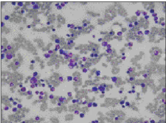
Fig. 3. Histologic finding. Atypical plasma cell infiltra- tion(×200).
Fig. 4. Bone Marrow Biopsy. Wright-Giemsa stain(×200).
Diffuse plasma cell distribution.
다발성 골수종과 고립형질 세포종은 유사한 현미경 소견 을 보인다. 형질세포의 미만성의 판 주위에 섬유간질은 거 의 없으며 형질세포를 따라 분지 하는 모세혈관을 관찰할 수 있다
9). 각각의 세포들은 정상 세포에서 보이는 핵의 편 심성 양상과 부분적인 염색질(핵의 주변으로 염색질이 뭉쳐 있는(chromatin beading)양상을 보인다. 특히 중요한 것 은 이핵 형태(binucleated)가 일반적이고 유사분열상과 어 느 정도의 핵다형성이 관찰된다는 것이다
9). 다수의 형질세 포 침윤을 보이는 대부분의 염증상태에서는 다른 백혈구들
도 관찰할 수 있고 간질은 섬유화 되어 있다. 그러나 악골에 서 형질 세포 침윤은 흔히 관찰되며 특히 치성 감염의 경우 에 반응성으로 잘 나타나므로 골수종을 감별하기란 어려운 편이다. 본 증례의 환자의 초기진단에서도 만성염증으로 진 단된 바 있다.
다발성 골수종은 면역 글로불린을 분비하는 형질세포의 파종성 질환으로 혈청의 면역글로불린양에 변화를 초래한 다
5). 감마 글로불린의 전체 양은 증가할 것이고 혈청 면역 전기영동에서 단일크론성 스파이크를 보여 단일 면역글로
Table 1. Diagnostic Criteria of Myeloma Abstracted from the Literature
11,13-21)Clinical or laboratory feature Additional findings required for diagnosis Reference
Marrow plasmacytosis Paraprotein 19
>5% Paraprotein>3g/dL 14, 15
≥10% Paraprotein with usual clinical features of myeloma 16
Osteolytic lesions with usual clinical features of myeloma 16 Paraprotein with at least one of the following: osteolytic lesions, 15 anemia, renal insufficiency, hypercalcemial, or marrow labeling
index>1%
Paraprotein, osteolytic lesions, plasmacytoma, or plasma cells in 11, 18 at least two peripheral blood smears
>10% Serum paraprotien>3.5g/dL, for lgG peak, >2.0g/dL for lgA peak, 17 or paraproteinuria≥1.0g/24hr
Paraprotein(IgG peak≤3.5g/dL, for igA peak≤2.0g/dL, or 17 paraproteinuria<1.0g/24hr) and osteolytic lesions
10-30% Paraprotein(IgG peak≤3.5g/dL, for igA peak≤2.0g/dL, or 17
paraproteinuria<1.0g/24hr) and normal IgM<50mg/dL, IgA<100mg/dL, or IgG<600mg/dL
Paraprotein and osteolytic lesions 13
>15% None 20
20% Osteolytic lesions 18
None 21
≥25% Generalized osteoporosis 11, 18
>30% Paraprotein and osteoporosis 15
Paraprotein(IgG peak≤3.5g/dL, IgA≤2.0g/DL, 17
paraproteinuria<1.0g/24hr), osteolytic lesions or plasmacytoma
none 18
bone if serum paraprotein>3.5g/dL for IgG peak, >2.0g/dL 17 Paraprotein and for IgA peak, or paraproteinuria≥1.0g/24hr
osteolytic lesion BM plasmacytosis>10% or plasmacytoma 11, 18
BM plasmacytosis≥10% or aggregates of plasma cells in marrow 15 biopsy
BM plasmacytosis>10% 11, 18
paraprotein or osteolytic lesions 19
paraprotein, osteolytic lesions, or plasma cells in at least two 11, 18
Plasmacytoma peripheral blood smears
paraprotein with usual clinical features of myeloma 16 osteolytic lesions with usual clinical features of myeloma 16 BM plasmacytosis>30%, paraprotein, osteolytic lesions,
or normal IgM<50mg/dL, IgA<100mg/dL, or IgG<600mg/dL 17
불린 클래스가 증가되었음을 나타낸다. 또한 골수종에서 면 역글로불린의 형태는 소변으로 배출되며 벤스-존스 (Bence-Jones)단백질 이라고 부른다. 이러한 면역 글로불 린 연결부위가 신장의 근심관을 손상시켜 신질환의 합병증 을 야기한다. 신질환을 야기하는 또다른 원인으로는 탈수, 과칼슘증, 고요산혈증, 감염, 신독성 약물의 사용등이다
5).
본 증례에서는 혈청 면역전기영동상에서 IaA, kappa type의 immunoglobulin이 나타났으나 소변에서는 관찰되 지 않았으며 신질환 또한 나타나지 않았다. 그러나 항생제 및 항암제를 장기간 복용하고 있으므로 신질환에 대한 지속 적인 평가 또한 필요할 것으로 생각된다.
다발성 골수종은 집중적인 화학요법에도 불구하고 예후가 좋지 않다
3,13). 질환 치료를 목적으로 세포분열을 정지시키 기 위해 다양한 약물군을 사용하거나 골수이식과 함께 전신 방사선 요법을 사용하기도 한다. 그러나 일단 multiple myeloma로 진단된 경우 systemic chemotherapy를 추천 한다. 일반적으로 60%의 multiple myeloma 환자가 치료 에 반응을 보이며 평균 생존기간은 2-3년이다
5). 본 증례의 환자는 VAD(vincristine-doxorubicin-dexamethasone) 으로 항암치료를 시작하였다. 이전에 multiple myeloma 환자에서는 mephalan과 prednisolone 투약을 이용한 항 암치료가 주가 되어왔으나
11)50%의 관해율과 24개월의 평 균 생존율을 보여 더 효과적인 항암제의 사용에 대한 연구 가 지속되어 왔으며 최근 vincristine과 doxorubicin을 지 속적으로 주입하며 dexamethasone을 경구투여하는 것에 대한 효과가 59%의 관해율을 보이며 관해까지의 시간이 짧고 평균 생존율이 1년 이상이라는 고무적인 결과를 보이 고 있다
12).
현재까지 multiple myeloma의 진단에 대한 표준법은 없 는 상태이며 Table 1에서와 같은 다양한 진단법
14-21)이 이용 되고 있다. 본원의 진단기준은 Table 2와 같이 지침을 세워 시행하고 있으며 본 증례의 환자는 본원의 진단기준에 의하 여 multiple myeloma로 진단되었다.
Ⅳ. 결 론
다발성 골수종의 진행은 심각한 이차특징(골파괴 병소, 빈혈, 면역결핍, 신장 손상)으로 나타나며 어떤 환자는 골수 강 외로 tumor가 진행된다
7). 또한 SPB, EMP는 진행되어 multiple myeloma로 진행될 가능성이 있다. 그러므로 모 든 plasmacytoma는 병소의 확장 및 전환여부를 평생 평가 할 필요가 있다. 그러기 위해 정기적인 bone marrow aspiration, CT 혹은 MRI 등을 이용한 skeletal survey, M protein의 존재여부 확진을 위한 serum & urine pro- tein electrophoresis등을 정기적으로 수행해야 할 필요가 있다. 따라서 본 증례에서도 구강내 발현된 병소는 절제술 로써 치유되었으나 전신적인 치료를 위해 항암 투약중이며 향후 지속적인 검사를 통하여 결과를 관찰하고자 한다.
참고문헌