CASE REPORT
pISSN 1225-7737/eISSN 2234-8042 http://medlib.yu.ac.kr/yujm YUJM 29(1):42-44, 201242
YUJM VOLUME 29, NUMBER 1, JUNE 2012교신저자: 박종원, 137-701, 서울시 서초구 반포대로 222 가톨릭대학교 의과대학 내과학교실
Tel: (02) 2258-6042, Fax: (02) 599-3589 E-mail: cwp@catholic.ac.kr
두개 내를 침범한 형질세포골수종 1예
이수현, 정윤영, 임예지, 고선영, 최유아, 김영운, 이성은, 박종원 가톨릭대학교 의과대학 내과학교실
A Case of Intracranial Involvement in Plasma Cell Myeloma
Su Hyun Lee, Yoon Yung Chung, Ye Jee Lim, Sun Young Ko, Yoo A Choi, Young Woon Kim, Sung Eun Lee, Chong Won Park
Division of Hematology, Department of Internal Medicine, The Catholic University of Korea, College of Medicine, Seoul, Korea
Plasma cell myelomas generally manifest as bone or soft-tissue tumors with variable mass effects, pain, and infiltrative behavior. Extramedullary involvement occurs most commonly in the spleen, liver, lymph nodes, and kidneys, but intracranial involvement in plasma cell myeloma is a rare extramedullary manifestation. These authors recently encountered a case of intracranial involvement of plasma cell myeloma. A 69-year-old man was hospitalized for headache and mental changes. Brain CT showed subdural hemorrhage caused by plasma cell myeloma. Plasma cell myeloma with intracranial involvement has poor prognosis, and the patient in this case died from acute complications, such as subdural hemorrhage. Based on this case report, it is suggested that more effective treatment regimens of plasma cell myeloma with intracranial involvement be developed.
Moreover, a screening method and decision on the appropriate time for intracranial involvement are needed for plasma cell myeloma patients.
Key Words: Intracranial neoplasm, Plasma cell myeloma
서 론
형질세포골수종은 형질세포의 악성 신생물로 보통 골수 및 골격계를 침범하며, 그 외 비장, 간, 림프절, 신장 등의 골수 외 장기에도 발생하지만, 두개 내 침범은 드물다. 만일 형질세포골수종이 두개 내를 침범한 경우 대개 두개강, 두 개골 기저, 코와 부비동을 구성하는 뼈를 침범하지만 뇌실 질, 경막 및 연수막의 침범은 매우 드물다.1-5
본 증례는 형질세포골수종으로 진단되어 전신항암요법 등의 치료 후에도 두개 내 형질세포골수종의 침범이 경막하 출혈 형태로 나타난 드문 증례이다. 본 증례와 같이 형질세
포골수종의 두개 내 침범은 불량한 예후를 보이며, 여러 종 류의 치료법이 시도되고 있지만, 효과는 명확히 알려져 있 지 않은 상태이다. 이에 진단 시기 및 치료 방법에 대한 정책 수립이 필요할 것으로 생각하여 본 증례를 보고하는 바이다.
증 례
69세, 남자가 갑자기 발생한 두통 및 의식저하를 주소로 내원하였다. 환자는 다발성 골침범을 보여 시행한 골수조직 검사에서 형질세포골수종 (IgG, Kappa type, stage IIIa) 진단 을 받은 후 총 4차례 vincristine, adriamycin, dexamethasone (VAD) 항암화학 요법 시행 후 완전 관해 판정받았으나, 4개 월 후 폐의 우측 상부에 형질세포골수종이 발생하여 병변부 의 방사성 치료와 3차례 bortezomib과 dexamethasone 병용 요법을 시행받았다. 5개월 후 골수조직 검사에서 재발 판정
두개 내를 침범한 형질세포골수종
YUJM VOLUME 29, NUMBER 1, JUNE 2012
43
Fig. 1. Brain CT(noncontrast).
A large amount of left subdural hemorrhage with heterogeneous high and isodensity, suggesting multi-stage hemorrhage. Associa- ted mass effect, resulting in mid- line shift and transtentorial her- niation.
Fig. 2. (A) Low-power microscopic view. The proliferation of small atypical cells is seen (H&E stain, × 100). (B) High-power microscopic view. The myeloma cells are round cells with eccent- rically located nuclei. The nuclei show hyperchromasia (H&E stain, × 400).
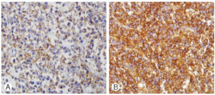
Fig. 3. (A) Plasma cells positive for CD56a (immunohistochemi- cal stain, × 400). (B) Plasma cells positive for CD138 (immuno- histochemical stain, × 400).
받고 thalidomide 투여 도중 내원 5일 전부터 갑자기 발생한 두통 및 의식저하로 응급실 내원하였다.
이학적 검사에서 혈압 125/84 mmHg, 맥박수 82회/분, 호 흡수 22회/분, 체온 36.8℃였다. 일반 혈액검사와 혈액 화학 검사 상 혈색소 8.6 g/dL, 백혈구 3,720/mm3, 혈소판 107,000/
mm3였으며, BUN과 Cr는 17.2 mg/dL와 3.85 mg/dL였다. 전 해질은 Na 139 mEq/L, K 4.1 mEq/L, Ca 8.8 mg/dL였고 간기 능 검사상 AST, ALT는 정상 수치였다. LDH는 1,793 U/L, 총단백은 6.9 g/dL, 알부민은 2.3 mg/dL였다. ESR, CRP는 각 각 120 mm/h, 0.87 mg/dL로 상승되어 있었다. 또한, 전기영 동검사 결과 M단백은 2.51 g/dL로 상승되었고, 혈청에서 유 리형 경쇄 비는 223.44로 확인되었다.
두통과 의식저하를 보여 시행한 뇌 단층촬영에서 급성 경 막하 출혈과 이로 인한 압박으로 뇌의 중심선 이동과 천막경 유탈출이 보여 (Fig. 1) 개두술 및 혈종제거 수술 중 혈종에 대한 흡인을 시행하였다. 두 검체 중 하나에서 형질세포가 보였고(Fig. 2A, 2B), 면역조직화학 염색에서 형질세포는 CD56a, CD138 양성으로 형질세포골수종에 합당하였다 (Fig. 3A, 3B). 하지만 수술 6일 후에도 의식이 회복되지 않았고, 결국 급성 신부전이 진행되어 사망하였다.
고 찰
형질세포골수종은 형질세포 질환으로 두개 내 형질세포종 의 발생은 드물며, 특히, 연수막 침범으로 인한 중추신경계 형질세포골수종은 약 1% 정도로 극히 드물다.1-5 윤 등4은 다발성 골수종의 중추신경계 침범 여부를 확인하기 위하여 시행한 뇌척수액의 세포학적 검사에서 1,438예 중 1예에서 만 골수종세포가 발견되었다. 현재까지 우리나라에서 중추 신경계 형질세포골수종의 증례보고는 4예로 매우 적다.3,4,6,7 형질세포골수종의 연수막 (leptomeningeal)의 전파는 정 확히 보고된 바가 없으나, 뇌 실질, 뇌수막 또는 골에 존재하 는 형질세포골수종으로부터 직접 파종된다는 설과 혈행성으 로 진행한다는 설이 있다.3,4 연수막으로 전이가 일어난 형질 세포골수종 환자의 부검 소견에서 거미막내 정맥으로 형질 세포가 침투된 소견과 함께 척수액 내로의 전이가 확인되어 혈행성으로 진행한다는 것으로 보고 있다.5 또한 형질세포골 수종은 매우 혈관이 발달된 종양으로 두개골 내 골조직이나 경막에 근접하지 않고, 실질 내 발생하는 것이 종종 보고되어 왔으며, 이는 출혈과 연관되기도 한다.2,5
형질세포골수종은 진단 후 병기에 따라 생존율은 수 개월 에서 10년 정도이나, 일반적으로 두개 내를 침범한 형질세포 골수종의 경우 진단 후 약 1개월에서 96개월 정도로 그 예후 는 매우 불량하다.2 혈액-뇌-장벽으로 항암제가 병변에 도달 하지 못하기 때문에 전신 항암 치료에 추가적 치료로 척수강 내 항암주사요법, 두부 방사선 치료가 시행되지만 그 효과는 매우 짧아 일시적인 반응을 보인다.5 형질세포골수종의 위치 가 적절하다면 수술적 제거 후 50 cGy 이상의 방사성 치료를 하거나 방사성 단독치료를 시행할 수 있다. 또 14 cGy의 저용 량 방사선학적 수술요법도 효과적인 치료가 될 수 있다. 다발 성 형질세포골수종이나 재발성, 악성 형질세포골수종의 경 우 항암 치료나 골수이식 등의 전신요법이 적합하다.2
이수현 등
44
YUJM VOLUME 29, NUMBER 1, JUNE 2012중추신경계 침범을 일으킨 형질세포골수종은 이처럼 예후 도 좋지 않을 뿐만 아니라, 본 증례처럼 뇌출혈이라는 급성 합병증을 일으킬 수도 있다. 또 한 문헌에 의하면 다발성 형질 세포골수종 환자에서 중추신경계를 침범하였을 때 병변이 척수인 경우 조기 진단을 하여 즉각적인 수술과 방사성 치료 로 50%의 완치와 30%의 부분 반응의 성과가 있었지만, 두개 내를 침범할 때 진단이 늦고 이로 인해 즉각적인 치료에 실패 하는 경우가 많았다.8 하지만 이와 관련된 증례가 적어 다발 성 형질세포골수종 환자에서 증상이 나타나기 전 중추신경 계 침범 여부 확인이 필요 한지, 필요하다면 어느 시기가 적절한지, 효과적인 항암제 및 방사선 치료, 수술적 치료, 척수강 내 항암주사요법, 골수 이식 등 각 치료의 효과와 예후에 대해서는 명확히 알려져 있지 않다. 이에 형질세포골 수종의 중추신경 침범에 대한 진단과 치료에 대한 추가적인 연구가 필요할 것으로 생각된다.
참고문헌