Effect on bone formation of the autogenous tooth graft in the treatment of peri-implant vertical bone defects in the minipigs
전체 글
수치
관련 문서
Conclusions : The mesiodens and the 3rd molar teeth are very similar to the inorganic component. These results provide reasonable rationale that mesiodens can be used
From the results of this study, we concluded that two different sized graft materials have positive effects on new bone formation.. Additionally, smaller
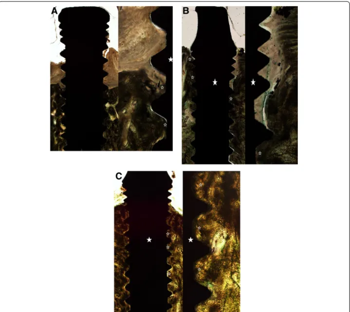
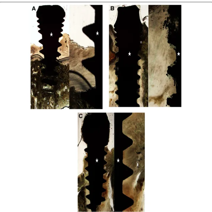
The grafted particulated tooth driven allografts were partially surrounded with new bone matrix and fairly supported graft associated new bone formation with some
The aim of this study was to compare the effect on bone regeneration relative to maintenance period of PTFE membrane in rabbit calcarial defects.. Eight adult
5) After GBR, the membrane was removed i n i ni ti al ti me, the usage of nonabsorbabl e membrane and autogenous bone resul ted i n the mostfavorabl e bone formati
success rates of dental implants placed at the time of or after alveolar ridge augmentation with an autogenous mandibular bone graft and titanium mesh: a 3-to
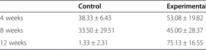
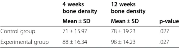
In 4-week group, the group filled with bone graft with decortication revealed larger new bone formation area than shown in the group that had a defect area
Biochemical markers of bone turnover can be classified according to the process that underlie in markers of bone formation, products of the osteoblast
