C A S E R E P O R T Open Access
Simultaneous gap arthroplasty and intraoral distraction and secondary contouring surgery for unilateral temporomandibular joint ankylosis
Aditi Sharma 1 , Jun-Young Paeng 1 , Tomohiro Yamada 2 and Tae-Geon Kwon 1*
Abstract
Background: Temporomandibular joint (TMJ) ankylosis can be accompanied by various degrees of functional and esthetic problems. Adequate mouth opening, occlusal stability, and harmonious facial form are the main goals of treatment for ankylosis. Distraction osteogenesis has proven to be an excellent treatment for lengthening the ramus-condyle unit. However, various timings for distraction have been suggested, and there is no consensus on selection criteria for performing the procedure in stages or simultaneously with other treatments.
Case presentation: In this case report, concomitant intraoral distraction and gap arthroplasty was planned to treat TMJ ankylosis and associated facial asymmetry. After gap arthroplasty and 23 mm of distraction, the ramus-condyle segment was successfully lengthened and mouth opening range was significantly increased. The resultant interocclusal space was stably maintained with an occlusal splint for 4 months after distraction. Finally, good occlusion was achieved after prosthetic treatment. The remaining mandibular asymmetry was corrected with osseous contouring and augmentation surgery. The mouth-opening range was maintained at 35 mm 24 months after treatment.
Conclusion: Gap arthroplasty with intraoral distraction as a one-stage treatment and subsequent contouring surgery can be applied to correct ankylosis with moderate malocclusion and facial asymmetry.
Keywords: Temporomandibular joint, Ankylosis, Intraoral distraction, Gap arthroplasty, Asymmetry
Background
Temporomandibular joint (TMJ) ankylosis is a fusion of the mandibular condyle to the base of the skull, which causes problems in speech, mastication, and facial ap- pearance, and sleep-disordered breathing. The main con- cept of ankylosis treatment includes complete resection of the ankylotic block, creation of a new joint lining with an interpositional substance, and reconstruction of the skeletal deformity. This protocol was fully demonstrated by Kaban et al. [1]. Costochondral grafts—which have the advantage of being an autogenous material with a cartilaginous articulating surface, allowing potential for growth and adaption—are commonly used to
reconstruct the mandibular condyle. However, recent analysis has shown that gap arthroplasty produces better postoperative maximal mouth opening than does costo- chondral graft [2]. Even after successful release of anky- losis, mandibular deformity may become aggravated after surgery because of the loss of ramus-condyle height. Total joint reconstruction or distraction osteo- genesis has therefore been suggested to reconstruct the ramus-condyle unit [3, 4].
Recently, distraction osteogenesis has proven to be an ideal treatment for TMJ ankylosis [5, 6]. However, the best timing for the distraction is still controversial. Some authors have suggested that distraction should be per- formed after gap arthroplasty [7–9], while others have performed distraction first, followed by gap arthroplasty at the time of distractor removal [10, 11]. Simultaneous gap arthroplasty and distraction osteogenesis for
* Correspondence: [email protected]
1
Department of Oral and Maxillofacial Surgery, School of Dentistry, Kyungpook National University, Daegu 700-421, Korea
Full list of author information is available at the end of the article
© 2016 Sharma et al. Open Access This article is distributed under the terms of the Creative Commons Attribution 4.0
International License (http://creativecommons.org/licenses/by/4.0/), which permits unrestricted use, distribution, and
reproduction in any medium, provided you give appropriate credit to the original author(s) and the source, provide a link to
the Creative Commons license, and indicate if changes were made.
nical details of simultaneous gap arthroplasty and distraction had not yet been fully documented, especially in terms of establishing occlusion during the treatment process.
In the reported case, the authors performed gap arthroplasty and intraoral distraction as a one-stage treatment, followed by mandibular contouring surgery and prosthetic treatment for unilateral TMJ ankylosis with facial asymmetry. The results demonstrate that stable outcomes after concomitant gap arthroplasty and distraction can be ensured by maintaining the occlusion with a splint until prosthetic treatment.
Case presentation
A 47-year-old woman presented with a complaint of facial asymmetry and limitation of mouth opening. The patient had a history of untreated chin trauma in adolescence.
Clinical examination showed maxillary canting and devi- ation of the mandibular dental midline and chin to the right. Maximal mouth opening was limited to 23 mm.
Radiological examination showed fibrous ankylosis with elongated coronoid process and shortened ramal height on the right side (Fig. 1a, b). A reverse L-shape osteotomy
achieved. A reverse L-shaped osteotomy line was made on the outer cortex of the ramal bone via a preauricular and intraoral approach. A 25-mm Ramus distractor (KLS Martin, Tuttlingen, Germany) was adapted to the ramus, and predrilling was performed on each side of the hori- zontal osteotomy line by using the angled screw driver, Angulus (KLS Martin, Tuttlingen, Germany). The de- signed osteotomy was created while maintaining the soft tissue attachment of the medial side of the proximal ramal segment. Finally, the distractor was fixed with miniscrews and its function was tested. After a 7-day latency period, the mandible was distracted to 23 mm at a rate of 0.5 mm × 2 times per day. During the distraction period, the patient performed mouth-opening exercises composed of voluntary, gentle mouth opening for 5 cycles (5 times/
cycle) per day. After the distraction period, lateral open bite was noted on the right side. The ledge of the distrac- tion device was removed. To maintain and stabilize the occlusion, a resin splint was applied for 4 months during the consolidation period until removal of the distractor (Fig. 2a, b). The occlusal splint was removed, and the remaining lateral open bite was closed by prosthetic treat- ment (Fig. 2c, d).
Fig. 1 a The panoramic radiograph showed right TMJ ankylosis and facial asymmetry. b Coronal CT image shows fibrous adhesion around the
right TMJ. c Reverse L-shape osteotomy line for distraction osteogenesis. d Distractor applied on rapid prototype model, and proposed line of
osteotomy (white dotted line) for ankylosis release. A 23-mm distraction of the condyle-ramus segment was planned
At the time of distractor removal, residual mandibular asymmetry was corrected by decortication and shaving of the right mandibular body, and augmentation of the left mandibular body using the decorticated bone from the contralateral side. After a 24-month follow-up period, mouth opening was maintained at 35 mm. Com- parison of pre- and postdistraction panoramic views showed a remarkable ramal height increase (Fig. 3).
Three-dimensional computed tomography images taken at each successive surgical step are shown in Fig. 4. The patient exhibited significantly improved symmetry and stable skeletal position after treatment (Fig. 5).
Discussion
There are several fundamental elements for successful treatment for TMJ ankylosis and related dentofacial de- formity: establishment of adequate mouth-opening range, complete removal of the ankylotic block to pre- vent reankylosis, and establishment of balanced facial appearance after surgery. To achieve these goals, various approaches have been suggested. After the introduction of distraction osteogenesis for mandibular ramus length- ening, some surgeons attempted simultaneous correc- tion of all deformities by performing distraction at the time of ankylotic mass removal. Critics of this approach have suggested that after release of the ankylotic block, changes in the mandibular position cannot be com- pletely controlled during the distraction period. During distraction, the condylar segment can move toward the condylar fossa and become positioned closer to the
articular surface, which can result in reankylosis if ad- equate physical therapy is not applied. However, it is not easy to perform active physical therapy during the dis- traction period because of postsurgical pain or discom- fort. For these reasons, a staged operation for TMJ ankylosis—comprising ankylosis release as the first sur- gery, and distraction as the second—has been proposed [7–9]. However, after reports of two successful cases of simultaneous gap arthroplasty and distraction [12, 13] in 1999, many authors subsequently reported similar suc- cesses [14, 15, 17]. Other surgeons have chosen to apply distraction as the first-stage surgery, followed by gap arthroplasty [10, 11]. As the distraction in such cases is applied to an ankylosed joint without mobility, it is easy to push the mandible forward during distraction. How- ever, performing minimal arthroplasty without vertical bony resection in the distracted ramus-condyle unit is not easy, and carries the risk of reankylosis.
Zhu et al. [16] proposed that one-stage surgical treat- ment is indicated for patients with mild-to-moderate preoperative malocclusion and skeletal deformities.
Staged treatment, on the other hand, is better suited to achieve a more stable postsurgical outcome in patients with severe dentofacial deformities. This is in accordance with our previous report showing the advantages of staged treatment; it promotes early postoperative en- gagement in active mouth-opening exercise, allows suffi- cient time to monitor malocclusion, and may reduce the chances of reankylosis [9]. The current patient had uni- lateral fibrous ankylosis on the right side (Sawhney’s
Fig. 2 Intraoral photographs. a, b Occlusal changes after distraction. Posterior lateral open bite was maintained for 4 months during the consolidation
period, until the final prosthetic treatment. c, d The occlusal splint was removed, and the remaining lateral open bite was closed by final prosthetic
treatment
It has been recommended that viability of the osteoto- mized segment can be maintained by preserving the in- tact medial pterygoid and masseter muscle during the distraction period [17], which is in accordance with our technique. During distraction of the ramus-condyle seg- ment, direct bone-to-bone contact should be avoided, especially in the case of bony ankylosis. Therefore, it is advisable to use interpositional grafts, which can serve as a physical barrier to reduce pressure on the recon- structed condyle. In our case report, an interpositional graft was not used because the ankylosis was of the fi- brous type.
Yoon et al. [19] reported simultaneous distraction and gap arthroplasty using an intraoral distractor via an extraoral approach. Xu et al. [17] reported use of a single preauricular incision for gap arthroplasty and distrac- tion, with a penetrating distraction port at the preauricu- lar incision. We positioned an intraoral distractor for lengthening the ramus-condyle unit via an intraoral ap- proach, and used a preauricular incision for ankylosis re- lease to minimize patient morbidity and extraoral scarring.
With this case report, we suggest that fibrous ankylosis with mild-to-moderate malocclusion and facial asym- metry can be successfully treated by simultaneous gap
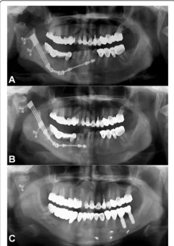
Fig. 3 Comparison of pre- and postdistraction panoramic views.
a Immediately after simultaneous gap arthroplasty and start of distraction osteogenesis. b After completion of a 23-day distraction period at a rate of 0.5 mm, two times per day). c After a 16-month follow-up period
Fig. 4 Three-dimensional computed tomography images taken at each successive surgical step. a Preoperative. b After gap arthroplasty and
distraction osteogenesis. c After mandibular contouring surgery to correct residual asymmetry of the mandible
arthroplasty and distraction as a single-stage treatment.
Residual skeletal asymmetry can subsequently be cor- rected by secondary contouring surgery. Occlusal stabil- ity plays a major role in promoting a favorable treatment outcome. In the present case, the definite occlusal in- stability after the distraction period was effectively man- aged by subsequent occlusal splint application and prosthetic treatment. If lateral and posterior open bite after distraction of the ramus-condyle unit cannot be managed by occlusal modifications such as splint appli- cation and subsequent orthodontic or prosthodontic treatment, adequate condyle-fossa relation and facial symmetry cannot be maintained. We therefore believe that in cases with severe joint ankylosis requiring complete removal of the condyle head and neck to the level of the coronoid notch, a staged operation is advan- tageous compared with single-stage distraction accom- panied by gap arthroplasty [9].
Conclusions
In this case report, we experienced a favorable outcome without evident relapse after 24 months of follow-up.
Therefore, we suggest that single- or multistage treat- ment can be applied depending on the degree of mal- occlusion or occlusal instability, which is a reflection of TMJ ankylosis severity. Gap arthroplasty with intraoral distraction as a one-stage treatment and subsequent contouring surgery can be applied to correct ankylosis with moderate malocclusion and facial asymmetry.
Consent
Written informed consent was obtained from the patient for the publication of this report and any accompanying images.
Competing interests
The authors declare that they have no competing interests.
Authors ’ contributions
TGK was the operator and revised the manuscript. AS, JYP, and TY drafted the manuscript. TGK carried out the operation and contributed to the conception of the report and critical revising. All authors read and approved the final manuscript.
Author details
1