∙ Received: December 28, 2010. Accepted: March 7, 2011.
∙ Corresponding author: Yong Joon Seong
Department of Nuclear Medicine, SMG-SNU Boramae Medical Center 39 Boramae-gil, Dongjak-gu, Seoul, 156-707, Korea
Tel: +82-2-870-2596, Fax: +82-2-870-2587 E-mail: [email protected]
Original Article 심근관류 SPECT의 분절별 관류 및 국소벽 운동에서 Wide Beam Reconstruction기법의 유용성 평가
서울특별시립 보라매병원 핵의학과 성용준⋅김태엽⋅문일상⋅조성욱⋅우재룡
The Evaluation of Usefulness of Wide Beam Reconstruction Method on Segmental Perfusion and Regional Wall Motion in Myocardial Perfusion SPECT
Yong Joon Seong, Tae Yeob Kim, Il Sang Moon, Seong Wook Cho and Jae Ryong Woo Department of Nuclear Medicine, SMG-SNU Boramae Medical Center, Seoul, Korea
Purpose: The aim of this study is to identify clinical usefulness of Wide Beam Reconstruction (WBR) which is called Xpress.cardiacTM to confirm the agreement between segmental perfusion and regional wall motion in myocardium compared to conventional OSEM method. Materials and Methods: Subjects were separated two groups. First group was composed of 20 normal control group. Second group was composed of 10 patients (abnormal group) who had coronary artery disease. Subjects underwent myocardial perfusion SPECT (201Tl rest and 99mTc-MIBI stress). Image acquisition and reconstruction were that rest stage was each step per 30, 15 seconds and stress stage was each step per 25, 13 seconds, OSEM and WBR methods were applied. Segmental perfusion and regional wall motion were applied 20-segment model of QPS, QGS algorithm in AutoQuant.
Status of perfusion was composed of 5 point scoring system (0=normal, 1=mild, 2=moderate, 3=severe hypokinesia, 4=dyskinesia). Status of regional wall motion was also composed of 5 point scoring (0=normal, 1=mild, 2=moderate, 3=severe hypokinesia, 4=dyskinesia). We evaluated the agreement between conventional OSEM and WBR through automatic quantification value. Results: The agreement of rest segmental perfusion between conventional OSEM and WBR in normal patients was 99% (396/400, k=0.662, p<0.0001) and one of rest regional wall motion was 83.8% (335/400, k=0.283), the agreement of stress segmental perfusion was 95.8%(383/400, k=0.656), one of stress regional wall motion was 87.3% (349/400, k=0.390). The match rate of rest segmental perfusion in abnormal patients was 83% (166/200, k=0.605, p<0.0001) and one of rest regional wall motion was 55.5% (111/200, k=0.385), the agreement of stress segmental perfusion was 79.5% (159/200, k=0.682), one of stress regional wall motion was 63.5% (127/200, k=0.486). Conclusion: Compared to conventional OSEM, WBR method had a good agreement of segmental perfusion in myocardium in normal and abnormal groups. However regional wall motion showed meaningful low agreement. Although WBR offers high resolution and contrast ratio, it is not useful method for gated myocardial perfusion SPECT. (Korean J Nucl Med Technol 2011;15(1):51-57)
Key Words : WBR (wide beam reconstruction), OSEM (ordered subset expectation
maximization), agreement
서 론
심근관류 SPECT에서 심근 내 분절별 관류와 국소벽 운동
기능 평가의 지표들은 관상동맥 질환의 진단 및 예후 예측에
있어 중요한 의미를 갖고 심근의 관류와 기능 지표를 전체적
또는 분절별로 자동 정량화할 수 있기 때문에 높은 재현성을
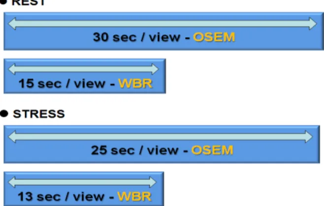
Fig. 1. Image acquisition and reconstruction were that rest stage was each step per 30, 15 seconds and stress stage was each step per 25, 13 seconds, OSEM-WBR methods were applied.
kappa coefficient (k) interpretation
< 0.00 no agreement
0.00 ~ 0.20 slight agreement
0.21 ~ 0.40 fair agreement
0.41 ~ 0.60 moderate agreement 0.61 ~ 0.80 substantial agreement 0.81 ~ 1.00 almost perfect agreement Table 1. Cohen's kappa coefficient of statistical measure
가진다 .
1-4)현재 이용되고 있는 재구성 알고리즘은 인접한 투
사각도를 갖는 투사데이터를 하나의 부분집합으로 구성하여 반복적 재구성방법 OSEM (ordered subsets expectation maximization)기법을 사용하여 영상을 처리하고 있다. 최근 핵의학 분야에서 다양한 소프트웨어 프로그램들이 개발되어 영상 획득시간을 짧게 하여 동등한 영상의 질을 만들어 내거 나 동일한 영상획득으로 영상의 질을 높일 수 있는 재구성기 법의 활발한 개발로 심근관류 SPECT에서도 영상의 질은 유 지하며 검사시간을 줄일 수 있는 UltraSPECT사에서 개발한 광대역 재구성 (wide beam reconstruction, WBR) 기법인 Xpress.cardiac
TM프로그램의 적용이 기존 OSEM 기법과 심 근 내 분절별 관류와 국소벽 운동에서의 일치율을 평가하여 WBR 기법의 임상적 유용성을 알아보고자 하였다.
5)실험재료 및 방법
관상동맥질환의 병력이 없고 핵의학전문의에 의한 판독 상 이상 소견이 없는 정상군 20명(남자 7명, 여자 13명)과 관 상동맥질환을 진단받은 비정상군 10명(남자 6명, 여자 4명) 을 대상으로, 평균연령은 63±11세 (37-85세)였다. 대상환자 모두는 심근관류 SPECT를 실시했던 환자로서 후향적으로 분석하였다.
INFINIA 감마카메라(GE, Israel)를 이용하여 환자는 6시 간 이상 금식한 후 휴식기 영상은
201Tl 74 MBq (3 mCi)를 정맥주사하고 10분 후에 투사영상 당 30초로 영상을 획득하 고 곧바로 투사영상 당 15초로 영상을 획득하였고 약물 부하 는 아데노신을 분당 0.14 mg/kg을 6분 동안 정맥주사하면서 3분 뒤
99mTc-MIBI 555 MBq (15 mCi)를 주사하고 2-3시간 후 부하기 영상은 투사영상 당 25초로 영상을 획득하고 곧바 로 투사영상 당 13초로 영상을 획득하였다. 직각으로 된 두 개의 검출기에 LEHR 조준기가 장착된 상태에서 우전사위 45˚부터 좌후사위 45˚까지 3˚간격으로 회전하며 step- and-shoot방식으로 영상을 얻었고 게이트 영상을 위하여 심 전도 상 R-R 간격을 16프레임으로 나누고 각 프레임 당
±20%의 범위에서 영상을 얻었으며, 에너지 창은
201Tl은 72, 167 keV에,
99mTc은 140 keV에 중심을 두고 ±10% 창을 열었 다. 영상 재구성은 휴식기 30초/부하기 25초 영상을 OSEM 기법으로 , 휴식기 15초/부하기 13초 영상은 WBR 기법을 적 용하였고 (Fig. 1) AutoQuant
TM소프트웨어에서 제공하는 자 동화 알고리즘을 이용하여 자동 정량분석하여 평가하였고 좌심실은 20분절 모델을 적용하였다.
정상군과 비정상군에서 관류상태는 동위원소의 섭취정도 에 따라 5등급(0=정상, 1=경도 결손, 2=중등도 결손, 3=심한 결손, 4=섭취 없음)으로 분류하였고 심근의 국소벽 운동은 5 등급(0=정상운동, 1=경도 운동저하, 2=중등도 운동저하, 3=
심한 운동저하, 4=무운동 및 이상운동)으로 분류하여 심근 내 분절별 관류와 국소벽 운동의 반정량값을 이용해 기존 OSEM기법과 WBR 기법에서의 일치율을 평가하였다.
일치한분절수 ※일치율 = × 100 총분절수
SPSS version 12.0 (SPSS Inc. USA)에서 일치도 통계는 OSEM 기법과 WBR 기법간의 정량적 지표들이 확률적으로 우연에 의해 일치할 확률을 제거한 Cohen's kappa를 사용하 여 분석하였고 분절별 관류백분율 정량값의 비교를 위해 대 응표본 t-검증을 시행하였다(Table 1).
결 과
총 30명의 환자를 휴식기 2회, 부하기 2회씩 촬영하여 얻
OSEM score WBR score
0 1 2 3 4 Total
0 138 9 1 1 0 149
1 5 10 1 4 1 21
2 1 0 3 2 0 6
3 2 3 1 11 1 18
4 1 0 0 1 4 6
Total 147 22 6 19 6 200
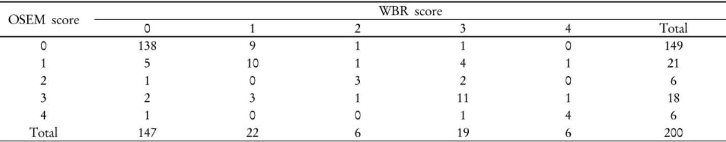
Table 2. Segmental score agreement between OSEM and WBR for assessment of segmental perfusion in abnormal group with rest gated myocardial perfusion SPECT (agreement 83%, 166/200 segments, k=0.605, p<0.0001)
OSEM score WBR score
0 1 2 3 4 Total
0 70 10 9 4 1 94
1 9 7 5 2 3 26
2 6 7 8 3 3 27
3 0 3 7 15 6 31
4 0 3 0 8 11 22
Total 85 30 29 32 24 200
Table 3. Segmental score agreement between OSEM and WBR for assessment of wall motion in abnormal group with rest gated myocardial perfusion SPECT (agreement 55.5%, 111/200 segments, k=0.385, p<0.0001)
OSEM score WBR score
0 1 2 3 4 Total
0 100 11 3 0 0 114
1 3 25 2 2 1 33
2 1 1 8 4 0 14
3 0 1 2 18 4 25
4 0 0 0 6 8 14
Total 104 38 15 30 13 200
Table 4. Segmental score agreement between OSEM and WBR for assessment of segmental perfusion in abnormal group with stress gated myocardial perfusion SPECT (agreement 79.5%, 159/200 segments, k=0.682, p<0.0001)
OSEM score WBR score
0 1 2 3 4 Total
0 79 10 10 2 0 101
1 5 7 5 4 1 22
2 3 2 13 11 2 31
3 0 0 3 13 13 29
4 0 0 0 2 15 17
Total 87 19 31 32 31 200
Table 5. Segmental score agreement between OSEM and WBR for assessment of wall motion in abnormal group with stress gated myocardial perfusion SPECT (agreement 65.5%, 127/200 segments, k=0.486, p<0.0001)
은 데이터에서 심근 내 분절별 관류와 국소벽 운동 상태를 5등급으로 분류한 정량적 지표들을 이용하여 구한 일치율을 평가하고 Cohen's kappa 계수도 높을 경우 일치율이 높다고 보았다 .
비정상군을 대상으로 한 기존 OSEM 기법과 WBR 기법 에서의 일치율은 휴식기 시 분절별 관류에서 총 200분절 중
166분절에서 일치하여 83% (166/200, k=0.605, p<0.0001)의 높은 일치율을 보였고 국소벽 운동에서는 55.5% (111/200, k=0.385, p<0.0001)로 의미 있는 낮은 일치율을 보였고 부하 기 시 분절별 관류에서 79.5% (159/200, k=0.0.682, p<0.0001) 의 높은 일치율을 보였고 국소벽 운동에서는 63.5%
(127/200, k=0.486, p<0.0001)의 의미 있는 낮은 일치율을 보
OSEM score WBR score
0 1 2 3 4 Total
0 392 2 0 0 0 394
1 2 4 0 0 0 6
2 0 0 0 0 0 0
3 0 0 0 0 0 0
4 0 0 0 0 0 0
Total 394 6 0 0 0 400
Table 6. Segmental score agreement between OSEM and WBR for assessment of segmental perfusion in normal group with rest gated myocardial perfusion SPECT (agreement 99%, 394/400 segments, k=0.662, p<0.0001)
OSEM score WBR score
0 1 2 3 4 Total
0 320 17 17 0 0 354
1 9 9 5 0 0 23
2 17 0 6 0 0 23
3 0 0 0 0 0 0
4 0 0 0 0 0 0
Total 346 26 28 0 0 400
Table 7. Segmental score agreement between OSEM and WBR for assessment of wall motion in normal group with rest gated myocardial perfusion SPECT (agreement 83.8%, 335/400 segments, k=0.283, p<0.0001)
OSEM score WBR score
0 1 2 3 4 Total
0 368 4 3 0 0 375
1 3 9 3 0 0 15
2 2 2 6 0 0 10
3 0 0 0 0 0 0
4 0 0 0 0 0 0
Total 373 15 12 0 0 400
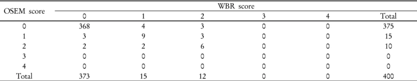
Table 8. Segmental score agreement between OSEM and WBR for assessment of segmental perfusion in normal group with stress gated myocardial perfusion SPECT (agreement 95.8%, 383/400 segments, k=0.656, p<0.0001)
OSEM score WBR score
0 1 2 3 4 Total
0 333 15 16 0 0 364
1 6 9 3 0 0 18
2 6 5 7 0 0 18
3 0 0 0 0 0 0
4 0 0 0 0 0 0
Total 345 29 26 0 0 400
Table 9. Segmental score agreement between OSEM and WBR for assessment of wall motion in normal group with stress gated myocardial perfusion SPECT (agreement 87.3%, 349/400 segments, k=0.390, p<0.0001)
였다 (Table 2-5).
정상군을 대상으로 한 기존 OSEM 기법과 WBR 기법에 서의 일치율은 휴식기 시 분절별 관류에서 총 400분절 중 396분절에서 일치하여 99% (396/400, k=0.662, p<0.0001)의 높은 일치율을 보였고 국소벽 운동에서는 83.8% (335/400, k=0.283, p<0.0001)로 의미 있는 낮은 일치율을 보였고 부하
기 시 분절별 관류에서 95.8% (383/400, k=0.656, p<0.0001) 의 높은 일치율을 보였고 국소벽 운동에서는 87.3%
(349/400, k=0.390, p<0.0001)의 의미 있는 낮은 일치율을 보
였다(Table 6-9). 정상군의 분절별 관류에서의 동위원소의 섭
취정도는 거의 모든 분절에서 정상 스코어 ‘0’이기 때문에 일
치율이 높은 것으로 생각되어 OSEM 기법과 WBR 기법간의
Segment Method Mean Standard deviation t-value p-value
apical anterolateral OSEM 85.60 6.59
1.633 0.119
WBR 83.15 6.71
apical inferolateral OSEM 86.05 6.34
1.841 0.081
WBR 83.20 5.19
apical inferior OSEM 79.15 7.53
1.814 0.086
WBR 77.15 7.30
apical inferoseptal OSEM 80.00 7.93 2.040 0.055
WBR 76.70 8.16
apical anteroseptal OSEM 80.85 6.43
1.652 0.115
WBR 78.75 8.94
apical anterior OSEM 77.40 6.96
1.704 0.105
WBR 75.70 6.06
mid anterolateral OSEM 84.10 5.55
-1.240 0.230
WBR 85.70 7.17
mid inferolateral OSEM 79.25 6.52 0.832 0.416
WBR 78.45 7.52
mid inferior OSEM 73.25 7.06
1.013 0.324
WBR 71.75 8.20
mid inferoseptal OSEM 70.65 7.46
-0.073 0.942
WBR 70.80 8.92
mid anteroseptal OSEM 74.90 6.68 0.930 0.364
WBR 73.55 6.64
mid anterior OSEM 76.20 5.69 1.047 0.308
WBR 75.05 6.96
basal anterolateral OSEM 60.55 9.20
-0.957 0.351
WBR 62.95 7.51
basal inferolateral OSEM 58.85 7.36
-1.071 0.298
WBR 60.45 5.53
basal inferior OSEM 55.25 7.73 0.778 0.446
WBR 54.00 5.16
basal inferoseptal OSEM 52.45 7.96 0.970 0.344
WBR 50.75 5.27
basal anteroseptal OSEM 51.75 10.55
-0.203 0.842
WBR 52.20 7.07
basal anterior OSEM 59.80 8.21
-0.469 0.644
WBR 60.70 6.11
anteroapical OSEM 78.45 6.10 2.376 0.115
WBR 76.35 6.82
inferoapical OSEM 77.70 6.46 3.826 0.079
WBR 74.85 6.78
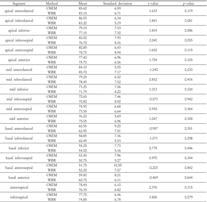
Table 10. Comparative analysis between OSEM and WBR for segmental perfusion measurements in normal group with rest gated myocardial perfusion SPECT
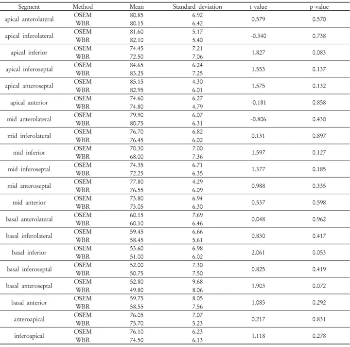
각 분절별 관류백분율의 정량값들을 이용하여 대응표본 t-검 증을 하였고 모든 분절에서 통계적으로 유의미한 차이를 보 이지 않았다 (p>0.05, Table 10,11).
고찰 및 결론
본 연구는 최근 핵의학 영상장비 및 소프트웨어의 활발한 개발에 따라 영상 획득시간을 짧게 하여 동등한 영상의 질을
만들어 내거나 동일한 영상획득으로 영상의 질을 높일 수 있
는 재구성기법인 UltraSPECT사의 WBR 기법이 심근 관류
SPECT에서도 적용할 수 있는지를 알아보기 위해 연구되었
다. 관상동맥 질환의 진단 및 예후 예측에 있어 중요한 의미
를 갖는 심근 내 분절별 관류와 국소벽 운동 기능 평가의 지
표들을 이용한 WBR 기법은 기존 OSEM 기법과 비교하여
정상/비정상군 모두에서 심근 내 분절별 관류의 일치율은 높
았지만 국소벽 운동에서는 의미 있는 낮은 일치율을 보였다.
Segment Method Mean Standard deviation t-value p-value
apical anterolateral OSEM 80.85 6.92
0.579 0.570
WBR 80.15 6.42
apical inferolateral OSEM 81.60 5.17 -0.340 0.738
WBR 82.10 5.40
apical inferior OSEM 74.45 7.21 1.827 0.083
WBR 72.50 7.06
apical inferoseptal OSEM 84.65 6.24
1.553 0.137
WBR 83.25 7.25
apical anteroseptal OSEM 85.15 4.30
1.575 0.132
WBR 82.95 6.01
apical anterior OSEM 74.60 6.27 -0.181 0.858
WBR 74.80 4.79
mid anterolateral OSEM 79.90 6.07 -0.806 0.430
WBR 80.75 6.31
mid inferolateral OSEM 76.70 6.82
0.131 0.897
WBR 76.45 6.02
mid inferior OSEM 70.30 7.00
1.597 0.127
WBR 68.00 7.36
mid inferoseptal OSEM 74.35 6.71 1.377 0.185
WBR 72.25 6.35
mid anteroseptal OSEM 77.80 4.29
0.988 0.335
WBR 76.55 6.09
mid anterior OSEM 73.80 6.94
0.537 0.598
WBR 73.05 6.30
basal anterolateral OSEM 60.15 7.69
0.048 0.962
WBR 60.10 6.46
basal inferolateral OSEM 59.45 6.66 0.830 0.417
WBR 58.45 5.61
basal inferior OSEM 53.60 6.98 2.061 0.053
WBR 51.00 6.02
basal inferoseptal OSEM 52.00 7.30
0.825 0.419
WBR 50.75 7.50
basal anteroseptal OSEM 52.80 9.68
1.903 0.072
WBR 49.80 8.06
basal anterior OSEM 59.75 8.05 1.085 0.292
WBR 58.55 7.56
anteroapical OSEM 76.05 7.07
0.217 0.831
WBR 75.70 5.23
inferoapical OSEM 76.10 6.23
1.118 0.278
WBR 74.50 6.13
Table 11. Comparative analysis between OSEM and WBR for segmental perfusion measurements in normal group with stress gated myocardial perfusion SPECT
WBR 기법은 높은 해상도와 대조도를 제공할 수 있다고 하 나 심근관류 SPECT에서의 적용은 유용성이 떨어진다고 사 료되며 추가적인 실험과 연구가 필요할 것이다 .
요 약
광대역 재구성(wide beam reconstruction, WBR) 기법인 Xpress.cardiac
TM프로그램을 적용하여 기존 OSEM (ordered
subsets expectation maximization) 기법과 심근 내 분절별 관 류와 국소벽 운동에서의 일치율을 확인하여 WBR 기법의 임 상적 유용성을 알아보고자 하였다.
관상동맥질환의 병력이 없고 핵의학 전문의에 의한 판독
상 이상소견이 없는 총 20명(남7명, 여자13명: 정상군)과 관
상동맥질환을 진단받은 총 10명(남6명, 여자4명: 비정상군)을
대상으로 휴식기
201Tl/부하기
99mTc-MIBI 심근관류 SPECT
를 실시하였다.
영상 획득과 재구성은 휴식기 시 투사영상당 30초, 곧바로 15초씩 영상을 얻고 부하기 시 투사영상 당 25초, 곧바로 13 초씩 영상을 얻어 OSEM과 WBR 기법을 적용하였고 심근 내 분절별 관류과 국소벽 운동은 AutoQuant 프로그램의 QPS/QGS 알고리즘의 20분절 모델을 적용하였다. 관류상태 는 5등급(0=정상, 1=경도, 2=중등도, 3=심한 결손, 4=섭취 없 음), 국소벽 운동은 5등급(0=정상, 1=경도, 2=중등도, 3=심한 운동저하 , 4=무운동)으로 분류한 반정량값을 이용해 기존 OSEM 기법과 WBR 기법에서의 일치율을 평가하였다.
정상군에서 기존 OSEM 기법과 WBR 기법에서의 일치율 은 휴식기 시 분절별 관류에서 99% (396/400, k=0.662,
p<0.0001), 국소벽 운동에서 83.8% (335/400, k=0.283), 부하기 시 분절별 관류에서 95.8% (383/400, k=0.656), 국소벽 운동 에서 87.3% (349/400, k=0.390)의 일치율을 보였다. 비정상군 에서 휴식기 시 분절별 관류에서 83% (166/200, k=0.605), 국 소벽 운동에서 55.5% (111/200, k=0.385), 부하기 시 분절별 관류에서 79.5% (159/200, k=0.682), 국소벽 운동에서 63.5%
(127/200, k=0.486)의 일치율을 보였다.
관상동맥 질환의 진단 및 예후 예측에 있어 중요한 의미를 갖는 심근 내 분절별 관류와 국소벽 운동 기능 평가의 지표
들을 이용한 WBR 기법은 기존 OSEM 기법과 비교하여 정 상․비정상군 모두에서 심근 내 분절별 관류의 일치율은 높았 지만 국소벽 운동에서는 의미 있게 낮은 일치율을 보였다.
WBR 기법은 높은 해상도와 대조도를 제공할 수 있다고 하 나 심근관류 SPECT에서의 적용은 유용성이 떨어진다고 사 료된다.
REFERENCES
1. 고창순 외. 제 3판 핵의학. 고려의학 2008;349-367
2. 안병철. 정량적 게이트 심근관류 SPECT. 대한핵의학회지 2003;38(4).
3. Paeng JC, Lee DS, Cheon GJ, Lee MM, Chung JK, Lee MC.
Reproducibility of an automatic quantitation of regional myo- cardial wall motion and systolic thickening on gated Tc-99m- sestamibi myocardial SPECT. J Nucl Med 2000;34:487-495.
4. UltraSPECT R&D Team, Haifa, Israel. Wide beam reconstruction:
Technology and applications (White paper). 2009 www.ultraspect.
com
5. UltraSPECT Inc, Haifa, Israel. Wide beam reconstruction: Short- Acquisition & High-Resolution Applications, Operation menual.
2009 www.ultraspect.com