*Corresponding author,
Phone: 82-54-820-5909; Fax: 82-54-820-6252;
E-mail: [email protected]
†This author contributed equally to this work.
Korean J Environ Agric (2011) Online ISSN: 2233-4173
Vol. 30, No. 3, pp. 346-356 http://dx.doi.org/10.5338/KJEA.2011.30.3.346 Print ISSN: 1225-3537
Characterization and Antifungal Activity from Soilborne Streptomyces sp. AM50 towards
Major Plant Pathogens
Jong-Ok Jang,
2,6Jung-Bok Lee,
3†Beam-Soo Kim,
1Sun-Chul Kang,
4Cher-Won Hwang,
5Kee-Sun Shin
6and Gi-Seok Kwon
1*1Dept. of Bioresource Sciences, Andong National University, Andong 760-749, South Korea, 2Dept. of Food Science and Technology Chungnam National University, Deajeon 350-764, South Korea, 3Dept. of Optometry, Kundong University, Andong, 760-833, South Korea, 4Dept. of Biotechnology, College of Engineering, Daegu University, Gyungsan, 712-714, Korea,
5School of Global Leadership, Handong Global University, Pohang, 791-708, Korea, 6Biological Resource Center, Korea Research Institute of Bioscience and Biotechnology (KRIBB), Daejeon 305-600, South Korea.
Received: 14 September 2011 / Accepted: 23 September 2011
ⓒ 2011 The Korean Society of Environmental Agriculture
346
Abstract
BACKGROUND: Chemical fungicides not only may pollute the ecosystem but also can be environmentally hazardous, as the chemicals accumulate in soil. Biological control is a frequently-used environment-friendly alternative to chemical pesticides in phytopathogen management. However, the use of microbial products as fungicides has limitations. This study isolated and characterized a three-antifungal-enzyme (chitinase, cellulase, and β-1,3-glucanase)-producing bacterium, and examined the conditions required to optimize the production of the antifungal enzymes.
METHOD AND RESULTS: The antifungal enzymes chitinase, cellulase, and β -1,3-glucanase were produced by bacteria isolated from an sawmill in Korea. Based on the 16S ribosomal DNA sequence analysis, the bacterial strain AM50 was identical to Streptomyces sp. And their antifungal activity was optimized when Streptomyces sp. AM50 was grown aerobically in a medium composed of 0.4% chitin, 0.4%
starch, 0.2% ammonium sulfate, 0.11% Na
2HPO
4, 0.07%
KH
2PO
4, 0.0001% MgSO
4, and 0.0001% MnSO
4at 30℃. A culture broth of Streptomyces sp. AM50 showed antifungal activity towards the hyphae of plant pathogenic fungi, including hyphae swelling and lysis in P. capsici, factors that may contribute to its suppression of plant pathogenic fungi.
CONCLUSION(S): This study demonstrated the multi- antifungal enzyme production by Streptomyces sp. AM50 for the biological control of major plant pathogens. Further studies will investigate the synergistic effect, to the growth regulations by biogenic amines and antifungal enzyme gene promoter.
Key Words: Antifungal activity, Biological control, Biogenic amines, Chitinase, Cellulase, Streptomyces sp.
Introduction
Chemical fungicides not only may pollute the ecosystem but also can be environmentally hazardous, as the chemicals accumulate in soil. Furthermore, the repeated use of such agrichemicals has encouraged the development of agrichemical resistance in the target organisms. Despite the controversial problems caused by the use of synthetic fungicides, fungicides will be increasingly used in agriculture industry in the
Open Access
Research Article
time to come, provide that safer and ecologically friendly fungicides become available. The requirements for the fungicides to be practically used in the fields are excellent potency against a multiplicity of pathogens and safety, not only for humans, and host plants but also for the environmental system.
Biological control is a frequently-used environment- friendly alternative to chemical pesticides in phytopathogen management. However, the use of microbial products as fungicides has limitations.
Several species of Serratia sp., Pseudomonas sp., Bacillus sp., and Streptomyces sp. have already been widely used for the biocontrol of plant pathogens (Sindhu et al ., 2001; Merav et al ., 2003). Yet, the synthesis of cell-wall-degrading enzymes is generally subject to induction and/or catabolic repression (Sachslehner et al ., 1998). Nonetheless, these hydrolytic enzymes are considered key in the lysis of the cell walls of higher fungi.
The antifungal activities of chitinase and cellulase often result in the lysis of fungal hyphae. As such, this ability to lyse fungal hyphae presents the possibility that living fungal hyphae, rather than chitin, may form the actual growth substrate for chitinolytic soil bacteria (Wirtse et al ., 1998). β- 1,3-Glucanase is already known to act as a synergist for the antifungal activity of chitinase (Marrianne et al ., 1993), which may facilitate the generation of fungal protoplasts that are effective biocontrol agents against many phytopathogenic fungi (Kitamoto et al ., 1998; Elad et al ., 2005).
Streptomyces spp. are major producer of chitinase in soil and use chitin as their carbon source. Various studies on the action of one or two complex antifungal enzymes, such as chitinase, cellulase, and β-1,3-glucanase, have revealed the mechanisms by which these enzymes degrade and/or lysephytopathogenic fungi hyphae.
However, this study isolated and characterized Streptomyces sp. AM50, a three-antifungal-enzyme (chitinase, cellulase, and β- 1,3-glucanase)-producing bacterium, and examined the conditions required to optimize the production of the antifungal enzymes.
Materials and Methods
Chemicals
The chitin used in the experiments was purchased from ACROS Organics Co. (New Jersey, USA), while the N- acetyl- D- glucosamine, carboxymethy cellulose (CMC), and laminarin were purchased from Sigma
Chemical Co. (St. Louis, USA). All other chemicals used in this study were also purchased from Sigma (St. Louis, USA).
Microorganisms
Cultures of the fungi Phytophthora capsici (KACC 40157), Colletotrichum gloeosporioides (KACC 40003), and Rhizoctonia solani AG-2-2 (KACC 40132) were obtained from the Korea Agricultural Culture Collection (KACC), and maintained on agar plates composed of a potato dextrose agar (PDA) at 28℃.
Preparation of colloidal chitin
Commercial chitin 15 g, was hydrolyzed with 150 mL of HCl for 12 hrs at 4℃ and filtered to remove the HCl-dissolved chitin. Cold distilled water was added to the filtrate, which was then stirred until the formation of a white suspension. This suspension was centrifuged at 10,000 rpm for 10 min and washed with distilled water, resulting in a pH of 5. The pH was then adjusted to 7 using 5 N KOH and centrifuged at 10,000 rpm for 10 min.
Isolation of chitinase and cellulase-producing bacterium The bacteria were initially isolated from soils Kawn- Geo Sawmill, Andong, Gyeongbuk, Korea. Approximately 1g of sample material was added to 20mL of liquid basal minimal medium (BMM) [0.2% (NH
4)
2SO
4, 0.11%
Na
2HPO
4, 0.07% KH
2PO
4, 0.0001% MgSO
4, and 0.0001%
MnSO
4] chitinase detection medium containing 0.4%
colloidal chitin and cellulase detection medium containing 0.4% CMC in 100mL Erlenmeyer flasks and incubated with shaking (180rpm) for 7days at 30℃ respectively.
Secondary enrichments were then established by inoculating 1mL of the primary enrichments into 20mL of a fresh liquid BMM containing 0.4% colloidal chitin and 0.4%
CMC respectively. After the samples were incubated at 30℃ for 7 days on a shaking incubator (180 rpm), they were spread onto a chitinase detection medium containing 0.4% colloidal chitin and cellulase detection medium containing 0.4% CMC. Chitinase was indicated by zones of clearing around colony and cellulase was yellow haloing by congo red staining.
16SrRNAgene sequences
To determine the 16S rRNA gene sequences of
strain AM50, DNA extraction, amplification,
purification, and sequencing were carried out as
described by Endo and Okada (2005). The 16S rRNA
gene sequences were aligned against representative reference sequences for members of the genus Streptomyces and related taxa using Clustal X, version 1.81 (Thompson et al ., 1997). The evolutionary distances were then calculated using the method of Jukes and Cantor (1969), while phylogenetic dendrograms and bootstrap analyses were performed using the Phylip 3.5c program package (Felsenstein, 1993).
Effect of culture conditions on production of antifungal enzymes
To determine the effect of the culture conditions, the growth was conducted in a BMM (basal minimal medium) containing 0.4% colloidal chitin. The main ingredients investigated included glucose, fructose, sucrose, soluble starch, and CMC at concentrations of 0.2, 0.4 and 0.8% as the carbon source. Meanwhile, each nitrogen sources used were peptone, tryptone, yeast extract, and ammonium sulfate at concentrations of 0.05, 0.1, and 0.2%.
Effect of temperature, aeration, and pH on production of antifungal enzymes
The antifungal-enzyme production was also examined in terms of the effect of the initial pH, which was adjusted between 3.0-11.0 using 1 N HCl and 1 N NaOH, various incubation temperatures at 20, 30, and 40℃, and the effect of oxygen using different agitation speeds of 0 rpm and 180 rpm.
Measurement of enzyme activity
The chitinase activity was measured with N- acetyl- D- glucosamine as the substrate. The reaction mixture, containing 100 μL of 1% N- acetyl- D- glucosamine, 200 μL of a 0.1 M phosphate buffer (pH 7.0), and 200 μL of the enzyme solution, was incubated at 40℃ for 30 min, then 0.1 N NaOH was added to stop the reaction. After centrifugation, 200 μL of the supernatant was mixed with a 3, 5-dinitrosalicylic (DNS) solution. The amount of reducing sugar produced was then measured using the 3,5-dinitrosalicylicreagent method (Miller, 1959), and the amount of reducing sugar calculated from standard curve concentrations of N- acetyl- D- glucosamine. One unit of enzyme activity was defined as 1 μg of N- acetyl- D- glucosamine released per milliliter of enzyme extract per hour.
Meanwhile, the cellulase activity was measured with 1% CMC as the substrate. The reaction mixture, containing 100 μL of 1% CMC, 200 μL of a 0.1 M
phosphate buffer (pH 7.0), and 200 uL of the enzyme solution, was incubated at 40℃ for 30 min. The amount of reducing sugar released was then calculated from a standard curve prepared with glucose, and one unit of cellulase defined as the amount of enzyme that released 1 μg of reducing sugar per minute.
Finally, the β- 1,3-Glucanase activity was measured with laminarin as the substrate. The reaction mixture, containing 100 μL of 0.1% laminarin, 200 μL of a 0.1 M acetic acid buffer (pH 4.0), and 200 μL of the enzyme solution, was incubated at 40℃ for 30 min.
All the experiments were carried out in triplicate, and although the values varied somewhat between the experiments, the trends were always similar.
Assay of activity against host cell wall and antifungal activity
The inhibition zone assay for antifungal activity was executed using the disc (8mm in diameter) method (Johnson and Cirl, 1972). Potato dextrose agar (PDA) plates were incubated at 28℃for 3-5 days until mycelial growth had enveloped the control, and the fungal species included P. capcisi, C. gloeosporioides, and R.
solani.
Effect of AM50 on hyphal morphology
P. capsici spores were grown in a V8 juice agar (V8 juice 20%, H
2O 80%, CaCO
34%, and agar 2%) medium, then1×10
5sproes/mL were transferred to test tubes containing 10 mL of a potato dextrose broth, followed by the addition of 100 μL of an AM50 culture broth. The resultant solution was incubated at 30℃, 3 days, and the hyphae observed under a light microscope.
Results
Selectionand identification of the antifungal-enzyme- producing bacterium
Antifungal enzyme-producing bacteria were identified
based on the production of clear zones around colonies
on a medium containing 0.4% colloidal chitin and/or a
CMC agar medium. From 10 sawmill-soil samples,
different bacteria were isolated and the largest zone
was then selected by the strain forming, the strain
AM50 was finally selected.
Streptomyces lanatus
NBRC 12787T(AB184845)Streptomyces fulvoviolaceus
NBRC 14148 (AB184573)Streptomyces misionensis
NBRC 13063T(AB184285)Streptomyces purpurascens
LMG 20526T(AJ781382)Streptomyces malachitofuscus
LMG 20067T(AJ781347)Streptomyces paradoxus
NBRC 14887T(AB184628)Streptomyces griseoflavus
NBRC 13044T(AB184274)Streptomyces althioticus
NBRC 12740 (AB184112)Streptomyces xylophagus
NBRC 13845T(AB184526)Streptomyces carpinensis
NBRC 14214T(AB184574)Streptomyces levis
NRRL B-16370T(DQ442517)Streptomyces griseus subsp. griseus
NBRC 13350 (AB045866)Streptomyces intermedius
DSM 40372T(Z76686)Streptomyces rutgersensis
DSM 40077T(Z76688)Streptomyces gougerotii
DSM 40324T(Z76687)Streptomyces owasiensis
NBRC 13832T(AB184515)Streptomyces hygroscopicus
subsp. hygroscopicusNBRC3401(AB184760)Streptomyces costaricanus
NBRC 100773T(AB249939)Streptomyces griseofuscus
NBRC 12870T(AB184206)Streptomyces graminearus
NBRC 15420T(AB184667)Streptomyces
sp. AM50 (EU100460)Streptomyces murinus
NBRC 14802T(AB184622)Streptomyces padanus
ATCC 25646 (AF455813)80 89
95
96
98 93
100
100 99
0.01
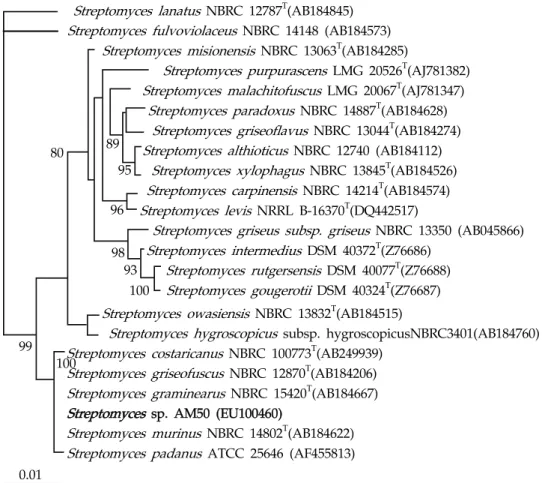
Fig. 1. Neighbor-joining tree based on almost-complete 16S rRNA gene sequences, showing phylogenetic relationships between strain AM50 and species from
Streptomyces
genus.Numbers at nodes indicate levels of bootstrap support (%) based on 1000 resambled datasets; only values above 70% are given. Bar, 0.01 substitutions per nucleotide position.
A comparative 16S rRNAgene sequence analysis revealed that strain AM50 was most closely related to members of the genus Streptomyces . Plus, in a neighbor-joining phylogenetic tree constructed on the basis of the 16S rRNA gene sequences, strain AM50 also fell within the radiation of the cluster comprising Streptomyces species. Strain AM50 exhibited 16S rRNA gene sequence similarity values of 100% with Streptomyces costaricanus NBRC 100773
T, Streptomyces griseofuscus NBRC 12870
T, Streptomyces graminearus NBRC 15420
T, Streptomyces murinus NBRC 14802
T, and Streptomyces padanus ATCC 25646 (Fig. 1).
Optimization of culture conditions for production of antifungal enzymes
Chitinase and/or cellulase production was found to be dependent on the carbon source used in the culture medium. To study the effect of carbon sources on the antifungal enzyme production by Streptomyces sp. AM50, growth was conducted in a minimal
medium containing various carbon sources, as shown in Fig. 2. The antifungal activity occurred at 0.4% starch which was antifungal enzymes (chitinase, cellulase, β -1,3-glucanase) production by Streptomyces sp. AM50 (Fig. 3). Then, carbon source concentration in the three enzymes and antifungal activity, three different proportions of starch 0.2% 0.4% and 0.8% were used the carbon source. The results were shown in Fig. 3.
chitinase production was highest (138.8 unit) when 0.8% was used as the source, but the other enzymes production were lowest. When 0.4% starch supplement, tree enzymes were evenly high (105.8 unit, 130.7 unit, and 44.3 unit), and antifungal activity was highest 36 mm at 9th day (Fig. 3).
When different nitrogen sources were tested, the results showed that chitinase and cellulase activities were higher with ammonium sulfate (Fig. 4 and 5).
The antifungal effect of optimal concentration was
0.2% and used in subsequent experiments (Fig. 5D).
Enzyme Activity (units/ml)
0 50 100 150 200 250 300 350
glucose fructose sucrose starch CMC
Enzyme Activity (units/ml)
0 10 20 30 40 50 60
glucose fructose sucrose starch CMC
Enzyme Activity (units/ml)
0 10 20 30 40 50
glucose fructose sucrose starch CMC Days after Incubation
1 3 5 7 9
clear zone(mm)
0 5 10 15 20 25 30 35
(A) (B)
(C) (D)
Fig. 2. Effect of carbon sources on chitinase (A), cellulase (B), and β-1, 3-glucanase (C) production by
Streptomyces
sp. AM50. (D) Inhibiting effect ofStreptomyces
sp. AM50 onP.
capsici
(●; 0.4% colloidal chitin minimal, ○; 0.4% colloidal chitin + 0.4% glucose, ▼; 0.4%colloidal chitin + 0.4% fructose, ▽; 0.4% colloidal chitin + 0.4% sucrose, ■; 0.4% colloidal chitin + 0.4% starch, □; 0.4% colloidal chitin + 0.4% CMC).
Days after Incubation
1 3 5 7 9 11
Enzyme Activity (units/ml)
0 20 40 60 80 100 120 140 160
Days after Incubation
1 3 5 7 9 11
Enzyme Activity (units/ml)
0 20 40 60 80 100 120 140 160
Days after Incubation
1 3 5 7 9 11
clear zone(mm)
0 10 20 30 40
Days after Incubation
1 3 5 7 9 11
Enzyme Activity (units/ml)
0 10 20 30 40 50
(A) (B)
(C) (D)
Fig. 3. Effect of starch (carbon source) on (A) chitinase, (B) cellulase, and (C) β-1, 3-glucanase production by
Streptomyces
sp. AM50. (D) Inhibiting effect ofStreptomyces
sp. AM50 onP.
capsici.
(●; chitin + 0.2% starch, ○; chitin + 0.4% starch, ▼; chitin + 0.8% starch).Enzyme Activity (units/ml)
0 5 10 15 20 25 30 35
peptone tryptone yeast extractammonium sulfate
Enzyme Activity (units/ml)
0 20 40 60 80 100
peptone tryptone yeast extract ammonium sulfate
Days after Incubation
1 3 5 7 9 11
clear zone(mm)
0 10 20 30 40
Enzyme Activity (units/ml)
0 5 10 15 20 25
peptone tryptone yeast extract ammonium sulfate
(A) (B)
(C) (D)
Fig. 4. Effect of nitrogen sources on (A) chitinase, (B) cellulase, and (C) β-1, 3-glucanase production by
Streptomyces
sp. AM50. (D) Inhibiting effect ofStreptomyces
sp. AM50 onP. capsici
(●; 0.2% peptone, ○; 0.2% tryptone, ▼; 0.2% yeast extract, ▽; 0.2% ammonium sulfate).Days after Incubation
1 3 5 7 9 11
Enzyme Activity (units/ml)
0 100 200 300 400
Days after Incubation
1 3 5 7 9 11
Enzyme Activity (units/ml)
0 50 100 150 200 250
Days after Incubation
1 3 5 7 9 11
clear zone(mm)
0 10 20 30 40
Days after Incubation
1 3 5 7 9 11
Enzyme Activity (units/ml)
0 100 200 300 400 500
(A) (B)
(C) (D)
Fig. 5. Effect of ammonium sulfate (nitrogen source) on (A) chitinase, (B) cellulase, and (C) β-1, 3-glucanase production by
Streptomyces
sp. AM50. (D) Inhibiting effect ofStreptomyces
sp. AM50 onP. capsici
(●; 0% ammonium sulfate, ○; 0.05% ammonium sulfate, ▼; 0.2% ammonium sulfate ▽;0.4% ammonium sulfate).
Optimization of temperature, pH, and aeration on production of antifungal enzymes
The effects of aeration and temperature of culture medium on the antifungal enzyme production was investigated. Fig. 6 showed the effect of temperature, aeration, and pH on the antifungal activity. The optimal conditions for antifungal enzymes production were a temperature of 30℃, aeration, and a pH from 7 to 9. The antifungal activity occurred at 20-40℃
which were in the range of the temperature (Fig. 6).
Production of chitinase, cellulase and/or β- 1,3- glucanase enzymes did definitely with in an initial
pH range 5-9 (data not shown).
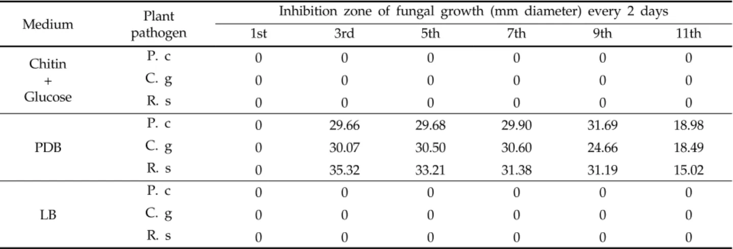
Specificity of antifungal activity and necessity of chitin The antifungal enzyme production by Streptomyces sp. AM50 was induced by the addition of colloidal chitin, glucoseor a common medium LB or PDB. As shown in data, the presence of chitin resulted in high antifungal enzyme activity and inhibited the growth of the fungal hyphae tips. While the addition of glucose, in contrast, decreased the antifungal activity compared to that with the chitin minimal medium and also resulted in LB medium.
0 10 20 30 40
1 3
5 7
9 11
2 1 4 3
6 5
clear zone(mm)
days 0
10 20 30 40
1 3
5 7
9 11
2 1 4 3
6 5
clear zone(mm)
days
0 10 20 30 40
1 3
57911
1 0 3 2
5 4 6
clear zone(mm)
day s
Fig. 6. Effect of shaking and temperature on producing of antifungal enzymes (A)
P. capsici
, (B)C. gloeosporioides
, and (C)R. solani
byStreptomyces
sp. AM50. (Y axis : 1 lane; 20℃, 180 rpm, 2 lane; 20℃, 0 rpm, 3 lane; 30℃, 180 rpm, 4 lane; 30℃, 0 rpm, 5 lane; 40℃, 180 rpm, 6 lane; 40℃, 0 rpm)Effect of Streptomyces sp. AM50 on P. capsici hyphal morphology
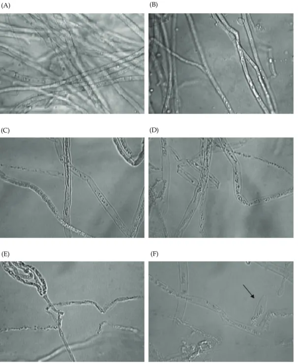
To investigate the effect of Streptomyces sp. AM50 culture broth (crude enzyme), morphological changes were observed in P. capsici mycelia. The tested fungus following co-culture with the filtered AM50 culture broth (crude enzyme) after 3 days in PDB medium was studies. As shown in Fig. 7 and 8, the control revealed normal hyphae (Fig 7A, B), whereas
abnormal hyphae were observed after treatment with the culture broth, including lysis or shrinking (Fig.
7C-E) of the hyphae tips, swelling (Fig. 7F) of the hyphae body, and arrested-growth of the P. capsici hyphae. Furthermore, when the P. capsici spore suspension and a 5-fold (v/v) amount of the culture broth were incubated simultaneously, the P. capsici mycelia could not grow.
(A) (B)
(C) (D)
(E) (F)
Fig. 7. Effect of antifungal activity from AM50 on morphology of
Phytophthora capsici
. (A) and (B) are normal mycelia ofP. capsici
under light microscope. (C), (D), and (E): Abnormal myceliallysis ofP. capsici
., (F): Abnormal swelling (arrow) ofP. capsici
Discussions
This study attempted to optimize the anti-fungal activity of Streptomyces sp. AM50 towards the plant pathogens P. capsici , C. gloeosporioides , and R.
solani . Various studies and case reports (Takahashi et al ., 1993; Chang et al ., 2001; Hassane et al ., 2001;
Wang et al ., 2002; Wichitra et al ., 2002; Daulagala and Allan, 2003) have already investigated the potent antifungal activity of Streptomyces sp., which are saprophytic soil bacteria (Akihiro et al ., 1998), the major producers of chitinase in soil, and use chitin as their source of carbon. Increased concern over the impact of chemical pesticides on the environment has resulted in the increased interest in biocontrol strategies for the management of the plant pathogens.
A large number of microorganisms, including fungi, bacteria, actinomycetes, and plant species, possess the ability to excrete cell-wall hydrolases, such as proteases, chitinase, cellulase, and β- 1,3-glucanase. These hydrolases play an important role in the reactions between biocontrol agents and pathogens by strongly inhibiting spore germination, tube elongation, and mycelial growth (Chernin and Chet, 2002). Most previous reports have only studied one or two antifungal enzymes, acting alone or synergistically. However, this study investigated three enzymes that inhibit the growth of plant pathogens in different ways, so be expected more effective fungicide use it.
Several mechanisms for the biocontrol of pathogenic fungi have already been proposed, including abnormal hyphae swelling (Akihiro et al ., 1993), lysis and degradation of the hyphae tips (Wang et al ., 1999), and antagonism towards spore germination or germ tube elongation (Marrianne et al ., 1993). The strain Streptomyces sp. AM50 showed abnormal swelling and lysis of the P. capsici hyphae tips. For instance, cellulose and/or β-glucansynthetase in P. capsici cells was inhibited by the Streptomyces sp. AM50 from enzyme or other unknown factors.
Microscopic observation of the processes that occurred after treatment with Streptomyces sp. AM50 also showed abnormal hyphae swelling, lysis, and complete degradation of the hyphae tips. While the hyphae tips of oomycetesare composed of glucan and cellulose, the hyphae tips of ascomycetes and basidiomycetes are composed of chitin, thereby explaining the broad anti-fungal activity of Streptomyces sp. AM50. Similar results were reported for the biocontrol fungus Trichoderma harzianum (Hassane et
al ., 2001) and Bacillus subtilis when used to inhibit Fusarium oxysporum (Phae et al ., 1992).
Previous reports onchitinase-producing microorganisms used chitin or colloidal chitin as the substrate for chitinase production andthe resulting antifungal activities (Wand et al ., 2002). However, this study showed that the fungal growth inhibition by Streptomyces sp. AM50 was not dependent on the presence of chitin. As such, while the chitinase synthesis and antifungal enzyme production by Streptomyces sp. AM50 were induced by chitin, the fungal growth was repressed by readily usable carbon sources, such as glucose (Kiyotaka et al ., 2000).
According to Sindhuand Dadarwal (2001), chitinase activity and cellulase activity are not always correlated with the inhibition of fungal growth, yet the present study found that chitinase, cellulase, and β-1,3- glucanase activity were all correlated with fungal growth inhibition.
Antifungal activity of Streptomyces sp. AM50 of the carbon catabolite repression, which can be exerted by many compounds metabolized via different routes.
In spite of their biochemical diversity, the presence of these molecules in a growth medium can lead to a decrease in the specific activities of enzymes involved in the catabolism of other carbon sources. Although this repression can potentially be performed at all levels of expression, the cells must contain a component that can sense the presence of such repressing compounds in the growth medium, giving rise to a signal that eventually results in repression of the enzyme activity (Kawakman et al ., 1994). In the bacterium Streptomyces sp. AM50, one or more antifungal enzymes were responsible for glucose. Thus, chitin+a glucose medium exhibited a high enzyme activity when compare with a chitin-only medium or PDB/LB (not data shown), yet the antifungal activities produced contrary results, demonstrating that the carbon source of Streptomyces sp. AM50 is important as regards inhibiting the growth of fungal hyphae. Thus, it is supposed that the antifungal enzyme promoter induced by chitin and other carbon sources was not subject to the catabolic repression of glucose.
Accordingly, this study demonstrated the multi- antifungal enzyme production by Streptomyces sp.
AM50 for the biological control of major plant pathogens.
Further studies will investigate the synergistic effect, to the
growth regulations by biogenic amines and antifungal
enzyme gene promoter.
Table 1. Antifungal activity of
Streptomyces
sp. AM50 with different mediaMedium Plant
pathogen
Inhibition zone of fungal growth (mm diameter) every 2 days
1st 3rd 5th 7th 9th 11th
Chitin + Glucose
P. c 0 0 0 0 0 0
C. g 0 0 0 0 0 0
R. s 0 0 0 0 0 0
PDB
P. c 0 29.66 29.68 29.90 31.69 18.98
C. g 0 30.07 30.50 30.60 24.66 18.49
R. s 0 35.32 33.21 31.38 31.19 15.02
LB
P. c 0 0 0 0 0 0
C. g 0 0 0 0 0 0
R. s 0 0 0 0 0 0
P. c: