Cytotoxicity of Dichloromethane Extracts of Asian Dust
Eun-Jung Park† · Dae-Seon Kim* · Seong-Do Yu* · Kwangsik Park College of Pharmacy, Dongduk Women’s University, Seoul 136-714, Korea
*Environmental Research Complex, Incheon 404-708, Korea (Received May 6, 2010/Revised July 5, 2010/Accepted August 5, 2010)
ABSTRACT
The appearance of Asian Dust (AD) originating from China and Mongolia during spring each year is a meteorological phenomenon periodically observed in extensive regions of East Asia. According to a previous epidemiological study, AD has adverse effects on both human beings and ecosystems. In this study, we collected total suspension particles (TSP) in the AD period and Non-AD (NAD) period. We extracted organic components from TSP using dichloromethane (DCM), and the polyaromatic hydrocarbons (PAHs) were analyzed. The DCM extracts contained PAHs such as benzo(b)fluoranthene, benzo[g,h,i]perylene, benzo(k)fluoranthene, benzo(a)pyrene, and pyrene. No significant difference was observed in cytotoxicity of the DCM extracts from AD versus NAD when tested on the human bronchial epithelial cells, BEAS-2B. We also examined the toxic mechanisms of AD extracts in cultured BEAS-2B cells and RAW264.7 cells, and in BEAS-2B cells observed increased levels of reactive oxygen species (ROS), decreased glutathione (GSH), and induced caspase-3 activity. Increased expression of oxidative stress-related and inflammation- related genes were also observed in BEAS-2B cells, while nitric oxide (NO) levels were increased in RAW264.7 cells. Taken together, the results suggest that in these cultured cells, AD may induce cytotoxicity through oxidative stress and pro-inflammatory signals.
Keywords: Asian Dust, PAHs, inflammation, BEAS-2B, RAW264.7
I. Introduction
The Asian Dust (AD) is meteorological phenomenon observed frequently in extensive regions of East Asia during the spring. Recently, it has also occurred irregularly during fall and winter.
The AD mainly originates from the Takla Makan desert and the Loess plateau areas of China, and the Gobi desert of Mongolia. Recently, the occurrences of AD are increasing, which may be due to the desertification in China, and industrialization, forest fires, and reckless defores- tation in Mongolia.1-3)
The AD particles are predominantly larger than 2 µm in aerodynamic diameter. Thus, TSP/PM10 ratios are almost constant, while the coarse/fine or TSP/PM2.5 ratios change noticeably between the
AD and non-AD periods.4,5) Because the AD originates in the desert, it is mainly composed of soil-based mineral components, but anthropogenic components (e.g., nitrate, sulfate, and heavy metals), could be attached during transportation. Therefore, the size and components of the AD differ considerably according to the origin, composition, transport pathway, and transport mechanism.6)
The AD may cause a variety of difficulties for humans. For example, PM 10 concentration was reported to be more than 3000 µg/m3 per hour in Korea during the time of the AD event in April of 2002.1,2) The social and economic damage (e.g., temporary closure of primary schools, semicon- ductor industries, and precision industries) was unavoidable.7) Furthermore, there were negative health consequences, such as increased mortality rates due to cardiovascular and respiratory problems, and increased hospital admissions by ischemic stroke, eye troubles, allergies, and asthma.8,9) Thus, many Koreans are worried about the possible
†Corresponding author : College of Pharmacy, Dongduk Women’s University
Tel: 82-32-560-7174, Fax: 82-32-568-2037 E-mail: [email protected]
adverse health effects of As dust event every spring. However, the mechanism of these adverse effects has not been clear.
In this study, we collected TSP (Total Suspension Particles) in the Asian Dust period (AD), as well as during the rainy season of 2007 (NAD), extracted organic components from TSP using DCM (dichloromethane), and then analyzed the organic components. Further, we compared cytoxicity of both extracts using the human bronchial epithelial cell, BEAS-2B, and investigated the toxic mechanism in BEAS-2B cells and RAW264.7 cells.
II. Materials and Methods
1. Particle collection and extraction
We collected TSPs during the Asian Dust period in 2007 (3 times; May 8, 13, and 25) and the rainy season (9 days; from July 10 to 18), on the roof of a university building, at a height of 12 meters above the ground, and pooled as Asian Dust and Non-Asian Dust, respectively. Sampling was performed using PTFE filters (ZefluorTM, 2.0 µm, PALL Life Sciences, NY, USA), the filter was cut into strips, and sonicated for 30 min in a glass bottle using DCM. Sonication in DCM was replicated 3 times. Finally, extracts were pooled, dried by nitrogen gas, and then, were solubilized in DMSO for toxicity tests.10,11)
2. Analysis of PAHs
We measured the concentration of PAHs (polycyclic aromatic hydrocarbons) including anthracene, fluoranthene, pyrene, benzo(a)anthra-
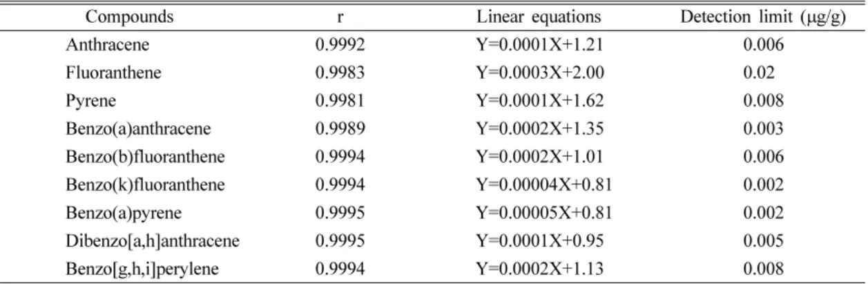
cene, benzo(b) fluoranthene, benzo(k)fluoranthene, benzo(a)pyrene, dibenzo[a,h]anthracene, and benzo[g,h,i]perylene in both extracts using GC- MS.12) Detection limits were suggested on Table 1.
3. Cell culture and cell viability test
BEAS-2B cells and RAW264.7 cells were maintained in DMEM/F12 and DMEM media (GIBCO Invitrogen, Seoul, Korea), containing 10% FBS, penicillin 100 IU/ml, and streptomycin 100 µg/ml, respectively. Cells were grown and maintained in 28 cm2 cell culture flasks at 37oC in a 5% CO2 humidified incubator.14-16)
Cell viability was also measured by the MTT assay. Cells were seeded on 96-well tissue culture plates with 3×10³-1×10⁴ cells per well. After 24- hour stabilization, AD extract (ADE) and NAD extract (NADE) were treated for 24, 48, 72, and 96 hours, respectively. At the end of exposure, MTT solution (Sigma-Aldrich, St. Louis, MO, USA) was added, and was incubated for 4 hours at 37oC. After solubilization with DMSO, the absorbance was quantified in 540 nm using the microplate spectrophotometer system (VersaMax, Molecular Devices, Sunnyvale, CA, USA). The viability of the treated group was expressed as the percentage of the control group, which was assumed to be 100%.13-16)
4. ROS generation
A fluorometric assay was performed using intracellular oxidation of DCFH-DA (dichloro- fluorescin diacetate, Sigma-Aldrich, St. Louis, MO, USA). Cells were treated with ADE of 25
Table 1. Detection limits for determination of polyaromatic hydrocarbon
Compounds r Linear equations Detection limit (µg/g)
Anthracene 0.9992 Y=0.0001X+1.21 0.006
Fluoranthene 0.9983 Y=0.0003X+2.00 0.02
Pyrene 0.9981 Y=0.0001X+1.62 0.008
Benzo(a)anthracene 0.9989 Y=0.0002X+1.35 0.003
Benzo(b)fluoranthene 0.9994 Y=0.0002X+1.01 0.006
Benzo(k)fluoranthene 0.9994 Y=0.00004X+0.81 0.002
Benzo(a)pyrene 0.9995 Y=0.00005X+0.81 0.002
Dibenzo[a,h]anthracene 0.9995 Y=0.0001X+0.95 0.005
Benzo[g,h,i]perylene 0.9994 Y=0.0002X+1.13 0.008
ug/ml and 50 ug/ml for 24 hours, and further incubated with 40 µM DCFH-DA for 15 min. At the end of DCFH-DA incubation, cells were observed using fluorescent microscope.14,15)
5. Measurements of GSH
The intracellular reduced glutathione (GSH) level was determined according to the method previously described.14,15) After harvest, cells were briefly treated for 24 hours with ADE of 25 ug/ml and 50 ug/ml, and pellet was lysed with 1%
perchloric acid. Cell lysates, KH2PO4/EDTA buffer, and o-phthaldialdehyde, were put in 96-black well plates, and incubated in the dark at room temperature for 30 minutes. Fluorescence was measured using the microplate spectrofluorometer (GeminiXPS, Molecular Devices, Sunnyvale, CA, USA), with excitation and emission wavelengths of 350 nm and 420 nm, respectively. Results were calculated as nmol of glutathione per mg of protein. Protein content of the cell lysates was calculated using a BCA protein assay kit (Pierce, Rockfold, IL, USA).
6. Measurement of caspase-3 activity
The activity of caspase-3 was determined using a colorimetric assay kit (R&D systems Inc., Minneapolis, MN, USA). Cells were briefly incubated with ADE of 25 ug/ml and 50 ug/ml for 24 hours. The enzyme activity of cell lysates was
tested by the addition of a caspase-specific peptide that was conjugated to the color reporter molecules p-nitroanaline (pNA). Protein content in the cell lysates was calculated using a BCA protein assay kit (Pierce, Rockfold, IL, USA).
7. Nitric oxide production
Cells were treated with ADE of 25 ug/ml and 50 ug/ml for 24 hours. NO (Nitric oxide) production by the extract was quantified spectrophotometrically, using Griess reagent. The absorbance was measured at 540 nm and the nitrite concentration was calculated using a calibration curve prepared using sodium nitrite as the standard.16)
8. Gene expression analysis
Cells were treated with ADE of 25 ug/ml and 50 ug/ml for 24 hours. The preparation of total RNA was performed using the RNAgents total RNA Isolation System (Promega, Madison, WI, USA).
Then, the RT-PCR reaction was performed with 1µg of total RNA, 20 µM oligo dT, and reaction mixture, which was provided by AccuPower RT/
PCR PreMix (Bioneer, Daejeon, Korea) at 42oC for 60 minutes. PCR was then carried out for 25- 30 cycles at 95oC for 1 min, 55oC for 1 min, and 72oC for 1 min. Amplified cDNA products were separated on 1.5% agarose gel by electrophoresis.
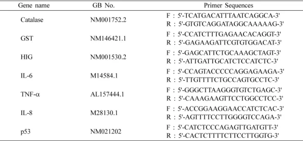
Table 2 reported the primer list used in this experiment.13-16)
Table 2. Primer sequences used in this study
Gene name GB No. Primer Sequences
Catalase NM001752.2 F : 5'-TCATGACATTTAATCAGGCA-3'
R : 5'-GTGTCAGGATAGGCAAAAAG-3'
GST NM146421.1 F : 5'-CCATCTTTGAGAACACAGGT-3'
R : 5'-GAGAAGATTCGTGTGGACAT-3'
HIG NM001530.2 F : 5'-GAGCATTCTGCAAAGCTAGT-3'
R : 5'-ATTGATTGCATCTCCATCTC-3'
IL-6 M14584.1 F : 5'-CCAGTACCCCCAGGAGAAGA-3'
R : 5'-TTGTTTTCTGCCAGTGCCTC-3'
TNF-α AL157444.1 F : 5'-GGGCTTAAGGGTGTCTGAGC-3'
R : 5'-CAAAGAAGTTCCTGGCCTCC-3'
IL-8 M28130.1 F : 5'-ACCGGAAGGAACCATCTCAC-3'
R : 5'-AGTTTTCCTTGGGGTCCAGA-3'
p53 NM021202 F : 5'-CATCTCCCAGAGTTGATGTT-3'
R : 5'-CACTCTTTTCTTCCTTGGTG-3'
9. Statistical analysis
The results of cell viability, GSH, NO and caspase-3 activity levels are presented as the mean±SD (standard deviation) from 4 repeated experiments. The values were compared using the student’s t-test, and levels of significance were represented for each result.
III. Results
1. The comparison of organic components in both periods
We collected a total TSP of 1,752 mg and 480 mg during the Asian Dust period and the rainy season in 2007. Table 3 showed concentration of PAHs (polyaromatic hydrocarbons) in DCM extracts.
The concentration of all PAHs was higher in ADE than NADE. Particularly, the gap of content of fluoranthene, pyrene, and benzo[g,h,i]perylene was marked.
2. The comparison of cytotoxicity of both extracts
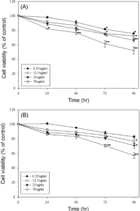
As shown in Fig. 1, both extracts dose- and time-dependently decreased cell viability. At 24 hours after exposure with a concentration of 6.25
~50µg/ml, cell viability was decreased to 98.0±
1.5%~82.2±0.2% and 100.8±1.4%~86.2±1.0 by ADE and NADE, respectively. At 96 hours after exposure with a concentration of 6.25~50 µg/ml, cell viability was decreased to 75.3±2.3%~52.2±
5.0% and 82.8±1.3%~57.1±3.8 by ADE and NADE, respectively.
Table 3. Concentration of polyaromatic hydrocarbon in extracts (ng/ml)
Name ADE NADE
Anthracene 5.444 2.164
Fluoranthene 35.292 <0.020
Pyrene 42.513 5.971
Benzo(a)anthracene 6.491 4.343
Benzo(b)fluoranthene 71.057 23.873 Benzo(k)fluoranthene 42.862 27.165
Benzo(a)pyrene 6.628 3.948
Dibenzo[a,h]anthracene 4.930 1.753 Benzo[g,h,i]perylene 56.775 2.640
Fig. 1. Comparison of cytotoxicity by ADE and NADE.
Experiments were performed independently four times using human bronchial epithelial cell line, BEAS-2B, and results were presented mean±SD relative to control group. (*p<0.05, **p<0.01).
(A) ADE, (B) NADE.
Fig. 2. The increase of ROS generation by ADE.
Experiments were performed using human bronchial epithelial cell line, BEAS-2B. (A) Control, (B) ADE 25 µg/ml, (C) ADE 50 µg/ml.
3. Increase of ROS generation by ADE We investigated intracellular oxidation of DCFH- DA to measure ROS generation by ADE. As shown in Fig. 2, fluorescent intensity was dose-dependently increased by ADE, but fluorescent intensity was very low compared to prior test results.13-15)
4. Decrease of intracellular GSH by ADE An increase of ROS in the cells was closely related to a decrease of intracellular GSH, antioxidants in the cells. In this study, intracellular GSH was decreased to 87.2±4.3% and 80.7±4.4%
at 24 hours after exposure with ADE of 25 µg/ml and 50 µg/ml, respectively (Fig. 3).
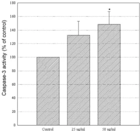
5. Increase of caspase-3 activity by ADE Increase of caspase-3 activity was included in the final step in apoptosis of the cells. In this study, caspase-3 activity was increased to 132.4±
20.7% and 148.4±18.8% of control by ADE of 25 µg/ml and 50 µg/ml, respectively (Fig. 4).
6. Increase of NO by ADE
NO acts as a second messenger in the inflammatory Fig. 3. The decrease of intracellular GSH by ADE.
Experiments were performed using human bronchial epithelial cell line, BEAS-2B. Results represent the average of three different experiments. GSH was calculated as nmol of glutathione per mg of protein and then was presented as the percent age of GSH level in control group (*p<0.01).
Fig. 4. The increase of caspase-3 activity by ADE.
Experiments were performed using human bronchial epithelial cell line, BEAS-2B. Data are represented as the percentage of caspase-3 activity of the control group (*p<0.05).
Fig. 5. The increase of NO by ADE. Experiments were performed using murine peritoneal macrophage cell line, RAW264.7. Cells were treated with ADE for 24 hrs. Data are represented as the percentage of the control group (**p<0.01). (A) Morphological change by ADE, (B) Increase of NO secretion by ADE.
process. In this study, the level of NO was increased to 119±2.8% and 151±7.5% of control by ADE of 25 µg/ml and 50 ug/ml, respectively (Fig. 5B).
7. Change of gene expression by ADE
To investigate the change of gene expression by ADE, we performed a PCR analysis. As shown in Fig. 6, ADE markedly increased the expression of both oxidative stress- (catalase, hypoxia inducible gene, and glutathione-S- transferase) and inflammatory- (TNF alpha, IL- 6, and IL-8) related genes. Furthermore, ADE increasd the expression of the DNA repair gene, p53.
IV. Discussion
The origin of Asian Dust, which generates in the
deserts of China and Mongolia, is sand, and consists primarily of particles greater than PM10.
Therefore, dust collected where the Asian Dust originates contained a large amount of soil-related components. Further, TSP and PM10 concentrations highly increased during Asian Dust events, while PM2.5 concentration barely changed.4)
Many epidemiological studies have suggested that both morbidity and mortality rates due to cardiovascular and respiratory increased during the Asian Dust events.17-19) The health effects of air pollutants depend on both the size and the chemistry of the particles, including gaseous pollutants, persistent organic pollutants, heavy metals, and particulate matter.20,21) In this study, we compared the concentration of PAHs in ADE and NADE. ADE more abundantly contained PAHs, such as anthracene, fluoranthene, pyrene, benzo [g,h,i]perylene, and benzo(a)pyrene. In a previous report, the concentrations of PAHs were also found to be higher during the time of the Asian Dust event than they were during non-Asian Dust events.6) This result suggests that PAHs were attached to Asian Dust particulates during long- range transport. Polycyclic aromatic hydrocarbons (PAHs) are organic compounds that contain two or more fused benzene rings. They come primarily from incomplete combustion of fossil fuels and are released into the atmosphere in both gaseous and aerosol phases, depending on their volatilities.
In addition, we compared the cytotoxicity using both extracts, but a large discrepancy in the cytotoxicity was not observed between ADE and NADE. This result was very different than that using water-soluble extracts taken from the same filters.22) We hypothesized that DCM extracts were lower contained gaseous material than water- soluble extracts, and toxicity of organic components was relatively lower than that of heavy metal.
Air pollutants act as pro-oxidants of lipids and proteins, or as free radicals generators, and promote oxidative stress and inflammatory responses.23) In our study, ADE induced the increase of ROS, the decrease of intracellular GSH, the increase of caspase-3 activity, and the increase of NO secretion.
In addition, ADE increased the expression of both oxidative stress-related genes (i.e., catalase, hypoxia inducible gene, and glutathione-S-transferase) and Fig. 6. The change of gene expression by ADE. Experi-
ments were performed using human bronchial epithelial cell line, BEAS-2B. Cells were treated with the indicated concentrations of ADE for 24 hrs. IL-6: Interleukin-6, HIG: hypoxia inducible gene, IL-8: Interleukin-8, TNF α: tumor necrosis factor α, GST: glutathione S-transferase.
inflammation-related genes (i.e., TNF-α, IL-6, and IL-8). In prior study, water extracts of ultrafine particulate pollutants were found to induce an increase of ROS in BEAS-2B cells,24) and PAHs induced IL-8 expression through nuclear factor-kB activation in A549 cells.25) Inflammatory markers were increased in the lungs and peripheral blood of rats, which was exposed through the concentrated Asian Dust extracts.26) IL-8, a member of a C-X-C chemokine family, is one of the most stable and potent chemotactics, and is produced by a variety of cell types (e.g., monocytes, epithelial and endothelial cells). It also plays an important role in the pathogenesis of airway inflammation.27,28)
Furthermore, ADE increased the expression of the DNA repair gene, p53. Benzo(a)pyrene is a kind of representative of toxicants, carcinogens, and mutagens. Molecular epidemiologists usually consider the spectrum of p53 mutations found in human tumors to be a signature of the corresponding environmental carcinogens.29) Our results are consistent with reports from Pavanello et al.,30) indicating that shorter telomere length in peripheral blood lymphocytes, predictive of lung cancer risk, is associated with chronic PAH exposure.
Consequently, we suggested that the Asian Dust may induce an inflammatory response through oxidative stress. We recommend further research on the subject in the future.
References
1. KMA (Korea meteorological administration), http://
www.kma.go.kr/dust/dust_03_04.jsp
2. KME (Ministry of Environment Republic Korea), 2002. http://w.me.go.kr Annual Report of Ambient Air Quality in Korea. 11-1480000-000532-10.
3. Lee, B. K., Lee, H. K. and Jun, N. Y. : Analysis of regional and temporal characteristics of PM10 dur- ing an Asian dust episode in Korea. Chemosphere, 63, 1106-1115, 2006.
4. Park, E. J., Kim, D. S. and Park, K. : Monitoring of ambient particles and heavy metals in a residential area of Seoul, Korea. Environmental Monitoring and Assessment, 137(1-3), 441-449, 2008.
5. Kim, K. H. and Kim, M. Y. : The effects of Asian Dust on particulate matter fractionation in Seoul, Korea during spring 2001. Chemosphere, 51, 707- 721, 2003.
6. Tamamura, S., Sato, T., Ota, Y., Wang, X., Tang, N.
and Hayakawa, K. : Long-range transport of poly- cyclic aromatic hydrocarbones(PAHs) from the eastern Asian continent to Kanazawa, Japan with Asian dust. Atmospheric Environment, 41, 2580- 2593, 2007.
7. KEI (Korea Environment Institute), http://sdocu.
synap.co.kr/preview/view.php?seq=1021069 8. Kwon, H. J., Cho, S. H., Chun, Y. S., Lagarde, F.
and Pershagen, G. : Effects of the Asian dust events on daily Mortality in Seoul, Korea. Environmental Research, 90(1), 1-5, 2002.
9. Lee, J. T., Son, J. Y. and Cho, Y. S. : A comparison of mortality related to urban air particles between periods with Asian dust days and without Asian dust days in Seoul, Korea, 2000-2004. Environ- mental Research, 105(3), 409-413, 2007.
10. Lauer, F. T., Mitchell, L. A., Bedrick, E., McDonald, J. D., Lee, W. Y., Li, W. W., Olvera, H., Amaya, M.
A., Berwick, M., Gonzales, M., Currey, R., Pin- gitore, N. E., Jr. and Burchiel, S. W. : Temporal-spa- tial analysis of U.S.-Mexco border environmental fine and coarse PM air sample extract activity in human bronchial epithelial cells. Toxicology and Applied Pharmacology, 238(1), 1-10, 2009.
11. Lin, C. C., Chen, S. J., Huang, K. L., Lee, W. J., Lin, W. Y., Tsai, J. H. and Chaung, H. C. : PAHs, PAH-induced carcinogenic potency, and particle- extract-induced cytotoxicity of traffic-related nano/
ultrafine particles. Environmental Science & Tech- nology, 42(11), 4229-4235, 2008.
12. http://www.epa.gov/glnpo/lmmb/methods/gcms5.2.
PDF, 1995. Standard operating procedure for the analysis of PAHs and Atrazine by GC/Ion Trap MS.
SOP# CH-IN-003.1
13. Park, E. J. and Park, K. : Gene expression profiles of cultures rat cardiomyocytes (H9C2) in response to arsenic trioxide at subcytotoxic level and oxi- dative stress, Journal of Health Science, 52(5), 512- 521, 2006.
14. Park, E. J. and Park, K. : Induction of reactive oxy- gen species and apoptosis in BEAS-2B cells by mercuric chloride. Toxicology in Vitro, 21(5), 789- 794, 2007.
15. Park, E. J., Choi, J., Park, Y. K. and Park, K. : Oxi- dative stress induced by cerium oxide nanoparticles in cultured BEAS-2B cells. Toxicology, 245(1-2), 90-100, 2009.
16. Park, E. J., Yi, J., Kim, Y., Choi, K. and Park, K. : Silver nanoparticles induce cytotoxicity by a Tro- jan-horse type mechanism. Toxicology in Vitro, 24(3), 872-878, 2010.
17. Kwon, H., Cho, S. H., Chun, Y., Lagarde, F. and Pershagen, G. : Effects of the Asian dust events on daily mortality in Seoul, Korea. Environmental Research, A90, 1-5, 2002.
18. Lee, J. T., Son, J. Y. and Cho, Y. S. : A comparison of mortality related to urban air particles between periods with Asian dust days and without Asian
dust days in Seoul, Korea, 2000-2004. Environ- mental Research, 105, 409-413, 2007.
19. Chen, Y., Sheen, P., Chen, E., Liu, Y., Wu, T., and Yang, C. : Effects of Asian dust storm events on daily mortality in Taipei, Taiwan. Environmental Research, 95, 151-155, 2004.
20. MacNee, W. and Donaldson, K. : Mechanism of lung injury caused by PM10 and ultrafine particles with special reference to COPD. The European Res- piratory Journal Supplement, 40, 47s-51s, 2003.
21. Oberdorster, G. : Significance of particle parameters in the evaluation of exposure-dose-response rela- tionships of inhaled particles. Inhalation Toxicology, 8, 73-89, 1996.
22. Park, E. J., Kim, D. S. and Park, K. : Monitoring of the distribution of ambient air particles in Seoul using a cascade impactor and the particle toxicity, Environmental Health & Toxicology, 25(2), 99-109, 2010.
23. Kampa, M. and Castanas, E. : Human health effects of air pollution. Environmental Pollution, 151(2), 362-367, 2008.
24. Li, N., Sioutas, C., Cho, A., Schmitz, D., Misra, C., Sempf, J., Wang, M., Oberley, T., Froines, J. and Nel, A. : Ultrafine particulate pollutants induce oxi- dative stress and mitochondrial damage. Environ- mental Health Perspectives, 111(4), 455-460, 2003.
25. Pei, X. H., Nakanishi, Y., Inoue, H., Takayama, K., Bai, F. and Hara, N. : Polycyclic aromatic hydro- carbons induce IL-8 expression through nuclear fac- tor kappaB activation in A549 cell line. Cytokine, 19(5), 236-241, 2002.
26. Lei, Y. C., Chan, C. C., Wang, P. Y., Lee, C. T. and Cheng, T. J. : Effects of Asian dust event particles on inflammation markers in peripheral blood and bronchoalveolar lavage in pulmonary hypertensive rats. Environmental Research, 95(1), 71-76, 2004.
27. Baggiolini, M. : Chemokines in pathology and med- icine. Journal of Internal Medicine, 250(2), 91-104, 2001.
28. Mukaida, N. : Interleukin-8: an expanding universe beyond neutrophil chemotaxis and activation. Inter- national Journal of Hematology, 72(4), 391-398, 2000.
29. Rodin, S. N. and Rodin, A. S. : Origins and selec- tion of p53 mutations in lung carcinogenesis. Sem- inars in Cancer Biology, 15(2), 103-112, 2005.
30. Pavanello, S., Pesatori, A. C., Dioni, L., Hoxha, M., Bollati, V., Siwinska, E., Mielzy ska, D., Bolognesi, C., Bertazzi, P. A. and Baccarelli, A. : Shorter teolmere length in peripheral blood lymphocytes of workers exposed to polycyclic aromatic hydrocar- bons. Carcinognensis, 31(2), 216-221, 2010.
n é