Kor. J. Hort. Sci. Technol. 28(6):996-1002, 2010
Determination of Chimera Types and Ploidy Level of Sports from ‘Campbell Early’ Grape (Vitis labruscana)
Jung Ho Noh1, Kyo Sun Park1, Hae Keun Yun2*, Gyung Ran Do1, Youn Young Hur1, Seung Hui Kim1, Han Chan Lee1, Myung Sang Ryou1, Seo Jun Park1, and Sung Min Jung1
1National Institute of Horticultural & Herbal Science, Suwon 440-706, Korea
2Department of Horticultural Science, Yeungnam University, Gyeongsan 712-749, Korea
Abstract. Flow cytometry (FCM) was used to measure the ploidy level of three different sports from ‘Campbell Early’ (Vitis labruscana) grape. Results of the study showed different ploidy levels. FCM analysis for ‘Campbell Early’ grape which contains 2C DNA diploid cells showed single peak around 35-40 while ‘Kyoho’ grape with 4C DNA tetraploid cells had a different level of 70-80. However, analysis of the sports displayed a histogram with 2 peaks containing both 2C and 4C nuclei. There was no difference in histograms of 2C DNA flesh and pericarp; on the other hand, 4C DNA flesh type of sports had a different histogram from that of the 2C DNA pericarp. Chromosome numbers of diploid (‘Campbell Early’), tetraploid (‘Kyoho’), and three sports were counted under the microscope. ‘Campbell Early’ and ‘Kyoho’ have 38 and 76 chromosomes, respectively. Three different sports are mixoploids with mixtures of diploid and tetraploid cells. Microscopic observations of shoot apical meristems in sports from ‘Campbell Early’ grape were carried out to determine the type of plant chimera. ‘Campbell Early’ grape (diploid) and ‘Kyoho’ grape (tetraploid) showed that both had 2 tunica layers covering corpus cells, while the three different sports had tunica layers showing mostly oblique division. Most cells from ‘Kyoho’ grape were larger than ‘Campbell Early’ grape. Cells from L-2 and L-3 layers of the three sports were similar to ‘Kyoho’ grape in size, although all cells in L-1 surface layer were uniform in size like ‘Campbell Early’ grape. Results of FCM analysis indicated that both normal and polyploid cells could be intermixed in sports and could become mixoploidy consisting of diploid and tetraploid. All sports used in the tests were periclinal chimera plants with two distinct L-1 and L-2 cell layers.
The result of this study suggests that all three sports which originated from ‘Campbell Early’ grape might be 2-4-4 type chimera formation.
Additional key words: flow cytometry, microscopic observation, mixoploidy, periclinal
*Corresponding author: [email protected]
※ Received 12 March 2010; Accepted 2 September 2010. We grateful thank to Kyung Uk Yi for the chromosome observation (Fig. 3).
Introduction
A few polyploid sports which were different from the ploidy level of original diploid varieties have been found in grape vineyards. These mutants have been identified not only through the larger size of grape berries but also by the significantly large, thick, and dark green leaves in contrast to the normal size of diploid variety (Ourecky et al., 1967).
Sports with characteristically large berry size could be one of the most useful genetic materials for tetraploid grape cultivars. The spontaneous polyploidy mutants have chimera with diploid epidermis and tetraploid internal tissues and they are rarely totally tetraploid (Dermen and Scott, 1962). Chimera formation is a very common phenomenon after a certain degree of mutation, whether induced naturally or artificially
(van Harten, 1998). Plant chimeras have genetically different cell groups within the shoot apical meristem (SAM) that give forms to the plant organs. It was reported that grape shoot apex has two tunica layers covering a 3-6 cell depth corpus (Branas, 1957; Chavan and Shah, 1955; Pratt, 1959;
Reichardt, 1955). Generally, dicotyledons have three germ layers, such as: L-1 (the epidermis), L-2 (sub-epidermal tissues which include gametes, leaf initials, and outer part of the cortex), and L-3 (ground tissue and vascular tissues).
Depending on where the polyploidization occurs in a plant, chimeras are distinguished into three different types: sectorial, periclinal, and mericlinal. Periclinal chimeras have a specific type of genetic mosaic in which one or two entire cell layers of the apical meristems are genetically distinct from the adjacent ones (Dermen, 1960). Varying ploidy levels of
chimerism in the cell layers of the grapevine shoot apical meristems was first detected in 1954. The cytochimera of grapevine apical meristems is composed of two genetically distinct tissue layers, L 1 and L 2 (Einset and Pratt, 1954;
Thompson and Olmo, 1963). Although the chimeric state of a few grapevine clones has been previously demonstrated by foreign scientists (Franks et al., 2002; Hocquigny et al., 2004; Thompson and Olmo, 1963), it has not been reported in Korea yet.
Ploidy level, especially that for sports in grapevines varies with chromosomes numbers (Dermen and Scott, 1962). To determine the ploidy level in grapevines through microscopic chromosome counting is complicated due to their high number and smallness in size (McNeilage and Considine, 1989).
Although microscopic chromosome counting for root tip cells had been conventionally conducted to determine the ploidy level (Bennett and Smith, 1976; Dolezel, 1991), this process is laborious and time-consuming (Costich et al., 1993; Dolezel, 1991).
Recently, flow cytometry (FCM) analysis of nuclear DNA content has become an important and extensively used tool to determine the ploidy level in a number of plant species such as Rhododendron spp. (De Schepper et al., 2001), Vaccinium spp. (Costich et al., 1993), and Actinidia spp. (Ollitrault- sammarcelli et al., 1994). The advantages of this method includes convenience, clarity, low cost and efficiency as com- pared to other conventional chromosome counting methods (Arumuganathan and Earle, 1991; Galbraith et al., 1983).
Likewise, the FCM technique can analyze a large number of samples within a short period of time.
This study determined and analyzed the ploidy level and chimera types of sports mutation (SM) of the ‘Campbell Early’ grapes grown in Korea through FCM technique.
Materials and Methods
Sample preparation
Grape cuttings of ‘Campbell Early’, ‘Kyoho’ and three different sports from ‘Campbell Early’ grape were obtained from the Vitis germplasm repository at the National Institute of Horticultural and Herbal Science (NIHHS) in Suwon, Korea for FCM and microscopic observation. The three sports were collected from the vineyard with ‘Campbell Early’ sports at Kimpo (KP-1) and Gapyeong (GP-1, GP-2) in the late 1990s. Dormant cuttings were collected in late autumn, stored at 4-5℃, and used for propagation in spring. Shoot apex and young leaves were collected from the rooted young plants while pericarp and flesh were collected from 8- years old grapevines maintained at the germplasm repository, NIHHS.
FCM analysis
Young leaves, pericarp and flesh tissues of the sports from
‘Campbell Early’ grape cultivar were used for flow cytometric measurement. An approximately 0.5 cm2 disc from each explant was chopped with a sharp razor blade, placed in a Ø55 plastic Petri dish containing HR-A nuclei extraction solution (Partec Nuclei Extraction Solution Kit, Germany) and then sieved- through a 50 µm celltrics disposable filter. The released nuclei suspension was stained with HR-B DAPI solution (Partec High Resolution Staining Kit, Germany). The ratio between HR-A and HR-B solution staining was 1:4 in volume. Five leaf samples were individually collected from each grape cultivar of ‘Campbell Early’ (2x = 38) and ‘Kyoho’ (4x = 76) and were then analyzed using PA Ploidy Analyzer, Partec to get the standard peak of diploid and tetraploid cells. The standard peak recorded was about channel 480-490 relative fluorescent intensity.
Chromosome count
Chromosome preparation and staining were performed as described by Yahata et al. (2006) with modifications. Appro- ximately 1.5 mm long root-tips were excised from young root, immersed in 2 mM 8-hydroxyquinoline at 4℃ for 12 h in dark, and then fixed. The fixed root-tips were digested with an enzyme solution containing 2% Cellulase (Sigma 038K1298), 1% Macerozyme R-200 (Yakult Honsha Co., Ltd., Japan) and 0.3% Pectolyase Y-23 (Kyowa Chemical Products Co., Ltd., Japan) in 10 mM citrate at 37℃ for 45 to 55 minutes. The digested samples were transferred onto slide glasses, smeared with a drop of fixative solution using a fine-pointed forceps, and air dried. Chromosomes were stained with a 2% Giemsa solution for 45 min, rinsed with distilled water, air dried, observed, and photographed under an optical microscope.
Microscopic observation
Cross sections of the apical meristem were observed under light microscope (Lee and Kwon, 2010; Luft, 1973). A 1 mm3 sample was fixed with 2.5% glutaraldehyde in 0.1M sodium phosphate buffer (pH 7.2) for 2 hrs at 4℃. Specimens were then rinsed and post-fixed with 1% osmium tetroxide (in the above buffer) for 2hrs, at 4℃, and kept overnight in phosphate buffer. After fixation, specimens were completely dehydrated using ethyl alcohol with varying grade series (40, 60, 80, 90, 95, and 100% in distilled water, v/v). The pro- cessed tissues were subjected to three changes of propylene oxide, with 15, 15, 30 min. per change, and gradually infil- trated at three hours each with 30, 50, and 100% propylene oxide embedding media, Epon. Specimens were kept overnight in 100% Epon before polymerization at 60℃ for 72hrs,
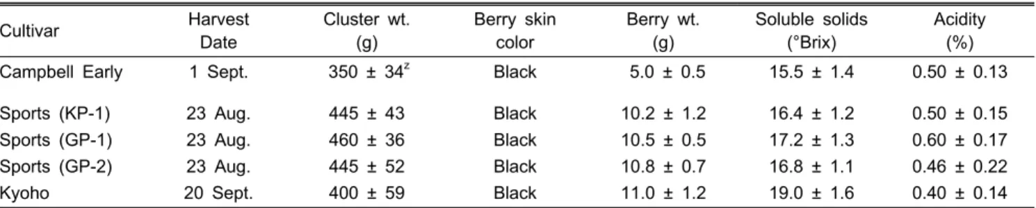
Table 1. Fruit characteristics of grape cultivar ‘Campbell Early’, ‘Kyoho’, and sports from ‘Campbell Early’.
Cultivar Harvest
Date
Cluster wt.
(g)
Berry skin color
Berry wt.
(g)
Soluble solids (°Brix)
Acidity (%)
Campbell Early 1 Sept. 350 ± 34z Black 5.0 ± 0.5 15.5 ± 1.4 0.50 ± 0.13
Sports (KP-1) Sports (GP-1) Sports (GP-2) Kyoho
23 Aug.
23 Aug.
23 Aug.
20 Sept.
445 ± 43 460 ± 36 445 ± 52 400 ± 59
Black Black Black Black
10.2 ± 1.2 10.5 ± 0.5 10.8 ± 0.7 11.0 ± 1.2
16.4 ± 1.2 17.2 ± 1.3 16.8 ± 1.1 19.0 ± 1.6
0.50 ± 0.15 0.60 ± 0.17 0.46 ± 0.22 0.40 ± 0.14
zMean ± SD (n = 3).
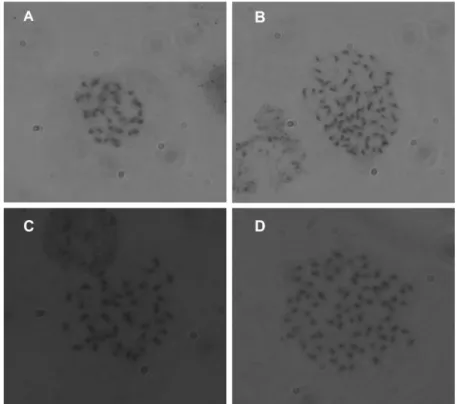
Fig. 1. FCM analysis of DAPI-stained nuclei in leaves of control and different three sports from ‘Campbell Early’ grape. A:
‘Campbell Early’ (2x), B: ‘Kyoho’ (4x), C: KP-1 (2x + 4x), D:
GP-1 (2x + 4x), E: GP-2 (2x + 4x). The ploidy level of each peak is indicated on the top.
sectioned (1,500 nm), with periodic acid staining, and viewed under a Carl Zeiss Axioskop 2 light microscope.
Results
Fruits of sports (KP-1, GP-1, and GP-2) have black skin when fully ripened. Berries were big with average weight of 10.5 g, which is 5.5 g heavier than that of ‘Campbell Early’, and had round shapes. When the grapes are fully ripened, the aroma of sports is similar to that of ‘Campbell Early’. The sports ripen between Aug. 20 and Aug. 25 in Suwon, Korea, which is 7 days earlier than ‘Campbell Early’
and approximately 30 days earlier than ‘Kyoho’. The mean total soluble solids content (TSS) of sports is 16.8 °Brix, which is 1 to 2 °Brix higher than ‘Campbell Early’. The soluble solids content is a little higher than ‘Campbell Early’, but the pH is similar (Table 1). The skin is medium in thickness, not as tough as ‘Campbell early’, and do not firmly stick to the flesh. It has soft flesh texture and juicy. Overall, the three sports were similar to ‘Kyoho’ in terms of berry size and shape, but have similar taste to that of ‘Campbell Early’.
‘Campbell Early’ grape which has diploid DNA cells showed single peak around channel 35-40 (Fig. 1A). ‘Kyoho’
grape with 4C DNA exhibited single peak at delayed position as compared with diploid channel level (Fig. 1B). From the histogram, shown on Fig. 1C to 1E, the sports from ‘Campbell Early’ grape displayed two peaks which contain both the 2C and 4C nuclei.
The FCM analyses for flesh and pericarp tissues of the
‘Campbell Early’ grape fruit displayed clear histograms for all the 2C DNA patterns (Fig. 2A) as well as the 4C DNA patterns in ‘Kyoho’ grape (Fig. 2B). FCM analyses for ‘Campbell Early’ and ‘Kyoho’ grapes showed no difference between flesh and pericarp with both having a clear histogram for the 2C DNA and 4C DNA, respectively. On the other hand, sports divided into two types of parts showed histograms with 2C DNA and 4C DNA. Analyses of sports pericarp exhibited a diploid similar to ‘Campbell Early’ grape while
analyses of sports flesh showed tetraploid similar to ‘Kyoho’
grape (Figs. 2C, D, and E).
Chromosome numbers of the experimental plants were determined under a microscope. ‘Campbell Early’, well known
Fig. 2. Comparison of ploidy levels on pericarp and flesh of fruits by FCM analysis in control and three sports from ‘Campbell Early’ grape. On the left line is histogram of pericarp ploidy level, and flesh in right line. A: ‘Campbell Early’, B: ‘Kyoho’, C:
‘KP-1’, D: ‘GP-1’, E: ‘GP-2’. The ploidy level of each peak is indicated on the top.
Fig. 3. Comparison of chromosome. A: 2x = 38 chromosome of a ‘Campbell Early’, B: 4x = 76 chromosome of a ‘Kyoho’, C: 2x
= 38 chromosome of a sport, D: 4x = 76 chromosome of a sport. Chromosome counts were made under the microscope in root-tip.
Fig. 4. Longitudinal section of shoot apical meristem domes of control with normal constitution. Above ink dotted line is tunica layer and inside the dotted line is corpus layer. A: ‘Campbell Early’, B: ‘Kyoho’. Bars represent 5 µm size.
Fig. 5. Longitudinal section of shoot apical meristem domes of three sports from ‘Campbell Early’ grape with 2-4-4 chimeral constitution.
A: ‘KP-1’, B: ‘GP-1’, C: ‘GP-2’. Above solid line is tunica layer, and the lower part is corpus layer. (dotted-arrow) L-1, above ink dotted line, is 2x and layers inside the dotted line are 4x. Bars represent 5 µm size.
as diploid, has 2x = 38 chromosomes (Fig. 3A) and ‘Kyoho’
grape, belonging to tetraploid, has 4x = 76 chromosome (Fig.
3B). In contrast, sports are mixoploids, mixture of both diploid cells (2x = 38) (Fig. 3C) and tetraploid cells (4x = 76) (Fig. 3D).
Genetically, both diploid ‘Campbell Early’ and tetraploid
‘Kyoho’ grapes have two tunica layers covering a corpus cell in depth (Figs. 4A and B). However, the 3 different sports which originated from ‘Campbell Early’ grape had a tunica
layer with oblique division which is laid under L-1 mutated (Figs. 5A, B, and C). Both ‘Campbell Early’ and ‘Kyoho’
grapes had uniform tissues in ploidy while in terms of cell size, ‘Kyoho’ grape was larger than ‘Campbell Early’ grape (Fig. 4). For the sports, a difference in dome cell size was observed between the outmost and inner layers of the apical meristem (Fig. 5). Without exception, cell size in all tissues below the surface layer were similar to tetraploid ‘Kyoho’
grape (inside the dotted line); however, cell size in tissues in the outmost layer were similar to diploid ones ‘Campbell Early’ grape (above dotted line). All sports used for the tests showed to be periclinal chimera plants, composed of two distinct cell layers (L-1 and L-2).
Discussion
FCM has been proven to be a simple technique used to detect the ploidy level in grapevine, as previously reported by Lodhi and Reisch (1995). The method has been effective and faster than the conventional way of counting chromosomes or measuring stomata length (Loureiro et al. 2005; O’Brien et al., 1996; Ollitrautt-Sammarcelli et al., 1994; Ozaki et al., 1988; Pinheiro et al., 2000; Sgorbati et al., 1986; Tosca et al., 1995; Vainola, 2000). Maertens (1990) was able to identify the ploidy level of cytochimeras fruit mutations in his preliminary work in pear using FCM. This technique, which was also used in this study to detect ploidy level of three sports from ‘Campbell early’ grape, revealed that all the sports used showed distinct 2 peak histograms for both 2C and 4C ploidy levels, between the fruits flesh and pericarp tissues, respectively. The observed discrepancy in the location of ploidy peaks' for the leaves and fruits was attributed to the difference of sample type or cell activity.
Although chromosome counting under a microscope is inefficient (Costich et al., 1993; Dolezel, 1991), we carried out a microscopic observation for a definite decision on chromosome numbers of these FCM analysis materials. The results appear to support the FCM analysis results.
Results of the study indicated that cells can be intermixed with different ploidy and could become mixoploidy, consisting of diploid and tetraploid in sports from ‘Campbell Early’
grape. However, although 2-4-4 chimera of three sports was identified through FCM analysis, further study is needed to determine the chimera type with histological differences in cell size using the microscope.
Previous works on grapevines reported typical tunica and corpus in shoot apical meristem having 2 tunica layers covering 3-6 corpus cell layers in depth (Chavan and Shah, 1955;
Pratt, 1959; Thompson and Olmo, 1963). The tunica consists of small populations of undifferentiated meristematic cells (Fletcher, 2002).
Similarly, this study revealed that original grapevines had two normal tunica layers covering corpus cells. However, the three different sports which originated from ‘Campbell Early’ grape had tunica layer of oblique structure. The tunica layer of sports showed not only a general cell arrangement of tunica layer but also a cell arrangement of corpus. Actually, L-2 layer of sports had the cell which divided in all directions.
Pratt (1959) pointed out that the types of chimeras described in grapes were not consistent with this type of shoot apex.
Therefore, further study should be undertaken to provide a clear morphology of the single tunica layer in ploidy chimera grapevine. The sports used in this experiment showed different cell sizes between the outmost and inner layers. All sports considered in this study were periclinal chimera plants, composed of two distinct cell layers (diploid L-1 and tetraploid L-2). Thus, the larger size berry exhibited by the sports as compared to the original plant could be due to the tetraploid mutation which occurred from the corpus layer of the fruit flesh tissue. Einset and Barbara (1951) reported that most sports were found to be chimeras, when a number of large- fruited natural sports of grapes were studied cytohistologically.
The results of microscopic observations greatly supported FCM data that all the three sports which originated from
‘Campbell Early’ grape could be 2-4-4 type in chimera formation.
Literature Cited
Arumuganathan, K. and E.D. Earle. 1991. Estimation of nuclear DNA content of plants by flow cytometry. Plant Mol. Biol.
Rep. 9:229-241.
Bennett, J. and B. Smith. 1976. Nuclear DNA amounts in angio- sperms. Phil. Trans. Roy. Soc. Lond. Ser. 274:227-274.
Branas, J. 1957. Sur quelques donnees ontogeniques. Progr. Agric.
Viticole. 148:58-67.
Chavan, A.R. and J.J. Shah. 1955. Origin and development of the vegetative axillary bud in Vitisrepens. W & A Indian Sci.
Cong. Asso. Proc. 42:225.
Costich, D.E., R. Ortiz, T.R. Meagher, L.P. Bruederle, and N.
Vorsa. 1993. Determination of ploidy level and nuclear DNA content in blueberry by flow cytometry. Theor. Appl. Genet.
86:1001-1006.
Dermen, H. 1960. Nature of plant sports. Amer. Hort. Mag.
39:123-173.
Dermen, H. and D.H. Scott. 1962. Potential in colchiploid grape.
Economic Botany 16:77-85.
De Schepper, S., L. Leus, E. Mertens, E. Van Bockstaele, M.
De Loose, P. Debergh, and J. Heursel. 2001. Flow cytometric analysis of ploidy in Rhododendron (subgenus Tsutsusi).
HortScience 36:125-127.
Dolezel, J. 1991. Flow cytometric analysis of nuclear DNA content in higher plants. Phytochem. Anl. 2:143-154.
Einset, J. and L. Barbara. 1951. Chimeral sports of grape. J.
Hered. 42:158-162.
Einset, J. and C. Pratt. 1954. “Giant” sports of grapes. Proc. Amer.
Soc. Hort. Sci. 63: 251-256.
Fletcher, J.C. 2002. Coordination of cell fate decisions and cell proliferation in the shoot apical meristem of angiosperm plants. BioEssays. 24:27-37.
Franks, T., R. Botta, and M.R. Thomas. 2002. Chimerism in grapevines: implications for cultivar identity, ancestry and genetic improvement. Theor. Appl. Genet. 104:192-199.
포도 ‘캠벨얼리’ 품종에서 발생한 아조변이체의 배수성 및 키메라 형태 검정
노정호1ㆍ박교선1ㆍ윤해근2*ㆍ도경란1ㆍ허윤영1ㆍ김승희1ㆍ이한찬1ㆍ류명상1ㆍ박서준1ㆍ정성민1
1국립원예특작과학원, 2영남대학교 원예학과 (*교신저자)
초 록. ‘캠벨얼리’ 아조변이체의 배수성을 flow cytometry(FCM)를 이용하여 검정하였다. 2배체 포도인 ‘캠벨얼리’와 4배체 포도인 ‘거봉’과는 달리 ‘캠벨얼리’에서 발생한 아조변이 3계통의 경우, 잎에서 2배체와 4배체 peak가 동시에 나타났다.
또한 ‘캠벨얼리’ 포도의 과육과 과피의 배수성은 2배체 peak를 보인 반면, ‘캠벨얼리’ 아조변이체는 과피의 배수성은 2배체로, 과육의 배수성은 4배체로 검정되었다. 정확한 염색체 숫자를 알아보기 위해 ‘캠벨얼리’ 유래 아조변이체, ‘캠벨어리’, ‘거봉’
포도의 근단 조직 염색체 숫자를 현미경 검경하였다. ‘캠벨얼리’와 ‘거봉’은 각각 38, 76개의 염색체로 나타난 반면 아조변이체
는 2x=38, 4x=76개의 염색체가 혼재한 것으로 나타났다. ‘캠벨얼리’ 아조변이체의 키메라 형태를 알아보기 위하여 신초 정단 분열조직을 현미경 관찰하였다. 대조구로 이용된 2배체 ‘캠벨얼리’와 4배체 ‘거봉’ 포도는 2개의 tunica 층이 corpus 층을 덮고 있었으며, 반면에 아조변이체의 경우 첫번째 tunica 층이 독특하게 분할되어 있었다. ‘거봉’ 포도의 정단분열 조직의 세포 크기는 대체로 ‘캠벨얼리’ 포도보다 더 컸다. 아조변이체의 L-2, L-3 층을 이루고 있는 세포 크기는 4배체인
‘거봉’ 세포와 유사하였으며 가장 바깥쪽 L-1층을 이루고 있는 세포의 크기는 ‘캠벨얼리’ 포도와 유사한 크기였다. 따라서 본 시험의 결과 ‘캠벨얼리’ 포도 품종에서 발생한 아조변이체들은 2-4-4형 키메라의 가능성이 높은 것으로 판단되었다.
추가 주요어 : flow cytometry, 조직검경, mixoploidy, 주연 키메라 Galbraith, D.W., K.R. Harkins, J.M. Maddox, N.M. Ayres, D.P.
Sharma, and E. Firozabady. 1983. Rapid flow cytometric analysis of the cell cycle in intact plant tissues. Science 220:1049-1051.
Hocquigny, S., F. Pelsy, V. Dumas, S. Kindt, M.-C. Heloir, and D. Merdinoglu. 2004. Diversification within grapevine cultivars goes through chimeric states. Genome 47:579-589.
Lodhi, M.A and B. Reisch. 1995. Nuclear content of Vitis species, cultivars, and other genera of the Vitaceae. Theor.
Appl. Genet. 90:11-16.
Loureiro, J., G. Pinto, T. Lopes, C. Dolezel, and C. Santos. 2005.
Assessment of ploidy stability of somatic embryogenesis process in Quercus suber L. using flow cytometry. Planta 221:815-822.
Luft, J.H. 1973. Compounding of Luft’s epon embedding medium for use in electron microscopy with reference to anhydride:
Epoxide ratio adjustment. Mikroskopie 29:337-342.
Lee, B.H. and Y.H. Kwon. 2010. Anatomical changes and ano- thocyanin contents of the exocarp by ethyl oleate treatment on
‘Merlot’ grapes. Kor. J. Hort. Sci. Technol. 28:370-373.
Maertens, P. 1990. Les nouvelles varietes de poires belges: des mutants diploides? Le Fruit Belge. 431:217-222.
McNeilage, M.A. and J.A. Considine. 1989. Chromosome studies in some Actinidia taxa and implication for breeding. N.Z.J.
Bot. 27:71-81.
O’Brien, I.E.W., D.R. Smith, R.C. Gardner, and B.G. Murray.
1996. Flow cytometric determination of genome size in Pinus.
Plant Sci. 115:91-99.
Ollitrault-Sammarcelli, F., J.M. Legave, N. Michaux-Ferriere, and A.M. Hirsch. 1994. Use of flow cytometry for rapid determination of ploidy level in the genus Actinidia. Sci.
Hortic. 57:303-313.
Ourecky, D.K., C. Pratt, and J. Einset. 1967. Fruiting behavior
of large-berried and large-clustered sports of grape. Proc.
Amer. Soc. Hort. Sci. 91:217-223.
Ozaki, Y., K. Narikyo, M. Hiramatsu, K. Ureshino, and H. Okubo.
1988. Application of flow cytometry for rapid determination of ploidy levels in asparagus (Asparagus officinalis L.). J.
Fac. Agric. Kyushu Univ. 43:83-88.
Pratt, C. 1959. Radiation damage in shoot apices of Concord grape. Amer. J. Bot. 46:103-109.
Pinheiro, A.A, M.T. Pozzobon, and V.T.C. Carneiro. 2000. Dupli- cation of the chromosome number of diploid Brachiaria brizantha plants using colchicine. Plant Cell Rep. 19:274-278.
Reichardt, A. 1955. Experimentelle untersuchungen über den effect von röntgenstrahlen in der vegetativen vermehrung einer alten rebensorte. Die Gartenbauwissenschaft. 20:355-413.
Sgorbati, S., M. Levi, E. Sparvoli, F. Trezzi, and G. Lucchini.
1986. Cytometry and flow cytometry of 4’, 6-diamidino-2- phenyldole (DAPI) stained suspensions of nuclei released from fresh and fixed tissues of plants. Physiol. Plant. 68:471-476.
Thompson, M.M. and H.P. Olmo. 1963. Cytohistological studies of cytochimeric and tetraploid grapes. Amer. J. Bot. 50:901- 906.
Tosca, A., R. Pandolfi, S. Citterio, A. Fasoli, and S. Sgorbati.
1995. Determination of flow cytometry of the chromosome doubling capacity of oryzalin and colchicine in gynogenetic haploids of gerbera. Plant Cell Rep. 14:455-458.
Vainola, A. 2000. Polyploidization and early screening of Rhododendron hybrids. Euphytica. 112:239-244.
van Harten, A.M. 1998. Mutation breeding: Theory and Practical Applications. Cambridge University Press. p. 26-27.
Yahata, M., H. Kunitake, K. Yasuda, K. Yamashita, H. Komatsu, and R. Matsumoto. 2006. Production of sexual hybrid pro- genies for clarifying the phylogenic relationship between Citrus and Citropsis species. J. Amer. Soc. Hort Sci. 131:764-769.