Received: August 20, 2019 Revised: October 15, 2019 Accepted: November 13, 2019 CliniCAl
neurophysiology
Correspondence to Jung-Ju Lee
Department of Neurology, Nowon Eulji Medical Center, Eulji University College of Medicine, 68 Hangeulbiseok-ro, Nowon-gu, Seoul 01830, Korea
Tel: +82-2-970-8312 Fax: +82-2-970-8862 E-mail: [email protected]
Febrile Hashimoto’s encephalopathy mimicking infectious encephalitis
Jung-Ju Lee, Michelle Sojung Youn, Jong-Moo Park, Ohyun Kwon, Woong-Woo Lee, Kyusik Kang, Byung Kun Kim
Department of Neurology, Eulji Medical Center, Eulji University College of Medicine, Seoul, Korea
Hashimoto’s encephalopathy (HE) is a heterogeneous encephalopathy with diverse clinical presentations. Here we report on a 69-year-old woman who presented with confusion, apha- sia, fever, and focal ictal discharges. Cerebrospinal fluid analysis and a workup for other fever origins revealed no abnormality and a high level of thyroperoxidase antibody was detected, which findings led to a diagnosis of HE. The symptoms subsided after treatment. This study highlights the importance of considering HE in patients presenting with fever and abnormal EEG findings.
Key words: Electroencephalography; Encephalopathy; Fever
Hashimoto’s encephalopathy (HE) is a rare neurological disorder with clinical manifesta- tions that vary between patients. This clinical diversity makes diagnosis difficult, and leads to frequent misdiagnoses. Symptoms of HE include disorders of consciousness, impair- ment of cognitive function, confusion, stroke-like episodes, and seizures.1,2 HE can be di- vided into two subtypes: 1) HE with diffuse encephalopathy that shows encephalopathic symptoms without focal neurological abnormalities, and 2) HE with focal encephalopathy that shows focal neurological abnormalities. However, high antithyroid antibody levels and responsiveness to immunotherapy are common in both HE subtypes.2 Electroen- cephalography (EEG) findings also vary between individual patients and may also depend on the clinical stage of the disease.3 Here we describe a patient with HE who displayed symptoms mimicking infectious encephalitis such as fever, aphasia, and focal EEG abnor- mality. We also discuss the possible pathomechanisms and clinical significance of HE.
CASE
A 69-year-old woman with diabetes visited the emergency department of the author’s hospital with sudden-onset confusion and aphasia. These symptoms began during a
ORCID Jung-Ju Lee
https://orcid.org/0000-0001-7498-5656 Michelle Sojung Youn https://orcid.org/0000-0002-6774-3234 Jong-Moo Park
https://orcid.org/0000-0002-4199-3024 Ohyun Kwon
https://orcid.org/0000-0001-9806-814X Woong-Woo Lee
https://orcid.org/0000-0002-8767-7967 Kyusik Kang
https://orcid.org/0000-0002-4021-4439 Byung Kun Kim
https://orcid.org/0000-0003-1053-4682
telephone call with an acquaintance, where the patient suddenly forgot who she was talking to and responded in- adequately. The acquaintance recommended for the patient to consume orange juice, but after her symptoms had per- sisted for longer than 3 hours she decided to visit a hospital.
At presentation her vital signs were stable except for a fever of 39.1°C, while a neurological examination showed confu-
sion with impaired comprehension, repetition, and naming without other focal neurological abnormalities. Her body temperature fluctuated between 37.5°C and 39.0°C over a 10-hour period, which was intermittently controlled by administering acetaminophen. The confusion with aphasia persisted after admission.
Tests for the blood count, erythrocyte sedimentation rate,
Fig. 1. Brain fluid-attenuated inversion recovery magnetic resonance imaging (MRI) (A, B) and diffusion-weighted MRI (C, D) showing no abnormalities in the left frontotemporal area.
A B C D
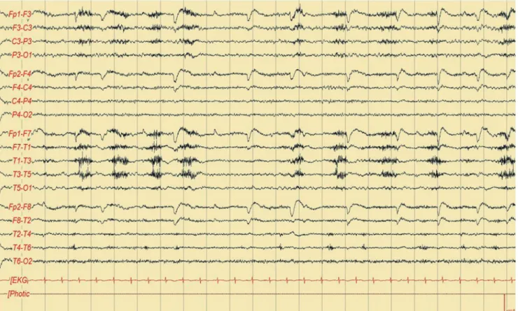
Fig. 2. Initial electroencephalography findings showing intermittent rhythmic and quasi-rhythmic slow waves in the left frontotemporal area with fluctuating amplitude and frequency. EKG, electrocardiography.
liver and kidney functions, coagulation screening, C-reactive protein, serum electrolytes, urinalysis, and antinuclear anti- body yielded normal results. Magnetic resonance imaging that included fluid-attenuated inversion recovery and diffu- sion-weighted imaging modalities revealed no significant abnormality (Fig. 1). EEG performed on the day after admis- sion showed intermittent rhythmic and quasi-rhythmic slow waves with a fluctuating frequency in the left hemisphere (Fig. 2).
Based on the above-described clinical and EEG manifes- tations, we performed a cerebrospinal fluid (CSF) test to exclude the possibility of infectious encephalitis. However, the CSF examination revealed no pleocytosis, and her CSF protein and glucose levels were within the reference limits (45 mg/dL and 64 mg/dL, respectively). Polymerase chain reaction analysis for Mycobacterium tuberculosis and herpes simplex virus also produced negative results. No specific inflammatory lesion or tumor was observed on computed tomography (CT) scans of her chest and abdomen, and no
bacterial or fungal growth was observed in the sputum, blood, urine, or CSF culture. While a thyroid function test showed no specific abnormality, the levels of antibodies against thyroperoxidase were significantly higher (>1,300 IU/
mL) than the normal value of <60 IU/mL.
Valproic acid was administered intravenously at a load- ing dose of 1,000 mg followed by a maintenance dose of 250 mg twice daily from the second day of admission. Her fever subsided after the antiepileptic treatment, while her cognitive symptoms improved considerably. However, mild confusion and incomprehension still persisted despite the disappearance of focal epileptic discharges in the EEG performed 4 hours after the initial EEG (Fig. 3). The patient’s score on the Korean version of the Mini Mental State Ex- amination (K-MMSE) was 16 on the third day of admission.
After administering oral prednisolone at 30 mg once daily from the fifth day of admission, she gradually recovered to her premorbid state. Her K-MMSE score was 26 on the tenth day. The aphasia and confusion had not recurred up to the
Fig. 3. Follow-up electroencephalography (EEG) performed at 4 hours after the initial EEG showing that the asymmetric rhythmic and quasi-rhythmic discharges observed in the initial EEG had nearly disappeared. EKG, electrocardiogram.
time of discharge at 3 months after initiating low-dose med- ication of prednisolone (5 mg once daily) and valproic acid (250 mg twice daily).
DISCUSSION
HE is a rare but very serious disease. Its incidence and preva- lence have not been elucidated due to diagnosis difficulties associated with the absence of specific clinical features and/
or neuroimaging findings.2 Despite high titers of antithyroid antibodies observed in patients, the thyroid function test shows little to no abnormality.1,2 Clinical suspicion is the only helpful method for an early diagnosis, which is crucial for preventing the significant neurological sequelae. Definitive diagnostic criteria for HE have not been established, but the following criteria are commonly applied: clouding of con- sciousness with reduced wakefulness, attention, or cognitive function; no evidence of CSF infection; and a high concen- tration of antithyroid antibodies (microsomal, thyroperoxi- dase, or thyroglobulin antibodies).2 Applying these criteria meant that the clinical and laboratory findings in our patient were compatible with HE.
Cases of fever caused by HE are rare. Fever may be caused by complications such as aspiration pneumonia or urinary tract infection in patients with altered mentality. However, despite our patient presenting with a fever, bacterial growth was not observed in the blood, sputum, or urine culture. Fur- thermore, no inflammatory lesion was observed on CT scans.
There is only one case report of febrile HE in the literature, and the underlying mechanism remains unclear.4 One possi- ble mechanism is that the increased tumor necrosis factor-a observed in autoimmune thyroiditis acts as an endogenous pyrogen in patients with HE.5 Another possible mechanism is that the fever is associated with complex partial seizures;
this is because the hypothalamic thermoregulatory center is thought to be involved during seizure propagation and may act as a symptomatogenic zone.6-8 The fever in our patient subsided after applying antiepileptic treatment. Therefore, while both mechanisms may contribute to febrile episodes, the latter mechanism seemed to be a more likely cause for the fever observed in our patient.
The EEG findings in our patient showed rhythmic and quasi-rhythmic slowing with fluctuation in the left hemi-
sphere. No definite evolution in the pattern (increase in volt- age with increase or decrease in frequency) or location was observed in the EEG of our patient. However, the fluctuation of rhythmic and quasi-rhythmic discharges was abolished after administering an antiepileptic drug, which meets the diagnostic EEG criteria for nonconvulsive status epilepticus (NCSE).9 Short-acting benzodiazepine is commonly used for detecting EEG changes and diagnosing NCSE. However, intravenous valproic acid can be substituted for benzodiaz- epine because its concentration peaks within a few minutes and so EEG changes can be detected rapidly after its admin- istration.
EEG findings may also lead to the misdiagnosis of HE as an infectious encephalitis such as herpes simplex encephalitis (HSE) due to focal epileptiform discharges with focal slowing typically being found in patients with HSE.10 However, unlike HSE, the EEG traces of HE are heterogeneous, with focal, generalized slowing, periodic discharges, or even triphasic waves typical of metabolic encephalopathy.4 Confirming a diagnosis of febrile HE showing focal EEG abnormalities therefore requires a CSF examination in order to exclude the possibility of HSE.
In conclusion, diagnosing febrile HE is challenging, and necessitates CSF analysis and antithyroid antibody tests to ensure accuracy. After excluding infectious encephalitis, the early administration of corticosteroids and controlling the seizures associated with HE are crucial interventions for suc- cessful treatment. Furthermore, the concentration of anti- thyroid antibodies should be checked when diagnosing HE, even in patients showing focal encephalopathy with fever.
Conflicts of Interest
The authors have no financial conflicts of interest.
REFERENCES
1. Brain L, Jellinek E, Ball K. Hashimoto’s disease and encephalopa- thy. Lancet 1966;2:512-514.
2. Chong JY, Rowland LP, Utiger RD. Hashimoto encephalopathy:
syndrome or myth? Arch Neurol 2003;60:164-171.
3. Henchey R, Cibula J, Helveston W, Malone J, Gilmore RL. Elec- troencephalographic findings in Hashimoto’s encephalopathy.
Neurology 1995;45:977-981.
4. Papathanasopoulos P, Mallioris K, Karanasios P, Dimopoulos D, Papapetropoulos T. Febrile Hashimoto’s encephalopathy. J Neu- rol Neurosurg Psychiatry 2000;68:795.
5. Kayser L, Broholm H, Francis D, Perrild H, Olsen BE, Bendtzen K, et al. Immunocytochemical localisation of tumour necrosis factor alpha in thyroid tissues from patients with neoplastic or autoim- mune thyroid disorders. Autoimmunity 1996;23:91-97.
6. Semel JD. Complex partial status epilepticus presenting as fever of unknown origin. Arch Intern Med 1987;147:1571-1572.
7. el-Ad B, Neufeld MY. Periodic febrile confusion as a presen- tation of complex partial status epilepticus. Acta Neurol
Scand 1990;82:350-352.
8. Matsuda N, Akanuma J, Shimizu S, Shibano K, Nakahara T, Sugi- yama Y, et al. Recurrent episodes of fever of unknown origin as temporal lobe epilepsy. Rinsho Shinkeigaku 2000;40:999-1002.
9. Leitinger M, Beniczky S, Rohracher A, Gardella E, Kalss G, Qerama E, et al. Salzburg Consensus Criteria for Non-Convulsive Status Epilepticus–approach to clinical application. Epilepsy Behav 2015;49:158-163.
10. Ch’ien LT, Boehm RM, Robinson H, Liu C, Frenkel LD. Character- istic early electro-encephalographic changes in herpes simplex encephalitis. Arch Neurol 1977;34:361-364.