© 2014 The Korean Ophthalmological Society
This is an Open Access article distributed under the terms of the Creative Commons Attribution Non-Commercial License (http://creativecommons.org/licenses /by-nc/3.0/) which permits unrestricted non-commercial use, distribution, and reproduction in any medium, provided the original work is properly cited.
451 Original Article
Changing Trends in Surgery for Retinal Detachment in Korea
Ga Eun Cho1, Seong Wook Kim2, Se Woong Kang1; Korean Retina Society
1Department of Ophthalmology, Samsung Medical Center, Sungkyunkwan University School of Medicine, Seoul, Korea
2Doctor Lee’s Clinic, Suwon, Korea
Purpose: To analyze trends in rhegmatogenous retinal detachment (RRD) surgery among the members of the Korean Retina Society from 2001 to 2013.
Methods: In 2013, surveys were conducted by email and post to investigate the current practice patterns re- garding RRD treatment. Questions included how surgeons would manage six cases of hypothetical RRD. Re- sults were compared to those reported in 2001.
Results: A total of 133 members (60.7%) in 2013 and 46 members(79.3%) in 2001 responded to the survey.
Preference for pneumatic retinopexy has decreased in uncomplicated primary RRD (p = 0.004). More re- spondents in 2013 selected vitrectomy as the primary procedure when mild vitreous hemorrhage (p = 0.001), myopia (p = 0.044) and history of successful scleral buckling on the fellow eye (p = 0.044) were added to the primary scenario. Vitrectomy was over twice as popular in cases of pseudophakic, macula-off RRD with pos- terior capsular opacity (p = 0.001).
Conclusions: For RRD with myopia, pseudophakia and media opacity, surgical interventions over the last de- cade have drastically shifted from scleral buckling and pneumatic retinopexy to vitrectomy.
Key Words: Rhegmatogenous retinal detachment, Pneumatic retinopexy, Scleral buckling, Surgery, Vitrectomy
Scleral buckling (SB) has been considered the standard of care for rhegmatogenous retinal detachment (RRD).
Use of vitrectomy has historically been limited to compli- cated retinal detachment cases [1], including those with vitreous hemorrhage, posteriorly located retinal breaks, gi- ant retinal tears and severe proliferative vitreoretinopathy.
Although vitrectomy was frequently performed with SB, previous studies showed vitrectomy alone was as effective
as vitrectomy with SB [2,3]. Growing evidence has ex- panded the indication for vitrectomy in RRD, such as in uncomplicated RRD without proliferative vitreoretinopa- thy or giant retinal tears [4,5].
A few randomized clinical trials have attempted to com- pare the outcomes of vitrectomy and SB in RRD in order to provide guidelines regarding indications for each technique [6-8]. Grey areas, however, still remain, necessitating a sur- geon’s judgment. The decision of which surgical technique to use depends on a variety of factors, including the loca- tion and number of retinal breaks, lens status, the patient’s compliance and the surgeon’s preference. Surveys have been used to study vitreoretinal surgeons’ RRD surgery preferences [9,10]. Vitrectomy, however, was not included as an option in previous studies, because the primary objec- tive of these studies was to evaluate the popularity of PR.
Received: January 28, 2014 Accepted: February 24, 2014
Corresponding Author: Se Woong Kang, MD. Department of Ophthal- mology, Samsung Medical Center, Sungkyunkwan University School of Medicine, #81 Irwon-ro, Gangnam-gu, Seoul 135-710, Korea. Tel: 82-2- 3410-3562, Fax: 82-2-3410-0712, E-mail: [email protected]
The results of this study were presented at the 109th annual meeting of the Korean Ophthalmological Society.
452
We assumed that recent improvements in instruments and sutureless vitrectomy would increase a surgeon’s pref- erence for vitrectomy in primary RRD repairs. We con- ducted a survey of the preferences for primary RRD sur- gery among Korean vitreoretinal specialists in 2001 and reported our results then [11]. In this current study, we re-surveyed the practice patterns of vitreoretinal special- ists for the repair of RRD in Korea in 2013, and analyzed changing trends in RRD surgery by comparing the results from the two surveys.
Materials and Methods
Survey
In 2013, a survey was conducted among the 219 mem- bers of Korean Retina Society via e-mail and post contain- ing the same two scenarios and six questions as the previ- ous survey that was administered in 2001 [11]. The primary scenario was adopted from studies performed in 1990 and 1997 in the USA [9,10].
1) The primary scenario
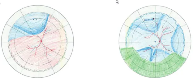
You are a 48-year-old ophthalmologist with a visual acu- ity of 20 / 20 in both eyes, emmetropia, and no known sys- temic or ophthalmic diseases. Localized RRD in your right eye developed with two small horseshoe tears at 11:30, and the macula was on, without signs of proliferative vitreoret- inopathy (Fig. 1A).
Question 1: The first question asked was “What kind of
surgical method would you like to have performed on you?” To further specify the preferences of surgical meth- od, the following clinical variables were added to the pri- mary scenario. Question 2: In addition to the primary sce- nario, what if there is lattice degeneration, combined with a horseshoe tear at 6 o’clock, in an attached retina, and a family history of rhegmatogenous retinal detachment?
Question 3: In addition to the primary scenario, what if there is a mild vitreous hemorrhage, and decreased visual acuity of 20 / 50? Question 4: In addition to the primary scenario, what if your refraction is -3.00 sphere in both eyes? Question 5: In addition to the primary scenario, what if you underwent a successful SB procedure in the other eye 15 years prior?
2) The second scenario
The second scenario was a case of pseudophakic RRD that developed two weeks prior in a 61-year-old woman who presented with decreased vision in her right eye. Her best corrected visual acuity was counting fingers in the right eye, and 20 / 20 in the left. On the ophthalmic exam, her right eye was pseudophakic, and there was total retinal detachment with lattice degeneration, accompanied by a horseshoe tear, and a retinal hole near 11:30. The inferior fundus was partially obscured by posterior capsular opaci- ty (Fig. 1B). The following question was presented: what kind of surgical method would you choose if this patient came to your clinic? (question 6).
Throughout these questionnaires, respondents were asked to choose one treatment among SB, PR, vitrectomy, or other, and were allowed to write specific procedures
Second scenario
*
2001 2013
Combination method Vitrectomy Pneumatic retinopexy Scleral buckling 37.0
13.0
50.0
15.0
77.5
7.5 Primary scenario
*
2001 2012 2001 2012
Mild vitreous hemorrhage
*
Myopia
*
Successful SB on fellow eye
*
Family history, additional tear
Combi Vit PR SB
34.8
2.2 17.4 4.3
76.1
6.0 2.2
19.6
78.3
9.0 7.5
83.5
2.2 21.7
76.1
15.8 14.3
69.9 45.1
7.5 46.6
9.8 4.3
10.9
84.8
6.0 12.0 6.0
75.9 15.8
65.2 74.4
A B
Fig. 1. Fundus drawings of the primary scenario (A) and the second scenario (B) presented in the questionnaire.
453 when they selected ‘other.’
Also, we asked the surgeon’s years since vitreoretinal fel- lowship and his or her average percentage of patients with vitreoretinal disease in their clinic. With these data, career index was obtained for each respondent. Career index was defined as years after fellowship multiplied by average per- centage of patients with vitreoretinal diseases in their clinic, which was divided by 100. There were 27 members who participated in the surveys for both 2001 and 2013. Subgroup analysis was performed to evaluate changes in their answers to the questionnaires over the 12-year period.
Statistical analysis
All the procedures were categorized into four surgical methods: SB, pneumatic retinopexy (PR), vitrectomy, and
the combination method (Combi), which is a combination of SB or encircling and vitrectomy. For example, SB with cryotherapy or laser treatment was regarded as SB, and vitrectomy with silicone oil or gas injection as vitrectomy.
The chi-square test and Fisher’s exact test, with a permuta- tion method for multiple comparisons, were used to ana- lyze the shift in the proportion of surgical procedures used between 2001 and 2013. The respondents were divided into younger/older generation and inexperienced/experienced surgeons by median years post fellowship and median val- ue of career index, respectively. Then, their answers for each question were compared. Asymptotic marginal-ho- mogeneity tests were used to analyze changes in responses in the subgroups of respondents who participated in both surveys.
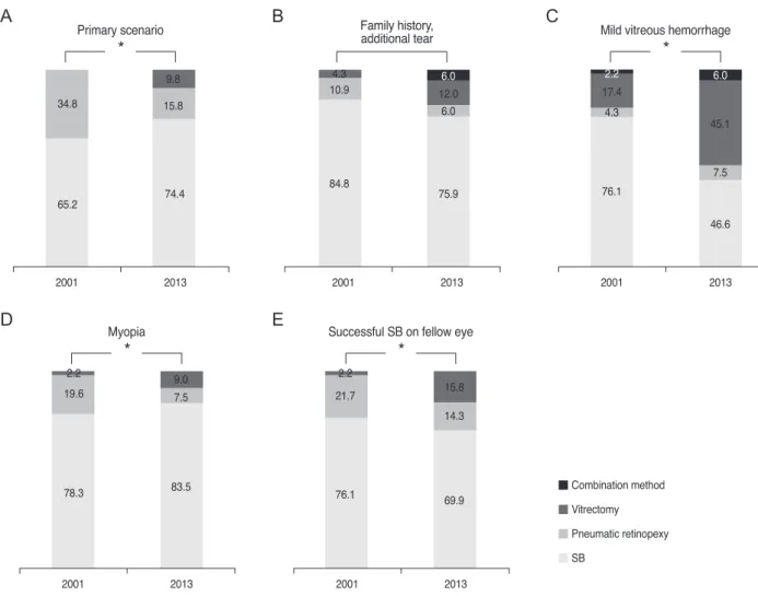
Fig. 2. Comparison of the results for questions 1 to 5 regarding the primary scenario (A-E) between 2001 and 2013. SB = scleral buckling.
*p < 0.05 by chi-square test.
Primary scenario
*
2001 2013 2001 2013
Mild vitreous hemorrhage
*
2001 2013
Myopia
*
2001 2013
Successful SB on fellow eye
*
2001 2013
Family history, additional tear
34.8
2.2 17.4
4.3
76.1
6.0
2.2 19.6
78.3
9.0 7.5
83.5
2.2 21.7
76.1
15.8 14.3
69.9
45.1 7.5 46.6
9.8 4.3
10.9
84.8
6.0 12.0
6.0
75.9 15.8
65.2 74.4
Combination method Vitrectomy Pneumatic retinopexy SB
A B C
D E
Results
Out of a total of 219 investigators who received the ques- tionnaire, 133 (60.7%) members responded, compared with 46 (79.3%) in 2001. Mean years since fellowship was lon- ger in 2013 (mean ± standard deviation, 12.9 ± 8.3) than in 2001 (10.0 ± 7.0, p = 0.024). The proportion (%) of patients with vitreoretinal disease was similar between both sur- veys (78.9 ± 2.4 in 2001 vs. 79 ± 2.2 in 2013, p = 0.973).
There was no significant difference in career index be- tween the two surveys (8.3 ± 6.9 in 2001 vs. 10.3 ± 7.7 in 2013, p = 0.091).
For the primary scenario, 74.4% chose SB, 15.8% chose PR and 9.8% chose vitrectomy in 2013. In the analysis of
the proportional change of each technique between the 2001 and 2013 surveys, the proportion of PR decreased significantly (34.8% in 2001 vs. 15.8% in 2013), compared to the proportional change of SB (p = 0.043) and vitrecto- my (p = 0.006) (Table 1 and Fig. 2A). The majority of re- spondents selected SB both in 2001 (84.8%) and 2013 (75.9%) when taking into account a family history of reti- nal detachment and lattice degeneration, accompanied by a horseshoe tear in an attached retina. Significant changes in techniques falling under the ‘other’ category were not detected between the results of 2001 and 2013 (Table 1 and Fig. 2B). The addition of a mild vitreous hemorrhage, how- ever, with decreased visual acuity to the level of 20 / 50, changed the preference of surgical method significantly
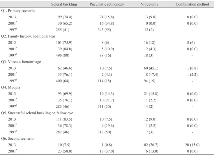
Table 1. Change of preference for method of rhegmatogenous retinal detachment surgery; comparison of four surveys conducted on 1997, 2001 and 2013
Scleral buckling Pneumatic retinopexy Vitrectomy Combination method Q1. Primary scenario
2013 99 (74.4) 21 (15.8) 13 (9.8) 0 (0.0)
2001* 30 (65.2) 16 (34.8) 0 (0.0) 0 (0.0)
1997† 255 (41) 343 (55) 12 (2) -
Q2. Family history, additional tear
2013 101 (75.9) 8 (6) 16 (12) 8 (6)
2001* 39 (84.8) 5 (10.9) 2 (4.3) 0 (0.0)
1997† 496 (80) 98 (16) 18 (3) -
Q3. Vitreous hemorrhage
2013 62 (46.6) 10 (7.5) 60 (45.1) 1 (0.8)
2001* 35 (76.1) 2 (4.3) 8 (17.4) 1 (2.2)
1997† 400 (64) 114 (18) 94 (15) -
Q4. Myopia
2013 93 (69.9) 19 (14.3) 21 (15.8) 0 (0.0)
2001* 35 (76.1) 10 (21.7) 1 (2.2) 0 (0.0)
1997† 285 (46) 311 (50) 14 (2) -
Q5. Successful scleral buckling on fellow eye
2013 111 (83.5) 10 (7.5) 12 (9.0) 0 (0.0)
2001* 36 (78.3) 9 (19.6) 1 (2.2) 0 (0.0)
1997† 282 (46) 312 (50) 17 (3) -
Q6. Second scenario
2013 10 (7.5) 1 (0.8) 102 (76.7) 20 (15.0)
2001* 23 (50.0) 17 (37.0) 6 (13.0) 0 (0.0)
Data are n (%); Q2: The case presented in survey of 1997† was primary scenario plus family history of retinal detachment and several lat- tice degenerations; Q3: The term “moderate hemorrhage” was used in survey in 1997†, while “mild hemorrhage was presented in surveys of 2001* and 2013; Q6: There was no question related to both pseudophakia and media opacity in surveys in 1997†.
*Data from Kang et al., Korea [11]; †Data from Benson et al., USA [10].
455 (Table 1 and Fig. 2C). Preference for SB was decreased
from 76.1% to 46.6%, and that for vitrectomy increased from 17.4% to 45.1%. The proportional change between these two methods was significant (p = 0.001). SB was consistently the most preferred surgical method in both 2001 and 2013, when a history of myopia or successful SB on the fellow eye was considered (Table 1, Fig. 2D and 2E).
Preference for vitrectomy over SB, however, increased with the presence of myopia (p = 0.044) and PR (p = 0.024).
The popularity of vitrectomy compared to PR increased significantly when factoring in a history of successful SB (p = 0.044).
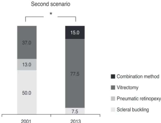
In 2001, 50.0% preferred SB, 37.0% preferred vitrecto- my, and 13.0% preferred PR for the second scenario. These results changed drastically in 2013 (Table 1 and Fig. 3):
77.4% selected vitrectomy and 15.1% selected Combi as the choice of treatment, while only 7.5% preferred SB and no one chose PR. A proportionate increase in vitrectomy (p <
0.001) and Combi (p = 0.001), compared to SB, was statis- tically significant.
The career index ranged from 0.75 to 40 among the 2013 participants. For the analysis, participants were divided into two groups by the median value (8.0) of career index.
Those with a career index less than or equal to 8.0 (less experienced group, n = 67) and those with a career index greater than the median (experienced group, n = 66) did not show significant differences in their choice of treat- ment to all six questions (Table 2). Also, when they were
divided into two groups (younger group, n = 67 vs. older group, n = 66) by the median years after fellowship, their surgical method preferences still did not differ significantly.
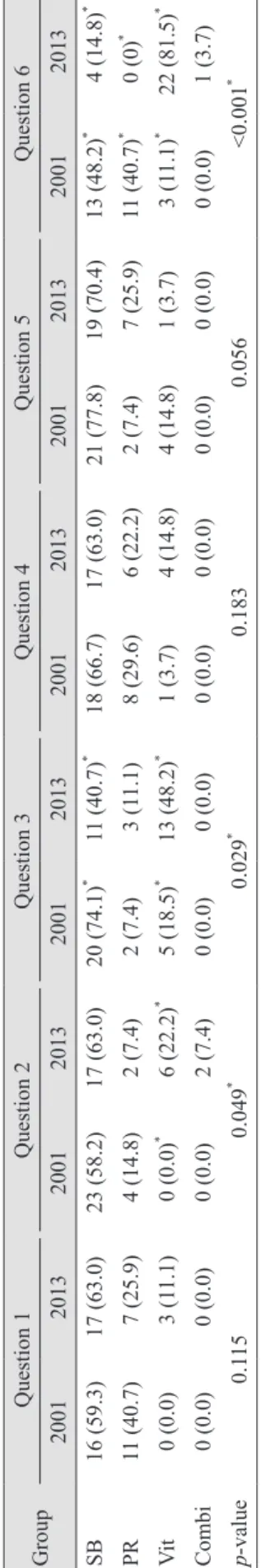
Twenty-seven members replied to the same question- naire in both 2001 and 2013. Changes in their answers for each question were analyzed using the year of survey as the variable. The 27 members had significant changes to their preferences for questions 2, 3 and 6 (Table 3). In 2013, they favored vitrectomy more often (questions 2, 3, and 6) and showed a decreased preference for SB (questions 3 and 6), and for PR (question 6), compared to 2001. Specifically, for question 6, total RRD with pseudophakia, 13 members selected SB in 2001, but in 2013, eight of them changed their choice to vitrectomy, one to Combi, and only four members adhered to SB. Likewise, all of the 11 members who selected PR in 2001 changed their choice to vitrectomy.
Discussion
In this study, we investigated the changing trends in managing primary RRD in Korea, using two surveys with identical questionnaires administered about a decade apart.
In 2013, more members of Korean Retina Society selected vitrectomy for treating primary RRD with the presence of myopia, mild vitreous hemorrhage and a history of suc- cessful SB on the other eye, compared to 2001. Further- more, significant intra-respondent change was also found among the 27 members who participated in both surveys.
The popularity of PR has noticeably decreased. The most prominent and significant change was that most members in 2013 chose primary vitrectomy in the second scenario, which involved a pseudophakic, macula-off RRD case with media opacity, whereas in 2001, the majority chose SB in the same case.
This study demonstrated the growing trend toward vit- rectomy as the preferred primary surgical method for pri- mary RRD. Previous studies conducted in other countries have also suggested the increasing popularity of vitrecto- my [12,13]. Minihan et al. [12] retrospectively compared RRD surgeries, performed 20 years apart, in a single cen- ter located in London, UK. They reported that 63% of pa- tients with primary RRD were treated by vitrectomy in 1999; in contrast, only one case was managed by vitrecto- my in 1979 and 1980. As there were extremely few cases in 1979 and 1980, a comparison of surgeries selected for
Second scenario
*
2001 2013
Combination method Vitrectomy Pneumatic retinopexy Scleral buckling 37.0
13.0
50.0
15.0
77.5
7.5
Fig. 3. Comparison of results for question 6. Preference of re- spondents changed significantly in 2013; the preference for vit- rectomy and combination method increased compared
to scleral buckling. *p < 0.05 by chi-square test.
Table 2. Comparison of surgical preference between two groups (inexperienced group, n = 67 vs. experienced group, n = 66) divided by the median value of career index GroupQuestion 1Question 2Question 3Question 4Question 5Question 6 InexperiencedExperiencedInexperiencedExperiencedInexperiencedExperiencedInexperiencedExperiencedInexperiencedExperiencedInexperiencedExperienced SB51 (76.1)48 (72.7)51 (76.1)50 (75.8)27 (42.3)32 (47.7)45 (67.2)48 (72.7)58 (88.0)51 (77.3)4 (6.0)6 (9.0) PR10 (14.9)11 (16.7)5 (7.5)3 (4.5)4 (6.0)6 (9.0)10 (14.9) 9 (13.6)4 (6.0)6 (9.1)0 (0.0)0 (0.0) Vit6 (9.0) 7 (10.6)6 (9.0)10 (15.2)35 (52.2)28 (41.8)12 (17.9) 9 (13.6)4 (6.0) 9 (13.6)53 (79.1)50 (75.8) Combi0 (0.0)0 (0.0)5 (7.4)3 (4.5)0 (0.0)1 (1.5)0 (0.0)0 (0.0)0 (0.0)0 (0.0)10 (14.9)10 (15.2) p-value* 0.9010.5720.4590.7520.2350.620 Data are n (%).
SB, scleral buckling; PR, pneumatic retinopexy; Vit, vitrectomy; Combi, combination method. * Chi-square test. Table 3. Change in preference of 27 members who participated in the survey of 2001 and 2013 GroupQuestion 1Question 2Question 3Question 4Question 5Question 6 200120132001201320012013200120132001201320012013 SB16 (59.3)17 (63.0)23 (58.2)17 (63.0)20 (74.1)* 11 (40.7)* 18 (66.7)17 (63.0)21 (77.8)19 (70.4)13 (48.2)* 4 (14.8)* PR11 (40.7)7 (25.9)4 (14.8)2 (7.4)2 (7.4)3 (11.1) 8 (29.6)6 (22.2)2 (7.4)7 (25.9)11 (40.7)* 0 (0)* Vit0 (0.0)3 (11.1)0 (0.0)* 6 (22.2)* 5 (18.5)* 13 (48.2)* 1 (3.7)4 (14.8)4 (14.8)1 (3.7) 3 (11.1)* 22 (81.5)* Combi0 (0.0)0 (0.0)0 (0.0)2 (7.4)0 (0.0)0 (0.0)0 (0.0)0 (0.0)0 (0.0)0 (0.0)0 (0.0)1 (3.7) p-value0.1150.049* 0.029* 0.1830.056<0.001* Data are n (%). SB, scleral buckling; PR, pneumatic retinopexy; Vit, vitrectomy; Combi, combination method. * p-value <0.05 by asymptotic marginal-homogeneity test.
457 RRD, with different clinical complexities, was not possi-
ble. Another study was a bi-center study that retrospective- ly investigated RRD surgeries performed in 2007 and 2008 in Vienna, Austria and New York, USA [13]. Al- though the authors attempted to assess the influence of Scleral Buckling versus Primary Vitrectomy in Rheg- matogenous Retinal Detachment study findings [6], the study period of only two years was not long enough to evaluate changes in general trends.
Recent mechanical and technical advances, such as su- tureless small gauge vitrectomy with high cutting rates, wide-angle viewing systems, and the introduction of per- fluorocarbon liquid have provided better views, easy re- moval of media opacity and intraoperative retinal reattach- ment with fewer complications, compared to techniques of the past decade. Also, under the current training system, retinal surgeons are given more exposure to, and are con- sequently more comfortable with vitrectomy than SB, as the indications for vitrectomy have been expanded to in- clude various vitreoretinal diseases other than RRD, such as diabetic retinopathy and epiretinal membrane, whereas SB is limited to RRD cases.
Especially in cases of RRD with pseudophakia, vitrecto- my was twice as popular. For question 6, 77.5% of respon- dents selected vitrectomy in 2013. When combined with the 15% that chose the Combi, a total of 92.5% answered that they would perform vitrectomy as the primary sur- gery. Since the first survey was conducted in 2001, several studies supporting vitrectomy as the primary treatment modality in pseudophakic RRD have been reported [6-8].
A multicenter randomized study by Heimann et al. [6]
compared the results of SB and vitrectomy in primary RRD. Their results indicated that SB resulted in better vi- sual outcomes than vitrectomy in phakic RRD; however, vitrectomy achieved a superior anatomical success rate and similar final visual acuity in pseudophakic/aphakic RRD. This evidence may have influenced the preference for vitrectomy in the case of pseudophakic RRD noted in the current survey.
Popularity of PR decreased in most cases, including in the case of uncomplicated primary RRD that was tradi- tionally regarded as a good indication [14,15]. Although PR has advantages over vitrectomy and SB, such as a short duration of operation, cost-effectiveness and availability as an outpatient-based procedure, disadvantages of this pro- cedure are that it necessitates a second retinopexy proce-
dure, such as laser photocoagulation, and requires main- taining the correct position after the procedure for at least several days. Moreover, missed or new breaks [16], limited indication [17] and higher probability of needing a second operation [18], decreased its popularity.
Besides the reasons described above, the increase of vit- rectomy and decrease of PR may also be attributable to the national health care system called the National Health In- surance Program of South Korea. This system offers uni- versal coverage for all citizens in South Korea, and health care providers are reimbursed on a fee-for-service basis. In tertiary hospitals of South Korea, the fee for vitrectomy is approximately double that for SB and quintuple for PR.
Therefore, the possibility that this payment system may affect surgeons’ decisions regarding surgical method can- not be excluded. Nonetheless questions regarding the pri- mary scenario were based on the assumption that, “if RRD happened on your eye...” so that respondents would answer based on their medical and scientific background solely, and ignore economic incentives.
This study has several limitations. First, there was a se- lection bias of respondents, because 20.7% and 39.3% of members did not reply to the survey in 2001 and 2013, re- spectively. In addition, the cases presented for the first sce- nario were either primary uncomplicated RRD or RRD combined with only a few clinical variables; however, in a real clinical situation, surgeons commonly encounter com- binations of multiple clinical variables in an eye with RRD. Finally, the preference of contemporary colleagues does not provide any evidence supporting one specific sur- gical modality over the other, and must be interpreted cau- tiously.
In conclusion, preference for vitrectomy in primary RRD has increased drastically among the members of the Korean Retina Society, especially when combined with myopia, media opacity and pseudophakia, over the course of the last decade.
Conflict of Interest
No potential conflict of interest relevant to this article was reported.
Acknowledgements
We are very grateful to the members of the Korean Ret- ina Society, especially, Jeeyun Ahn (Seoul National Uni- versity), Ik Soo Byon (Pusan National University), Jeong Hun Bae (Sungkyunkwan University), Sun Ryang Bae (The Catholic University of Korea), Kwangyul Chang (Guri St. Mary’s Eye Clinic), Moohwan Chang (Dankook University), Woohyok Chang (Yeungnam University), Hee Seung Chin (Inha University), Han Joo Cho (Konyang University), Hee Yun Cho (Hanyang University), Ho Kyun Cho (Chung-Ang University), Sung Won Cho (Konyang University), Young Jae Cho (Ansan Yonsei Eye Clinic), Young Wook Cho (Daegu Fatima Hospital), Kyung Seek Choi (Soonchunhyang Univerisity), Young Kwang Chu (Siloam Eye Hospital), Eun Jee Chung (NHIS Ilsan Hospi- tal), Hum Chung (Seoul National University), Hyewon Chung (Konkuk University), Seungmin Ha (Kyungdae Eye Clinic), Yong Seop Han (Gyeongsang National Uni- versity), Kuhl Huh (Korea University), Young-Joon Jo (Chungnam National University), Chang Ryong Kim (Bora Eye Hospital), Chul Gu Kim (Konyang University), Ha- kyoung Kim (Hallym University), Hyun Jin Kim (Seoul Medical Center), Hyung Chan Kim (Konkuk University), Jae Suk Kim (Inje University), Jung-Yeul Kim (Chungnam National University), Jong Woo Kim (Konyang Universi- ty), June-Gone Kim (University of Ulsan), Kiseok Kim (Saevit Eye Hospital), Kwang Soo Kim (Keimyung Univ- erysity), Kyu Seop Kim (The Catholic University of Ko- rea), Sang Jin Kim (Sungkyunkwan University), Soonhy- un Kim (Nune Eye Hospital), Si Yeol Kim (Nune Eye Hospital), Tae Wan Kim (Seoul National University), Young Gyun Kim (Eulji University), Youngduk Kim (Bora Eye Hospital), Yong Baek Kim (Eyelove Eye Center), Yu Cheol Kim (Keimyung University), Yun Young Kim (Catholic University of Daegu), Yun Taek Kim (Ewha Womans University), Hyoung Jun Koh (Yonsei Universi- ty), No Hoon Kwak (EOS Eye Center), Hyung Woo Kwak (Kyung Hee University), Soon Il Kwon (Hallym Universi- ty), Christopher Seungkyu Lee (Yonsei University), Dae Yeong Lee (Gachon University), Dongcho Lee (Good Eye Clinic), Eun Koo Lee (Kong Eye Center), Jonghyun Lee (Inje University), Joo Yong Lee (University of Ulsan), Joo- Eun Lee (Inje University), Jun Gyo Lee (Seoul Eye Clinic), Ji Eun Lee (Pusan National University), Kihwang Lee (Ajou University), Mee Yon Lee (Chung-Ang University),
Sun Ho Lee (Jeju National University), Sung Jun Lee (Yonsei Bon Eye Clinic), Sung Jin Lee (Soonchunhyang University), Seung Woo Lee (Dongguk University), Seung Jun Lee (Kangwon National University), Tae Gon Lee (Konyang University), Phil Young Lee (Seoul Bohun Vet- erans Hospital), Homin Lew (Konyang University), Young Ju Lew (Konyang University), Yeon-Sung Moon (Inha University), Sungjin Na (Eyelove Eye Center), Dong Heun Nam (Gachon University), Dong Ho Park (Kyungpook Na- tional University), Ha-Sung Park (Happy Eye 21 Clinic), Hyun Jun Park (Pusan National University), Jung Min Park (Maryknoll General Hospital), Jung Hyun Park (Inje University), Jong Moon Park (Gyeongsang National Uni- versity), Jong Seok Park (Eulji University), Kyu Hyung Park (Seoul National University), Youngsook Park (HanGil Eye Hospital), Young-Hoon Park (The Catholic University of Korea), Tae Kwann Park (Soonchunhyang University), Young Jung Roh (The Catholic University of Korea), Min Sagong (Yeungnam University), JaePil Shin (Kyungpook National University), Minchul Shin (Hallym University), Joonhong Sohn (HanGil Eye Hospital), Ji Hun Song (Ajou University), Su Jeong Song (Sungkyunkwan University), Won Kyung Song (CHA University), Yumi Song (Bundang Jesaeng Medical Center), Se Joon Woo (Seoul National University), Ji-Wook Yang (The Catholic University of Ko- rea), Sung Jae Yang (University of Ulsan), Yun-Sik Yang (Wonkwang University), Jinseong Yoo (U-Jin St. Mary’s Eye Center), Hee Seong Yoon (Sungmo Eye Hospital), Young Hee Yoon (University of Ulsan), Ie Na Yoon (Yon- sei University), Yong Sung You (Nune Eye Hospital), Seung Young Yu (Kyung Hee University), Il Han Yun (Inje University), who shared their valuable experiences and opinions about retinal detachment surgeries by participat- ing in this survey.
References
1. Schwartz SG, Flynn HW. Primary retinal detachment:
scleral buckle or pars plana vitrectomy? Curr Opin Oph- thalmol 2006;17:245-50.
2. Escoffery RF, Olk RJ, Grand MG, Boniuk I. Vitrectomy without scleral buckling for primary rhegmatogenous reti- nal detachment. Am J Ophthalmol 1985;99:275-81.
3. Campo RV, Sipperley JO, Sneed SR, et al. Pars plana vit- rectomy without scleral buckle for pseudophakic retinal
459 detachments. Ophthalmology 1999;106:1811-5.
4. Gartry DS, Chignell AH, Franks WA, Wong D. Pars plana vitrectomy for the treatment of rhegmatogenous retinal de- tachment uncomplicated by advanced proliferative vitreo- retinopathy. Br J Ophthalmol 1993;77:199-203.
5. Azad RV, Chanana B, Sharma YR, Vohra R. Primary vit- rectomy versus conventional retinal detachment surgery in phakic rhegmatogenous retinal detachment. Acta Ophthal- mol Scand 2007;85:540-5.
6. Heimann H, Bartz-Schmidt KU, Bornfeld N, et al. Scleral buckling versus primary vitrectomy in rhegmatogenous retinal detachment: a prospective randomized multicenter clinical study. Ophthalmology 2007;114:2142-54.
7. Ahmadieh H, Moradian S, Faghihi H, et al. Anatomic and visual outcomes of scleral buckling versus primary vitrec- tomy in pseudophakic and aphakic retinal detachment: six- month follow-up results of a single operation: report no. 1.
Ophthalmology 2005;112:1421-9.
8. Brazitikos PD, Androudi S, Christen WG, Stangos NT. Pri- mary pars plana vitrectomy versus scleral buckle surgery for the treatment of pseudophakic retinal detachment: a randomized clinical trial. Retina 2005;25:957-64.
9. Snyder WB, Bloome MA, Birch DG. Pneumatic retinopexy versus scleral buckle: preferences of Vitreous Society members, 1990. Retina 1992;12:43-5.
10. Benson WE, Chan P, Sharma S, et al. Current popularity of pneumatic retinopexy. Retina 1999;19:238-41.
11. Kang SW, Kim SW; The Korean Retina Society. Preferenc- es for treatment modalities of simple rhegmatogenous teti- nal detachment in Korea. J Korean Ophthalmol Soc 2002;43:1179-85.
12. Minihan M, Tanner V, Williamson TH. Primary rheg- matogenous retinal detachment: 20 years of change. Br J Ophthalmol 2001;85:546-8.
13. Falkner-Radler CI, Myung JS, Moussa S, et al. Trends in primary retinal detachment surgery: results of a Bicenter study. Retina 2011;31:928-36.
14. Tornambe PE. Pneumatic retinopexy: the evolution of case selection and surgical technique: a twelve-year study of 302 eyes. Trans Am Ophthalmol Soc 1997;95:551-78.
15. Hilton GF, Tornambe PE, Grizzard WS. Results of a tem- porary balloon buckle in the treatment of 500 retinal de- tachments and a comparison with pneumatic retinopexy.
Am J Ophthalmol 1989;108:612-4.
16. Zaidi AA, Alvarado R, Irvine A. Pneumatic retinopexy:
success rate and complications. Br J Ophthalmol 2006;90:427-8.
17. Chan CK, Lin SG, Nuthi AS, Salib DM. Pneumatic reti- nopexy for the repair of retinal detachments: a comprehen- sive review (1986-2007). Surv Ophthalmol 2008;53:443-78.
18. Day S, Grossman DS, Mruthyunjaya P, et al. One-year out- comes after retinal detachment surgery among medicare beneficiaries. Am J Ophthalmol 2010;150:338-45.