Oncolytic Viruses - A New Era for Cancer Therapy
Daniel Ngabire1, Irvine Niyonizigiye2, Min-jae Kang2 and Gun-Do Kim2*
1Gene and Cell Therapy Research Center for Vessel-Associated Diseases, School of Medicine, Pusan National University, Yangsan 50612, Korea
2Department of Microbiology, College of Natural Sciences, Pukyong National University, Busan 48513, Korea
Received July 23, 2019 /Revised July 26, 2019 /Accepted July 26, 2019
In recent decades, oncolytic viruses (OVs) have extensively been investigated as a potential cancer drug. Oncolytic viruses have primarily the unique advantage in the fact that they can only infect and destroy cancer cells. Secondary, oncolytic viruses induce the activation of specific adaptive immunity which targets tumor-associated antigens that were hidden during the initial cancer progression. In 2015, one genetically modified oncolytic virus, talimogene laherparepvec (T-VEC), was approved by the American Food and Drug Administration (FDA) for the treatment of melanoma. Currently, vari- ous oncolytic viruses are being investigated in clinical trials as monotherapy or in combination with preexistent cancer therapies like immunotherapy, radiotherapy or chemotherapy. The efficacy of onco- lytic virotherapy relies on the balance between the induced anti-tumor immunity and the anti-viral response. Despite the revolutionary outcome, the development of oncolytic viruses for the treatment of cancer faces a number of obstacles such as delivery method, neutralizing antibodies and induction of antiviral immunity due to the complexity, variability and reactivity of tumors. Intratumoral admin- istration has been successful reducing considerably solid tumors with no notable side effects un- fortunately some tumors are not accessible (brain) and require a systemic administration of the onco- lytic viruses. In order to overcome these hurdles, various strategies to enhance the efficacy of oncolytic viruses have been developed which include the insertion of transgenes or combination with im- mune-modulatory substances.
Key words : Clinical trials, combination therapy, immunotherapy, oncolytic viruses, tumor microenvi- ronment
*Corresponding author
*Tel : +82-51-629-5618, Fax : +82-51-629-5619
*E-mail : [email protected]
This is an Open-Access article distributed under the terms of the Creative Commons Attribution Non-Commercial License (http://creativecommons.org/licenses/by-nc/3.0) which permits unrestricted non-commercial use, distribution, and reproduction in any medium, provided the original work is properly cited.
Journal of Life Science 2019 Vol. 29. No. 7. 824~835 DOI : https://doi.org/10.5352/JLS.2019.29.7.824
Oncolytic viruses (OVs)
The hypothesis for the usage of OVs for the treatment of cancer started back in the 1900s from anecdotal reports of cases of patients that were assumingly presenting tumor regression signs after viral infections [45]. The majority of patients had blood-related malignancy such as leukemia but the remission was short-lived (approximately one or two months) [30, 87]. These observations led to the hypothesis that under controlled condition, OVs might be developed as cancer drugs [34]. The invention of the electron micro- scope and the progress in molecular biology (genetics) al- lowed a better understanding of the virus life cycle and the beginning of investigations in animal models.
OVs, like most viruses, are the smallest known infectious
agents and are unable to survive on their own without in- fecting a living organism. Their major components are: a ge- netic material (DNA or RNA, double or single strands), and a capsid that envelops and protects the genetic material. In addition, some OVs possess an envelope that contains glyco- proteins and present various shapes.
Malignant cells undergo multiple transformations such as overexpression of cell surface receptors (such as epidermal growth factor receptor; EGFR) and aberrant signaling path- ways (such as Ras/mitogen-activated protein kinase (MAPK), phosphoinositide 3-kinase (PI3K)/AKT/mammalian target of rapamycin (mTOR), and vascular endothelial growth fac- tor (VEGF) pathways), or repress tumor suppressor genes (such as p53 and Rb) to acquire the hallmarks of cancer and overcome normal cellular restraints. The disruption in these pathways inhibits interferon (IFN) signaling which is the overall immune response to viral infection [46]. The IFN pathway is induced after recognition of (a) viral elements by toll-like receptors (TLRs), (b) dsRNA from RNA viruses by retinoic acid-inducible gene I (RIG-I) and melanoma dif- ferentiation-associated protein 5 (MDA5), and (c) viral DNA by the DNA sensor protein cyclic GMP-AMP synthase - Review -
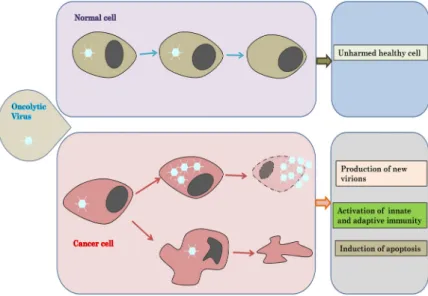
Fig. 1. Principle of oncolytic virotherapy. Oncolytic viruses are natural or programmed can- cer-killing viruses. The infection of nor- mal leads to the activation of antiviral pathway such as Type 1 interferons which will block the virus replication. In cancer cells, antiviral and cell proliferation path- ways are altered and oncolytic viruses uti- lize these disruptions to specifically target and infect only cancer cells which lead to the lysis of infected cancer cells and the expansion of oncolytic viruses to other tu- mor cells.
(cGAS) which activates the stimulator of interferon genes (STING) protein. IFNs prevent the viruses from infecting ad- ditional cells, inhibit protein synthesis and activate immune response through inflammation [26, 53].
The aberrations mentioned above are exploited by OVs and allow them to specifically replicate and kill only cancer cells. These proprieties make OVs ideal candidates for the development of specific anticancer products [1, 21, 80].
Immune system and OVs
OVs have two major mechanisms of eliminating cancer cells that involve direct cell lysis and destruction of intra- tumoral blood vessels or the activation of adaptive im- munity.
Once cancer cells are infected with OVs, they can initiate the immune response by IFN through the interaction be- tween released antigens and TLRs. The viral proteins or tu- mor-associated antigens will interact with TLRs on the cell surface or will be detected by intracellular components of TLRs [84].
The TLR pathways initiate the host antiviral responses via downstream proteins like TNF-associated factor 3 (TRAF3), IFN-related factor 3 (IRF3), IRF7 and RIG-I.
After cell death, tumor lysed cells release tumor-asso- ciated antigens that can flow through blood circulation and be captured by antigen-presenting cells (APCs), such as den- dritic cells (DCs) and macrophages, and presented to lymphocytes. Additionally, viral pathogen-associated molec- ular patterns (PAMPs) and danger-associated molecular pat- terns (DAMPs) such as high mobility group box 1 (HMGB1)
protein, ATP, calreticulin, heat shock proteins, and uric acid are also released [39].
Tumor-associated antigens together with cytokines and DAMPs molecules can initiate an innate immune response with myeloid cells and adaptive immune response by the presentation of antigens to lymphocytes. This particular property is beneficial in cancer treatment as cold tumors can be activated in hot tumors. The activation of both innate and adaptive immune response provides a specific answer to dis- tant tumors or metastasis. PAMPs and DAMPs can directly activate natural killer (NK) cells that will then specifically target and kill tumor cells even with a low expression of major histocompatibility complex (MHC) class I molecules which is the case in most cancers. Tumor-specific CD8+ have demonstrated in preclinical studies the ability to mediate tu- mor rejection.
The stimulation of long-lasting antitumor immune re- sponse likely plays a pivotal role in the duration and extent of clinical responses. With this increased appreciation for the role of immune stimulation in OV efficacy, many oncolytic viruses are now being designed to express transgenes encod- ing immune stimulatory cytokines, such as granulocyte- macrophage colony-stimulating factor (GM-CSF), to enhance OV immunogenicity [44].
Barriers to oncolytic virotherapy
Neutralizing antibodies and complement
For the viruses to initiate a potential antitumor response, they need to reach the tumor site. Blood is the efficient ve- hicle for OVs, therefore, intravenous therapy (IV) is the most
Fig. 2. Mechanisms of oncolytic viruses. Oncolytic viruses enter the cell through the interaction with cell receptors that are usually overexpressed in cancer. Some viruses utilize more than one specific receptor but different viruses can also share one receptor.
Other viruses enter the cell via endocytosis through fusion of membranes. Once in the cell, viruses take advantage of aberrant pathways to replicate. CD, Cluster of differentiation; CAR, Coxsackievirus-adenovirus receptor; ICAM-1, Intracellular adhesion molecule 1; LDLR, Low density lipoprotein receptor; HSV, Herpes simplex virus; HVEM, Herpesvirus entry mediator; SARs, Sialic acid receptors; SLAM, Signaling lymphocyte activation molecule; JAK, Janus kinase; STAT, Signal transducer and activa- tor of transcription; Rb, Retinoblastoma; PKR, Protein kinase R; RIG-Ⅰ, Retinoic acid-inducible gene Ⅰ; TK, Thymidine kinase.
privileged route for the administration of OVs and can reach the metastatic site. Unfortunately, the majority of patients, having been exposed to the viral family naturally or by vac- cination, present preexistent neutralizing antibodies and the OVs are rapidly recognized and eliminated by the circulat- ing antibody without or with help of complement molecules [22].
In absence of complement, recent studies have demon- strated that the neutralization of OVs was relatively weak almost absent but even in the absence of neutralizing anti- bodies, IV delivery of oncolytic viruses, such as herpes sim- plex virus (HSV) and vaccinia virus (VV), has been shown to be inhibited by antiviral activity present in serum as a result of the activation of the complement system. The com- plement system acts as the first line of innate immune de- fense, opsonizing and neutralizing foreign pathogens, target- ing them for phagocytosis, and clearing them from the circu- latory system. Antibody-mediated complement activation enhances the neutralizing capacity of antibodies, making
complement of particular relevance for OVs in which preex- isting immunity may be prevalent [89].
Tumor heterogeneity
The increasing resistance to cancer therapeutics, especially targeted therapy, is often attributed to the complexity within tumors and represents a major obstacle in the successful treatment of cancer.
The location of tumors in the brain limits the efficacy of brain tumors treatment due to the blood-brain barrier. One of the most investigated strategies is to develop oncolytic viruses with a natural tropism for the central nervous system (CNS) and some viruses such as poliovirus, parvovirus H1, and reovirus have shown the ability to reach glioblastoma multiforme (GBM) in clinical trials [20, 82].
Genetic alterations that occur in tumors present a problem for oncolytic virotherapy. The tumor microenvironment is a very complex structure in which stromal cells (endothelial cells, adipocytes, cancer associated fibroblasts; CAFs, and
Fig. 3. The induction of immune response by onco- lytic viruses. The cell lysis of cancer cells by oncolytic viruses is followed by the release of neo-oncolytic viruses, tumor-associated anti- gens (TAAs), danger-associated molecular patterns (DAMPs) and pathogen-associated molecular patterns (PAMPs). The new onco- lytic viruses can infect other existing cancer cells and continue the cycle of cancer cells elimination. TAAs, DAMPs and PAMPs are processed by antigen presenting cells (APCs) and initiate adaptive immune response by CD8+T lymphocytes against cancer cells.
immune infiltrating cells) play an important role in the re- sponse to immunotherapy and oncolytic virotherapy. These cells are also responsible for creating physical barriers such as dense fibrotic capsules, necrosis, and acidosis, which can impede viral delivery into the tumor [60].
Strategies to enhance oncolytic virotherapy
Combination therapy
Conventional therapy such as chemotherapy and radio- therapy can be combined with oncolytic virotherapy.
Cyclophosphamide, used both in chemotherapy and im- munosuppression (B- and T-lymphocytes), is an alkylating agent regularly combined with OVs. Other immunosuppress- ants such as paclitaxel and temozolomide inhibit regulatory T cells (Treg) activity in a dose-dependent manner [27].
Various animal studies demonstrated a remarkable recov- ery in the treatment of tumor-bearing mice with OV com- bined with immune checkpoints inhibitors (ICIs) [51]. ICIs are a relatively new class of immunotherapies that aim to overcome tumor-induced immune suppression and evasion caused by the expression of immune checkpoints. Monoclo- nal antibodies that inhibit programmed cell death protein 1 (PD-1), its ligand (PD-L1), and cytotoxic T lymphocyte anti- gen 4 (CTLA-4) have demonstrated remarkable responses in melanoma, non-small cell lung cancer (NSCLC), renal cell carcinoma, bladder cancer, head and neck cancer, Hodgkin lymphoma, Merkel cell carcinoma, and likely other cancers as well [24, 81].
Chimeric antigen receptor-T (CAR-T) therapy has given promising results in blood-related cancers like leukemia and lymphoma but has a limited response in solid tumors, there-
fore, the combination with OVs is believed to improve the outcome of treated patients. In CAR-T therapy, T cells from patients are genetically modified to express chimeric antigen receptor (CAR) that specifically recognizes a tumor-associated antigen (TAA) then the engineered T cells are transfused back to the patients [48, 79].
Transgene arming
A large number of genes has been used in vitro experi- ments to change the OV genetic material and improve their therapeutic activity. These genes include regulatory cyto- kines, proapoptotic genes, extracellular matrix degradation enzymes, immune checkpoints inhibitors monoclonal anti- bodies and antiangiogenic proteins [32].
The FDA approved HSV talimogene laherparepvec (T- VEC) for the treatment of melanoma was engineered from HSV-1 with the deletion of the ICP34.5 gene involved in vi- rus replication and ICP47 gene in immune evasion. The ad- ditional modification was the insertion of GM-CSF, a cyto- kine and growth factor that stimulates the differentiation and maturation of granulocytes and monocytes [55, 73].
OVs can also be armed with bispecific antibody (BiTE) where one arm of the antibody can bind to a cancer cell via a tumor-specific antigen and the other arm interact with CD8+ T lymphocytes [18, 88].
Safety concerns
Although OVs have proven to have an excellent safety record in the clinic, there remain a number of unique chal- lenges in the clinical development of oncolytic viruses. The ability of OVs to actively replicate raises a number of con- cerns and therefore requires regulations before being dis-
tributed for treatment. The overall concern in the develop- ment of OVs is the environmental shedding of the virus from patients receiving treatment and the risk of trans- mission to the general population. The shedding from the patient’s body through one or all of the following routes:
blood, fecal, urine, saliva, or wounds/sores on the skin is taken into consideration. Shedding is a considerable bio- safety concern as it raises the possibility of transmission of OV products from treated to untreated individuals [7]. As such, the FDA has established guidelines as to how and when data regarding shedding should be collected during preclinical and clinical development and the way it is to be used to assess the potential for transmission to untreated individuals. Some viruses such as HSV, vesicular stomatitis virus (VSV), and reovirus display minimal shedding, where- as other viruses including adenovirus and VV are known to be more problematic.
OVs in clinical development
Herpes viruses (HSV)
HSV-1, enveloped double-stranded DNA virus of the Herpesviridae family, was the first virus to be genetically modified to treat cancer after the deletion of thymidine kin- ase (TK). HSV-1 replicates in the nucleus and is highly pathogenic to human as it can affect peripheral nerves through surface nectins (nectin 1 and nectin 2) of neurons.
Due to this risk, further engineering modifications resulted in the deletion of the ICP34.5 gene (Table 1) responsible for the neurovirulence and blocks protein kinase R (PKR)-IFN antiviral response. T-VEC is an oncolytic HSV-1 approved for the treatment of melanoma. ICP34.5 was deleted in T-VEC together with US11 which inhibits the phosphor- ylation of PKR and therefore stops the induction of apoptosis in infected cancer cells [42]. In addition to the deletion, the GM-CSF gene has been inserted in T-VEC which enhance the antitumor immune response. Since the approval of T-VEC by the FDA in 2015, other engineered HSV-1 are be- ing evaluated in clinical studies. Seprehvir (HSV1716), in which ICP34.5 was deleted, is being investigated (Table 1) in patients with hepatocellular carcinoma (HCC), GBM, mes- othelioma, and neuroblastoma [54]. G207 contains a deletion of the ICP34.5 gene with disruption of UL39 and is being evaluated in GBM [61]. For OrienX010, in addition to ICP34.5 gene deletion and GM-CSF insertion, ICP47 gene was de- leted (Table 1). ICP47 gene blocks the presentation of HSV-1
viral proteins, therefore, its deletion allows the control of the infection [56]. Different from other oncolytic HSV-1 vi- ruses, HF-10 is a natural mutant that conserved both copies of ICP34.5 genes but with the deletion of UL56. HF-10 is currently in clinical trials for breast cancer, melanoma and pancreatic cancer [38].
Measles virus (MV)
MVs, single-stranded RNA viruses of the Paramyxovir- idae family, infect cells through the interaction of the enve- lope protein, hemagglutinin (H) proteins, and the host cel- lular molecules, CD46 [5]. They initiate fusogenic syncytia that lead to cell death and result in the release of intracellular danger signals to activate the immune system. A sodium/io- dide membrane protein (SLC5A5) has been inserted in the measles virus encoding thyroidal sodium iodide symporter (MV-NIS) OV and is currently being investigated in clinical studies (Table 1). I131-label with sodium iodide allows to per- form easy monitoring and at the same time allows to con- duct radiotherapy [28, 66].
Newcastle disease virus (NDV)
NDVs, double-stranded RNA viruses also of Paramyx- oviridae family, are bird viruses that infect cells through he- magglutinin-neuraminidase (HN) protein on the plasma membrane or via direct endocytosis. PV701 and NDV-HUJ OVs from NDV have shown the capacity to selectively mul- tiply in cancer cells and induce immune activation. They are currently in clinical trials (Table 1). Both administrated to patients via IV, PV701 was evaluated in patients with un- responsive solid tumors while NDV-HUJ is being inves- tigated in patients with recurrent GBM [11].
Vesicular stomatitis virus (VSV)
VSV, single-stranded RNA viruses of Rhabdoviridae family, entry cells using low-density lipoprotein (LDL) re- ceptor and are also dependent on the aberrant IFN signal- ing pathway. The low preexistence of neutralizing anti- bodies allows the IV delivery of the OVs [6]. In clinical tri- als, VSV- IFNβ and VSV-IFNβ-NIS after infection produce human IFNβ which protects non-neoplastic cells but in- crease the immune response towards cancer cells. In addi- tion to IFNβ, VSV-IFNβ-NIS oncolytic virus expresses so- dium iodide symporter for tumor-monitoring and for radio- therapy [71, 93].
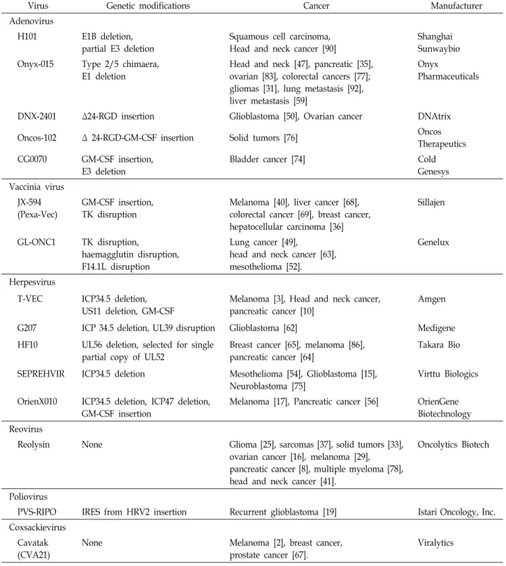
Table 1 Clinical trials of oncolytic virus
Virus Genetic modifications Cancer Manufacturer
Adenovirus
H101 E1B deletion, partial E3 deletion
Squamous cell carcinoma, Head and neck cancer [90]
Shanghai Sunwaybio Onyx-015 Type 2/5 chimaera,
E1 deletion
Head and neck [47], pancreatic [35], ovarian [83], colorectal cancers [77];
gliomas [31], lung metastasis [92], liver metastasis [59]
Onyx
Pharmaceuticals
DNX-2401 Δ24-RGD insertion Glioblastoma [50], Ovarian cancer DNAtrix
Oncos-102 Δ 24-RGD-GM-CSF insertion Solid tumors [76] Oncos
Therapeutics CG0070 GM-CSF insertion,
E3 deletion
Bladder cancer [74] Cold
Genesys Vaccinia virus
JX-594 (Pexa-Vec)
GM-CSF insertion, TK disruption
Melanoma [40], liver cancer [68], colorectal cancer [69], breast cancer, hepatocellular carcinoma [36]
Sillajen
GL-ONC1 TK disruption,
haemagglutin disruption, F14.1L disruption
Lung cancer [49],
head and neck cancer [63], mesothelioma [52].
Genelux
Herpesvirus
T-VEC ICP34.5 deletion, US11 deletion, GM-CSF
Melanoma [3], Head and neck cancer, pancreatic cancer [10]
Amgen
G207 ICP 34.5 deletion, UL39 disruption Glioblastoma [62] Medigene
HF10 UL56 deletion, selected for single partial copy of UL52
Breast cancer [65], melanoma [86], pancreatic cancer [64]
Takara Bio
SEPREHVIR ICP34.5 deletion Mesothelioma [54], Glioblastoma [15], Neuroblastoma [75]
Virttu Biologics
OrienX010 ICP34.5 deletion, ICP47 deletion, GM-CSF insertion
Melanoma [17], Pancreatic cancer [56] OrienGene Biotechnology Reovirus
Reolysin None Glioma [25], sarcomas [37], solid tumors [33],
ovarian cancer [16], melanoma [29], pancreatic cancer [8], multiple myeloma [78], head and neck cancer [41].
Oncolytics Biotech
Poliovirus
PVS-RIPO IRES from HRV2 insertion Recurrent glioblastoma [19] Istari Oncology, Inc.
Coxsackievirus Cavatak (CVA21)
None Melanoma [2], breast cancer,
prostate cancer [67].
Viralytics
Adenovirus
Adenoviruses, naked double-stranded DNA viruses of Adenoviridae family, possess a large genome that allows performing numerous modifications. Infection by adenovi- ruses can occur in both human and animals, therefore, the preexistence of neutralizing antibodies is frequent and limit
the efficacy of oncolytic adenovirus. Adenoviruses are one of the most investigated viruses in OV drug development.
Adenoviruses infect host cells through coxsackievirus-ad- enovirus receptor and in the early stages produce E1A and E1B that that specifically target p53 (tumor suppressor) and retinoblastoma protein (pRb). Modifications in adenoviruses
include manipulation of these early genes (E1A and E1B) expression and the insertion transgenes [91].
H101, approved in China for treatment of head and neck cancer, has both genes E1A and E1B deleted. In CG0070, OV investigated for bladder cancer, E1A gene is under the control of E2F-1 promoter which is dependent on Rb.
DNX-2401 contains a 24 bp Arg-Gly-Asp (RGD) binding mo- tif in the E1A gene which suppresses the ability to bind to Rb. For ONCOS-12, in addition to the insertion of the RGD motif, GM-CSF was adding for the enhancement of the im- mune response. DNX-204 is being investigated for glio- blastoma (Table 1) and ovarian cancer while clinical trials for Oncos-12 are conducted in solid tumors [14].
Vaccinia viruses (VVs)
VVs, enveloped double-stranded DNA virus of the Poxviridae family, infect host cells by endocytosis and pos- sess a high tropism specific for tumor cells. VVs have al- ready proved to successfully activate immune response dur- ing the fight against smallpox which led to the eradication of the disease. In order to increase the cancer cell selectivity, TK gene, B18r, and VGF are usually modified [13]. Currently, three oncolytic VVs are in clinical investigations [12].
JX-594 (PexaVec) is Wyeth strain in which TK was dis- rupted and GM-CSF was inserted [69, 70]. GL-ONC1 con- tains the disruption of TK, HA, and F14.5L [63]. In TG6002, an enzyme, FCU1, which can convert a nucleoside analog, 5-flucytosine (5-FC), into a chemotherapeutic drug, 5-fluo- rouracil (5-FU), was inserted [23]. The combination therapy has proven to improve the antitumor efficacy of VV. The deletion of TK limits the viruses’ replication to dividing cells which express high levels of human TK.
Coxsackievirus
Coxsackievirus, naked single-stranded RNA virus of Picornaviridae family, replicates in the cytoplasm and infect cancer cells through intracellular adhesion molecule-1 (ICAM-1) and decay-accelerating factor (DAF). One partic- ular coxsackievirus was developed for the treatment of can- cer, it’s coxsackievirus A21 (Table 1) also known as Cavatak.
Cavatak possesses a natural tropism for cancer cells with ICAM-1 and DAF overexpression such as multiple myeloma, melanoma, and breast cancer. Cavatak doesn't contain any modification and has a natural ability to enhance the im- mune response by the DAMPs [4]. Cavatak has shown to increase the infiltration of NK cells and CD8+T cells in tu-
mors together with increased levels of IFNs [67].
Reovirus
Reovirus, naked double-stranded RNA virus of Reovir- idae family, replicates in the cytoplasm of infected host cells.
Cancer cells with mutant Ras pathway are most likely to be infected as the PKR pathway is often blocked. This unique natural tropism led to many clinical investigations in numer- ous cancers such as gliomas, melanoma, ovarian cancer, pan- creatic cancer, multiple myeloma head, and neck cancer and colorectal cancer. The major obstacle for the use of reovirus as an OV is that 100% of participants in clinical trials pre- sented preexistent neutralizing antibody. Reolysin (Table 1) is an oncolytic reovirus with no genetic modification that has proven to be efficient as a monotherapy or in combina- tion with chemotherapy and radiotherapy [57, 58].
Retrovirus
Retrovirus, enveloped single-stranded RNA of Retrovir- idae family, uses its enzyme, reverse transcriptase, to pro- duce DNA from RNA. Toca-511 is an oncolytic retrovirus in which a gene coding for cytosine deaminase has been inserted. cytosine deaminase can convert 5-FC into a chemo- therapeutic drug, 5-FU [72]. Retroviruses are different from other OVs as they do not induce cell lysis but replicate se- lectively in mitotic cells. In clinical trials, patients with high-grade glioma presented an overall survival of 13.6 months [43].
Poliovirus
Polioviruses, naked single-stranded RNA viruses of Picornaviridae family, are very pathogens to humans but on- ly 1% of infected persons develop poliomyelitis. Polioviruses infect host cells through CD155 and replicate in the cyto- plasm [9]. Due to the pathogenic potency, polioviruses have to be attenuated. In PVS-RIPO (Table 1), internal ribosome entry site (IRES) site of polioviruses is replaced with IRES from human rhinovirus type 2 (HRV2). This engineering in- creases the selectivity of PVS-RIPO for GBM and activates T cells [85].
Conclusion and future perspectives
The approval by the FDA of T-VEC in 2015 triggered a high interest in the development of OV for cancer therapy.
Despite decades of investigations, there are a number of ob-
stacles delivery methods due to neutralizing antibodies and to the location of tumors. The delivery system is the main issue as most of the oncolytic viruses today are delivered locally. This system works with skin tumor or accessible tu- mors in the abdomen but poses a problem in some tumors like brain tumors and blood-related cancer. A second ob- stacle is that oncolytic virotherapy relies on the activation of innate and adaptive immune responses which are usually absent or suppressed. This activation, though beneficial for specific immune response, contributes to the elimination of OVs, therefore, significant cancer cells lysis. The currently available data about oncolytic virotherapy are just the tip of the iceberg. Despite the existing hurdles, oncolytic viruses are set to be the next generation of drugs in cancer therapy.
Acknowledgement
This work was supported by a research grant of Pukyong National University (2019 year).
References
1. Alemany, R. 2013. Viruses in cancer treatment. Clin. Transl.
Oncol. 15, 182-188.
2. Andtbacka, R. H. I., Curti, B. D., Kaufman, H., Daniels, G.
A., Nemunaitis, J. J., Spitler, L. E., Hallmeyer, S., Lutzky, J., Schultz, S. M., Whitman, E. D., Zhou, K., Karpathy, R., Weisberg, J. I., Grose, M. and Shafren, D. 2015. Final data from CALM: A phase II study of Coxsackievirus A21 (CVA21) oncolytic virus immunotherapy in patients with advanced melanoma. J. Clin. Oncol. 33, doi:10.1200/jco.2015_
suppl.9030.9030-9030.
3. Andtbacka, R. H., Kaufman, H. L., Collichio, F., Amatruda, T., Senzer, N., Chesney, J., Delman, K. A., Spitler, L. E., Puzanov, I., Agarwala, S. S., Milhem, M., Cranmer, L., Curti, B., Lewis, K., Ross, M., Guthrie, T., Linette, G. P., Daniels, G. A., Harrington, K., Middleton, M. R., Miller, W. H. Jr, Zager, J. S., Ye, Y., Yao, B., Li, A., Doleman, S., Vander Walde, A., Gansert, J. and Coffin, R. S. 2015. Talimogene laherparepvec improves durable response rate in patients with advanced melanoma. J. Clin. Oncol. 33, 2780-2788.
4. Annels, N. E., Arif, M., Simpson, G. R., Denyer, M., Moller- Levet, C., Mansfield, D. and Harrington, K. 2018. Oncolytic immunotherapy for bladder cancer using Coxsackie A21 virus. Mol. Ther. Oncolytics 9, 1-12.
5. Aref, S., Bailey, K. and Fielding, A. 2016. Measles to the rescue: A review of oncolytic measles virus. Viruses 8, doi:
10.3390/v8100294.
6. Barber, G. N. 2004. Vesicular stomatitis virus as an oncolytic vector. Viral Immunol. 17, 516-527.
7. Buijs, P. R., Verhagen, J. H., van Eijck, C. H. and van den Hoogen, B. G. 2015. Oncolytic viruses: from bench to bed-
side with a focus on safety. Hum. Vaccin. Immunother. 11, 1573-1584.
8. Carew, J. S., Espitia, C. M., Zhao, W., Kelly, K. R., Coffey, M., Freeman, J. W., Nawrocki, S. T. 2013. Reolysin is a novel reovirus-based agent that induces endoplasmic reticular stress-mediated apoptosis in pancreatic cancer. Cell Death Dis. 4, doi: 10.1038/cddis.2013.259.
9. Carpenter, A. B. 2019. Recombinant oncolytic poliovirus for glioblastoma: A current review of PVS (RIPO). Georgetown Med. Review 3, https://gmr.scholasticahq.com/article/7789.
10. Chang, K. J., Senzer, N. N., Binmoeller, K., Goldsweig, H.
and Coffin, R. 2012. Phase I dose-escalation study of talimo- gene laherparepvec (T-VEC) for advanced pancreatic cancer (ca). J. Clin. Oncol. 30, doi:10.1200/jco.2012.30.15_suppl.e14546.
11. Cheng, X., Wang, W., Xu, Q., Harper, J., Carroll, D., Galinski, M. S. and Jin, H. 2016. Genetic modification of on- colytic Newcastle disease virus for cancer therapy. J. Virol.
90, 5343-5352.
12. Chon, H. and Kim, C. 2018. Combination of oncolytic vacci- nia virus and immune checkpoint blockade overcomes re- sistance to immunotherapy in renal cell carcinoma. Proceed- ing of the American Association for Cancer Research Annual 10. Meeting. April 14-18. Chicago. U. S. A.
13. Chon, H. J., Lee, W. S., Yang, H., Kong, S. J., Lee, N. K., Moon, E. S. and Kim, J. H. 2019. Tumor microenvironment remodeling by intratumoral oncolytic vaccinia virus enhan- ces the efficacy of immune-checkpoint blockade. Clin. Cancer Res. 25, 1612-1623.
14. Chu, R. L., Post, D. E., Khuri, F. R. and Van Meir, E. G.
2004. Use of replicating oncolytic adenoviruses in combina- tion therapy for cancer. Clin. Cancer Res. 10, 5299-5312.
15. Cockle, J. V., Ilett, E., Scott, K., Bruning-Richardson, A., Picton, S., Short, S. C. and Melcher, A. 2014. Evaluating the effects of oncolytic Herpes Simplex Virus (SEPREHVIR) on paediatric high grade glioma viability, migration and in- vasion: Potential for clinical application. Hum. Gene Ther. 25, https://doi.org/10.1089/hum.2014.2538.abstracts.
16. Cohn, D. E., Sill, M. W., Walker, J. L., O'Malley, D., Nagel, C. I., Rutledge, T. L., Bradley, W., Richardson, D. L., Moxley, K. M. and Aghajanian, C. 2017. Randomized phase IIB eval- uation of weekly paclitaxel versus weekly paclitaxel with oncolytic reovirus (Reolysin®) in recurrent ovarian, tubal, or peritoneal cancer: An NRG oncology/gynecologic oncol- ogy group study. Gynecol. Oncol. 146, 477-483.
17. Cui, C., Lian, B., Chi, Z., Si, L., Sheng, X., Li, D., Li, S. M., Tang, B., Mao, L. L., Wang, X., Zhou, L., Yan, X., Bai, X.
and Guo, J. 2017. OrienX010 oncolytic viral therapy in phase Ic trial of intralesional injection in liver metastases among patients with stage IV melanoma after standard treatment.
J. Clin. Oncol. 35, doi:10.1200/jco.2017.35.15_suppl.e21013.
18. De Gruijl, T. D., Janssen, A. B. and van Beusechem, V. W.
2015. Arming oncolytic viruses to leverage antitumor im- munity. Expert Opin. Biol. Ther. 15, 959-971.
19. Desjardins, A., Gromeier, M., Herndon, J. E. 2nd., Beaubier, N., Bolognesi, D. P., Friedman, A. H., Friedman, H. S., McSherry, F., Muscat, A. M., Nair, S., Peters, K. B., Randaz-
zo, D., Sampson, J. H., Vlahovic, G., Harrison, W. T., Mc Lendon, R. E., Ashley, D. and Bigner, D. D. 2018. Recurrent glioblastoma treated with recombinant poliovirus. N. Engl.
J. Med. 379, 150-161.
20. Desjardins, A., Vlahovic, G. and Friedman, H. S. 2016.
Vaccine therapy, oncolytic viruses, and gliomas. Oncology 30, 211-218.
21. Farkona, S., Diamandis, E. P. and Blasutig, I. M. 2016.
Cancer immunotherapy: the beginning of the end of cancer?
BMC Med. 14, 73. doi: 10.1186/s12916-016-0623-5.
22. Ferguson, M. S., Lemoine, N. R. and Wang, Y. 2012. Syste- mic delivery of oncolytic viruses: Hopes and hurdles. Adv.
Virol. 2012, 1-14.
23. Foloppe, J., Kempf, J., Futin, N., Kintz, J., Cordier, P., Pichon, C. and Erbs, P. 2019. The enhanced tumor specificity of TG6002, an armed-oncolytic vaccinia virus deleted in two genes involved in nucleotide metabolism. Mol. Ther. Onco- lytics 27, 1-14.
24. Fonteneau, J. 2016. Oncolytic viruses and immune check- point inhibitors. Immunother. Open Acc. 2, doi: 10.4172/2471- 9552.1000e105.
25. Forsyth, P., Roldán, G., George, D., Wallace, C., Palmer, C.
A., Morris, D., Cairncross, G., Matthews, M. V., Markert, J., Gillespie, Y., Coffey, M., Thompson, B. and Hamilton, M. 2008. A phase I trial of intratumoral administration of reovirus in patients with histologically confirmed recurrent malignant gliomas. Mol. Ther. 16, 627-632.
26. Fukuhara, H., Ino, Y. and Todo, T. 2016. Oncolytic virus therapy: a new era of cancer treatment at dawn. Cancer Sci.
107, 1373-1379.
27. Fulci, G., Breymann, L., Gianni, D., Kurozomi, K., Rhee, S.
S., Yu, J. and Chiocca, E. A. 2006. Cyclophosphamide enhan- ces glioma virotherapy by inhibiting innate immune re- sponses. Proc. Natl. Acad. Sci. USA. 103, 12873-12878.
28. Galanis, E., Atherton, P. J., Maurer, M. J., Knutson, K. L., Dowdy, S. C., Cliby, W. A. and Block, M. S. 2015. Oncolytic measles virus expressing the sodium iodide symporter to treat drug-resistant ovarian cancer. Cancer Res. 75, 22-30.
29. Galanis, E., Markovic, S. N., Suman, V. J., Nuovo, G. J., Vile, R. G., Kottke, T. J., Nevala, W. K., Thompson, M. A., Lewis, J. E., Rumilla, K. M., Roulstone, V., Harrington, K., Linette, G. P., Maples, W. J., Coffey, M., Zwiebel, J. and Kendra, K. 2012. Phase II trial of intravenous administration of Reolysin® (Reovirus Serotype-3-dearing Strain) in patients with metastatic melanoma. Mol. Ther. 20, 1998-2003.
30. Gattiker, H. H., Wiltshaw, E. and Galton, D. A. G. 1980.
Spontaneous regression in non-Hodgkin's lymphoma.
Cancer 45, 2627-2632.
31. Geoerger, B., Grill, J., Opolon, P., Morizet, J., Aubert, G., Lecluse, Y., van Beusechem, V. W., Gerritsen, W. R., Kirn, D. H. and Vassal, G. 2003. Potentiation of radiation therapy by the oncolytic adenovirus dl1520 (ONYX-015) in human malignant glioma xenografts. Br. J. Cancer 89, 577-584.
32. Gesundheit, B. and Rosenzweig, J. P. 2017. Oncolytic viruses
—Genetically engineering the future of cancer therapy.
Front. Oncol. 7, 271. doi: 10.3389/fonc.2017.00271.
33. Gollamudi, R., Ghalib, M. H., Desai, K. K., Chaudhary, I., Won,g B., Einstein, M., Coffey, M., Gill, G. M., Mettinger, K., Mariadason, J. M., Mani, S. and Goel, S. 2010. Intrave- nous administration of Reolysin, a live replication com- petent RNA virus is safe in patients with advanced solid tumors. Invest. New Drugs 28, 641-649.
34. Hansen, R. M. and Libnoch, J. A. 1978. Remission of chronic lymphocytic leukemia after smallpox vaccination. Arch.
Intern. Med. (Chicago) 138, 1137-1138.
35. Hecht, J., Bedford, R., Abbruzzese, J. L., Lahoti, S., Reid, T. R., Soetikno, R. M., Kirn, D. H. and Freeman, S. M. 2003.
A phase I/II trial of intratumoral endoscopic ultrasound in- jection of ONYX-015 with intravenous gemcitabine in un- resectable pancreatic carcinoma. Clin. Cancer Res. 9, 555-561.
36. Heo, J., Breitbach, C. J., Moon, A., Kim, C. W., Patt, R., Kim, M. K., Lee, Y. K., Oh, S. Y., Woo, H. Y., Parato, K., Rintoul, J., Falls, T., Hickman, T., Rhee, B. G., Bell, J. C., Kirn, D.
H. and Hwang, T. H. 2011. Sequential therapy with JX-594, a targeted oncolytic poxvirus, followed by sorafenib in hep- atocellular carcinoma: preclinical and clinical demonstration of combination efficacy. Mol. Ther. 19, 1170-1179.
37. Hingorani, P., Zhang, W., Lin, J., Liu, L., Guha, C. and Kolb, E. A. 2011. Systemic administration of reovirus (Reolysin) inhibits growth of human sarcoma xenografts. Cancer 117, 1764-1774.
38. Hirooka, Y., Kasuya, H., Ishikawa, T., Kawashima, H., Ohno, E., Villalobos, I. B. and Kodera, Y. 2018. A phase I clinical trial of EUS-guided intratumoral injection of the oncolytic virus, HF10 for unresectable locally advanced pancreatic cancer. BMC Cancer 18, 596.
39. Howells, A., Marelli, G., Lemoine, N. R. and Wang, Y. 2017.
Oncolytic viruses—interaction of virus and tumor cells in the battle to eliminate cancer. Front. Oncol. 7, doi:10.3389/
fonc.2017.00195.
40. Hwang, T. H., Moon, A., Burke, J., Ribas, A., Stephenson, J., Breitbach, C. J., Daneshmand, M., De Silva, N., Parato, K., Diallo, J. S., Lee, Y. S., Liu, T. C., Bell, J. C. and Kirn, D. H. 2011. A mechanistic proof-of-concept clinical trial with JX-594, a targeted multi-mechanistic oncolytic poxvirus, in patients with metastatic melanoma. Mol. Ther. 19, 1913-1922.
41. Jaime-Ramirez, A. C., Yu, J. G., Caserta, E., Yoo, J. Y., Zhang, J., Lee, T. J., Hofmeister, C., Lee, J. H., Kumar, B., Pan, Q., Kumar, P., Baiocchi, R., Teknos, T., Pichiorri, F., Kaur, B.
and Old, M. 2017. Reolysin and histone deacetylase in- hibition in the treatment of head and neck squamous cell carcinoma. Mol. Ther. Oncol. 5, 87-96.
42. Johnson, D. B., Puzanov, I. and Kelley, M. C. 2015. Talimo- gene laherparepvec (T-VEC) for the treatment of advanced melanoma. Immunotheraphy 7, 611-619.
43. Jolly, D. J., Robbins, J. M., Ostertag, D., Ibañez, C., Kasahara, N., Gruber, H. and Chu, A. 2016. Ascending dose trials of a retroviral replicating vector (Toca 511) in patients with recurrent high-grade glioma: Clinical update, molecular analyses, and proposed mechanism of action. Mol. Ther. 24, S27.
44. Kaufman, H. L., Kohlhapp, F. J. and Zloza, A. 2015. Oncolytic
viruses: a new class of immunotherapy drugs. Nat. Rev.
Drug Discov. 14, 642-662.
45. Kelly, E. and Russell, S. J. 2007. History of oncolytic viruses:
genesis to genetic engineering. Mol. Ther. 15, 651-659.
46. Kirn, D. H. and McCormick, F. 1996. Replicating viruses as selective cancer therapeutics. Mol. Med. Today 2, 519-527.
47. Kirn, D., Hermiston, T. and McCormick, F. 1998. ONXY-015:
clinical data are encouraging. Nat. Med. 4, 1341-1342.
48. Kowalsky, S. J., Liu, Z., Feist, M., Berkey, S. E., Ma, C., Ravindranathan, R. and Bartlett, D. L. 2018. Superagonist IL-15-armed oncolytic virus elicits potent antitumor im- munity and therapy that are enhanced with PD-1 blockade.
Mol. Ther. 26, 2476-2486.
49. Krug, L. M., Zauderer, M. G., Adusumili, P. S., McGee, E., Sepkowitz, K., Klang, M., Yu, Y. A., Scigalla, P. and Rusch, V. W. 2015. Phase I study of intra-pleural administration of GL-ONC1, an oncolytic vaccinia virus, in patients with malignant pleural effusion. J. Clin. Oncol. 33, 7559-7559.
50. Lang, F. F., Conrad, C., Gomez-Manzano, C., Yung, W. K.
A., Sawaya, R., Weinberg, J. S., Prabhu, S. S., Rao, G., Fuller, G. N., Aldape, K. D., Gumin, J., Vence, L. M., Wistuba, I., Rodriguez-Canales, J., Villalobos, P. A., Dirven, C. M. F., Tejada, S., Valle, R. D., Alonso, M. M., Ewald, B., Peterkin, J. J., Tufaro, F. and Fueyo, J. 2018. Phase I study of DNX- 2401 (Delta-24-RGD) oncolytic adenovirus: replication and immunotherapeutic effects in recurrent malignant glioma.
J. Clin. Oncol. 36, 1419-1427.
51. LaRocca, C. J. and Warner, S. G. 2018. Oncolytic viruses and checkpoint inhibitors: Combination therapy in clinical trials.
Clin. Transl. Med. 7, 35, doi: 10.1186/s40169-018-0214-5.
52. Lauer, U. M., Schell, M., Beil, J., Berchtold, S., Koppenhöfer, U., Glatzle, J., Alfred Königsrainer, A., Möhle, R., Nann, D., Fend, F., Pfannenberg, C., Bitzer, M. and Malek, N. P. 2018.
Phase I study of oncolytic vaccinia virus GL-ONC1 in pa- tients with peritoneal carcinomatosis. Clin. Cancer Res. 24, 4388-4398.
53. Lawler, S. E., Speranza, M. C., Cho, C. F. and Chiocca, E.
A. 2017. Oncolytic viruses in cancer treatment: a review.
JAMA Oncol. 3, 841-849.
54. Learmonth, K., Braidwood, L., Woll, P. and Conner, J. 2015.
Immune responses following intrapleural administration of oncolytic SEPREHVIR in patients with malignant pleural mesothelioma. J. Immunother. Cancer 3, 335.
55. Liu, B. L., Robinson, M., Han, Z. Q., Branston, R. H., English, C., Reay, P. and Love, C. A. 2003. ICP34. 5 deleted herpes simplex virus with enhanced oncolytic, immune stimulat- ing, and anti-tumour properties. Gene Ther. 10, 292-303.
56. Liu, H., Yuan, S. J., Chen, Y. T., Xie, Y. B., Cui, L., Yang, W. Z. and Tian, Y. T. 2013. Preclinical evaluation of herpes simplex virus armed with granulocyte-macrophage col- ony-stimulating factor in pancreatic carcinoma. World J.
Gastroenterol. 19, 5138-5143.
57. Mahalingam, D., Fountzilas, C., Moseley, J., Noronha, N., Tran, H., Chakrabarty, R. and Sarantopoulos, J. 2017. A phase II study of REOLYSIN® (pelareorep) in combination with carboplatin and paclitaxel for patients with advanced
malignant melanoma. Cancer Chemother. Pharmacol. 79, 697- 703.
58. Mahalingam, D., Goel, S., Aparo, S., Patel Arora, S., Noronha, N., Tran, H. Chakrabarty, R., Selvaggi, G., Gutierrez, A., Coffey, M., Nawrocki, S. T., Nuovo, G. and Mita, M. M.
2018. A phase II study of pelareorep (REOLYSIN®) in com- bination with gemcitabine for patients with advanced pan- creatic adenocarcinoma. Cancers 10, doi: 10.3390/cancers 10060160.
59. Makower, D., Rozenblit, A., Kaufman, H., Edelman, M., Lane, M. E., Zwiebel, J., Haynes, H. and Wadler, S. 2003.
Phase II clinical trial of intralesional administration of the oncolytic adenovirus ONYX-015 in patients with hepatobili- ary tumors with correlative p53 studies. Clin. Cancer Res.
9, 693-702.
60. Mantovani, A., Romero, P., Palucka, A. K. and Marincola, F. M. 2008. Tumour immunity: Effector response to tumour and role of the microenvironment. Lancet 371, 771-783.
61. Markert, J. M., Medlock, M. D., Rabkin, S. D., Gillespie, G.
Y., Todo, T., Hunter, W. D. and Martuza, R. L. 2000.
Conditionally replicating herpes simplex virus mutant, G207 for the treatment of malignant glioma: results of a phase I trial. Gene Ther. 7, 867-874.
62. Markert, J. M., Liechty, P. G., Wang, W., Gaston, S., Braz, E., Karrasch, M., Nabors, L. B., Markiewicz, M., Lakeman, A. D., Palmer, C. A., Parker, J. N., Whitley, R. J., and Gillespie, G. Y. 2009. Phase Ib trial of mutant herpes simplex virus G207 inoculated pre-and post-tumor resection for re- current GBM. Mol. Ther. 17, 199-207.
63. Mell, L. K., Brumund, K. T., Daniels, G. A., Advani, S. J., Zakeri, K., Wright, M. E. and Szalay, A. A. 2017. Phase I trial of intravenous oncolytic vaccinia virus (GL-ONC1) with cisplatin and radiotherapy in patients with locoregion- ally advanced head and neck carcinoma. Clin. Cancer Res.
23, 5696-5702.
64. Nakao, A., Kasuya, H., Sahin, T. T., Nomura, N., Kanzaki, A., Misawa, M., Shirota, T., Yamada, S., Fujii, T., Sugimoto, H., Shikano, T., Nomoto, S., Takeda, S., Kodera, Y. and Nishiyama, Y. 2011. A phase I dose-escalation clinical trial of intraoperative direct intratumoral injection of HF10 onco- lytic virus in non-resectable patients with advanced pancre- atic cancer. Cancer Gene Ther. 18, 167-175.
65. Nakao, A., Kimata, H., Imai, T., Kikumori, T., Teshigahara, O., Nagasaka, T., Goshima, F. and Nishiyama, Y. 2004.
Intratumoral injection of herpes simplex virus HF10 in re- current breast cancer. Ann. Oncol. 15, 988-989.
66. Packiriswamy, N., Upreti, D., Zhou, Y., Dispenzieri, A., Peng, K. W. and Russell, S. J. 2017. T cell responses to tumor associated antigens in multiple myeloma patients treated with MV-NIS, an oncolytic measles virus. Blood 130, 3793.
67. Pandha, H., Harrington, K., Ralph, C., Melcher, A., Grose, M. and Shafren, D. 2015. Phase I/II storm study: Intrave- nous delivery of a novel oncolytic immunotherapy agent, Coxsackievirus A21, in advanced cancer patients. J. Im- munother. Cancer 3, doi: 10.1186/2051-1426-3-S2-P341.
68. Park, B. H., Hwang, T., Liu, T. C., Sze, D. Y., Kim, J. S.,
Kwon, H. C., Oh, S. Y., Han, S. Y., Yoon, J. H., Hong, S.
H., Moon, A., Speth, K., Park, C., Ahn, Y. J., Daneshmand, M., Rhee, B. G., Pinedo, H. M., Bell, J. C. and Kirn, D. H.
2008. Use of a targeted oncolytic poxvirus, JX-594, in pa- tients with refractory primary or metastatic liver cancer: a phase I trial. Lancet Oncol. 9, 533-542.
69. Park, S. H., Breitbach, C. J., Lee, J., Park, J. O., Lim, H. Y., Kang, W. K. and Patt, R. 2015. Phase 1b trial of biweekly intravenous Pexa-Vec (JX-594), an oncolytic and immunoth- erapeutic vaccinia virus in colorectal cancer. Mol. Ther. 23, 1532-1540.
70. Parviainen, S., Ahonen, M., Diaconu, I., Kipar, A., Siurala, M., Vähä-Koskela, M. and Hemminki, A. 2015. GMCSF- armed vaccinia virus induces an antitumor immune re- sponse. Int. J. Cancer 136, 1065-1072.
71. Patel, M. R., Jacobson, B. A., Ji, Y., Drees, J., Tang, S., Xiong, K. and Mesev, E. 2015. Vesicular stomatitis virus expressing interferon-β is oncolytic and promotes antitumor immune responses in a syngeneic murine model of non-small cell lung cancer. Oncotarget 6, 33165-33177.
72. Philbrick, B. D. and Adamson, D. C. 2019. Early clinical tri- als of Toca 511 and Toca FC show a promising novel treat- ment for recurrent malignant glioma. Expert Opin. Investig.
Drugs 28, 207-216.
73. Pol, J., Kroemer, G. and Galluzzi, L. 2016. First oncolytic virus approved for melanoma immunotherapy. Oncoimmunol.
5, doi: 10.1080/2162402X.2015.1115641.
74. Ramesh, N., Ge, Y., Ennist, D. L., Zhu, M., Mina, M., Ganesh, S., Reddy, P. S. and Yu, D. C. 2006. CG0070, a con- ditionally replicating granulocyte macrophage colony-stim- ulating factor–armed oncolytic adenovirus for the treat- ment of bladder cancer. Clin. Cancer Res. 12, 305-313.
75. Rampling, R., Cruickshank, G., Papanastassiou, V., Nicoll, J., Hadley, D., Brennan, D., Petty, R., MacLean, A., Harland, J., McKie, E., Mabbs, R. and Brown, M. 2000. Toxicity evalu- ation of replication-competent herpes simplex virus (ICP 34.5 null mutant 1716) in patients with recurrent malignant glioma. Gene Ther. 7, 859-866.
76. Ranki, T., Pesonen, S., Hemminki, A., Partanen, K., Kairemo, K., Alanko, T., Lundin, J., Linder, N., Turkki, R., Jager, E., Karbach, J., Wahle, C., Kankainen, M., Backman, C., Euler, M. E., Haavisto, E., Hakonen, T., Heiskanen, R., Jaderberg, M., Juhila, J., Priha, P., Suoranta, L., Vassilev, L., Vuolanto, A. and Joensuu, T. 2016. Phase I study with ONCOS-102 for the treatment of solid tumors–an evaluation of clinical response and exploratory analyses of immune markers. J.
Immunoth. Cancer 4, doi: 10.1186/s40425-016-0121-5.
77. Reid, T., Galanis, E., Abbruzzese, J., Sze, D., Andrews, J., Romel, L., Hatfield, M., Rubin, J. and Kirn, D. 2001. Intra-ar- terial administration of a replication-selective adenovirus (dl1520) in patients with colorectal carcinoma metastatic to the liver: a phase I trial. Gene Ther. 8, 1618-1626.
78. Sborov, D. W., Nuovo, G. J., Stiff, A., Mace, T., Lesinski, G. B., Benson, D. M., Efebera, Y. A., Rosko, A. E., Pichiorri, F., Grever, M. R. and Hofmeister, C. C. 2014. A phase I trial of single-agent reolysin in patients with relapsed multi-
ple myeloma. Clin. Cancer Res. 20, 5946-5955.
79. Scott, E. M., Duffy, M. R., Freedman, J. D., Fisher, K. D.
and Seymour, L. W. 2018. Solid tumor immunotherapy with T cell Engager-armed oncolytic viruses. Macromol. Biosci. 18, doi: 10.1002/mabi.201700187.
80. Shah, A. C., Benos, D., Gillespie, G. Y. and Markert, J. M.
2003. Oncolytic viruses: clinical applications as vectors for the treatment of malignant gliomas. J. Neurooncol. 65, 203- 226.
81. Sivanandam, V., LaRocca, C. J., Chen, N. G., Fong, Y. and Warner, S. G. 2019. Oncolytic viruses and immune check- point inhibition: The best of both worlds. Mol. Ther.
Oncolytics 13, 93-106.
82. Van Tellingen, O., Yetkin-Arik, B., De Gooijer, M. C., Wesseling, P., Wurdinger, T. and De Vries, H. E. 2015.
Overcoming the blood–brain tumor barrier for effective glioblastoma treatment. Drug Resist. Updat. 19, 1-12.
83. Vasey, P. A., Shulman, L. N., Campos, S., Davis, J., Gore, M., Johnston, S., Kirn, D. H., O'Neill, V., Siddiqui, N., Seiden, M. V. and Kaye, S. B. 2002. Phase I Trial of intra- peritoneal injection of the E1B-55-kd-gene–deleted ad- enovirus ONYX-015 (dl1520) given on days 1 through 5 ev- ery 3 weeks in patients with recurrent/refractory epithelial ovarian cancer. J. Clin. Oncol. 20, 1562-1569.
84. Vesely, M. D., Kershaw, M. H., Schreiber, R. D. and Smyth, M. J. 2011. Natural innate and adaptive immunity to cancer.
Annu. Rev. Immunol. 29, 235-271.
85. Walton, R. W., Brown, M. C., Sacco, M. T. and Gromeier, M. 2018. Engineered oncolytic poliovirus PVSRIPO subverts MDA5-dependent innate immune responses in cancer cells.
J. Virol. 92, doi: 10.1128/JVI.00879-18.
86. Watanabe, D., Goshima, F., Mori, I., Tamada, Y., Matsumoto, Y. and Nishiyama, Y. 2008. Oncolytic virotherapy for malig- nant melanoma with herpes simplex virus type 1 mutant HF10. J. Dermatol. Sci. 50, 185-196.
87. Wiernik, P. H. 1976. Spontaneous regression of hematologic cancers. Nat. Cancer Inst. Monogr. 44, 35-38.
88. Wing, A., Fajardo, C. A., Posey, A. D., Shaw, C., Da, T., Young, R. M. and Guedan, S. 2018. Improving CART-cell therapy of solid tumors with oncolytic virus–driven pro- duction of a bispecific T-cell engager. Cancer Immunol. Res.
6, 605-616.
89. Wong, H. H., Lemoine, N. and Wang, Y. 2010. Oncolytic viruses for cancer therapy: Overcoming the obstacles.
Viruses 2, 78-106.
90. Xia, Z. J., Chang, J. H., Zhang, L., Jiang, W. Q., Guan, Z.
Z., Liu, J. W., Zhang, Y., Hu, X. H., Wu, G. H., Wang, H.
Q., Chen, Z. C., Chen, J. C., Zhou, Q. H., Lu, J. W., Fan, Q. X., Huang, J. J. and Zheng, X. 2004. Phase III randomized clinical trial of intratumoral injection of E1B gene-deleted adenovirus (H101) combined with cisplatin-based chemo- therapy in treating squamous cell cancer of head and neck or esophagus. Ai Zheng 23, 1666-1670.
91. Yamamoto, M. and Curiel, D. T. 2010. Current issues and future directions of oncolytic adenoviruses. Mol. Ther. 18, 243-250.
초록:종양 용해성 바이러스–암 치료에서의 새 시대
다니엘 가비르1․이르빈 니요니지기에2․강민재2․김군도2*
(1부산대학교 혈관성 질환 유전자세포치료 연구센터, 2부경대학교 미생물학과)
최근 수십 년 간 종양 용해성 바이러스(Oncolytic viruses; OV)는 암 치료제로서의 잠재성에 의해 광범위하게 연구되어왔다. 종양 용해성 바이러스는 두 가지의 독특한 장점을 가지고 있는데, 첫째로 암세포만을 특이적으로 감염시키고 사멸시킬 수 있다는 것이고, 두 번째로는 암이 진행되는 초기 단계에 숨어서 인식되지 않는 상태인 종양 관련 항원들을 인식하는 특정한 적응 면역을 활성화 시키는 것이다. 2015년에는 유전자 변형 종양 용해성 바이러스인 Talminogene laherparepvec (T-VEC)이 미국 식약청(FDA)의 승인을 받았으며, 현재는 다양한 종양 용 해성 바이러스들이 단일로 사용되거나 기존의 암 치료 방법인 면역 치료법, 방사선 치료법, 화학 치료법과 함께 사용되어 임상 시험에서 활성이 연구되고 있다. 종양 용해성 바이러스 치료법의 효능은 항 종양 면역 활성과 항바 이러스 반응의 균형이 어느 정도인가에 의해 조절되기 때문에, 획기적인 성과에도 불구하고 암 치료를 위한 종양 용해성 바이러스의 개발은 전달 방법, 바이러스를 인식하는 신체 내 항체 및 종양의 복잡성, 가변성, 반응성에 따른 항바이러스의 면역 유도와 같은 다양한 장애물을 극복하여야 하는 문제가 있다. 종양 내에 직접 종양 용해성 바이러스를 투여하는 방법은 눈에 띄는 부작용이 없이 고형 종양을 줄이는 것에 성공하였으나, 아쉽게도 뇌종양 같은 일부 종양에는 사용할 수 없고 전신 투여가 필요한 단점이 존재한다. 이러한 장애물들을 극복하기 위해서 종양 용해성 바이러스의 효능을 높이기 위한 형질 전환 유전자의 삽입 혹은 면역 조절 물질과 바이러스를 조합하 는 등의 다양한 전략들이 개발되고 있다.
92. You, L., Yang, C. T. and Jablons, D. M. 2000. ONYX-015 works synergistically with chemotherapy in lung cancer cell lines and primary cultures freshly made from lung cancer patients. Cancer Res. 60, 1009-1013.
93. Zhang, L., Steele, M. B., Jenks, N., Grell, J., Suksanpaisan,
L., Naik, S. and Peng, K. W. 2016. Safety studies in tumor and non-tumor-bearing mice in support of clinical trials using oncolytic VSV-IFNβ-NIS. Hum. Gene Ther. Clin. Dev.
27, 111-122.