대한결핵협회 결핵연구원
이승헌, 박영길, 류성원, 심명섭, 류우진, 김희진
Detection of Clarithromycin-resistant Strains from Clinical Isolates of Mycobacterium abscessus
Seung Heon Lee, Ph.D., Young Kil Park, Ph.D., Sung Weon Ryo, M.S., Myung Sup Shim, B.S., Woo Jin Lew, M.D., Hee Jin Kim, M.D.
Korean Institute of Tuberculosis, Seoul, Korea
Background: Mycobacterium abscessus is the most pathogenic and drug-resistant rapid-growing mycobacterium.
Clarithromycin or azithromycin are the only regular oral antimycobacterial agents that have an effect on M.
abscessus. We tried to detect the clarithromycin-resistant strains from the clinical isolates of M. abscessus.
Methods: We tried to isolate the clarithromycin-resistant strains from 220 clinical isolates of M. abscessus by performing using reverse hybridization assay (RHA) and the broth microdilution test (BMT).
Results: Seven resistant strains (3.2%) from all the tested clinical isolates were detected by BMT. Three of these resistant strains were also detected by RHA and it was confirmed that they had point mutants.
Conclusion: These results showed that clarithromycin resistance in M. abscessus clinical isolates is related to a point mutation and other unknown mechanisms. (Tuberc Respir Dis 2008;64:422-426)
Key Words: Mycobacterium abscessus, Reverse hybridization assay, Broth microdilution test, 23S rRNA, Clari- thromycin
본 연구는 보건복지부 보건의료기술연구개발사업의 지원에 의하 여 이루어진 것임(01-PJ10-PG6-01GM03-0002).
Address for correspondence: Hee Jin Kim, M.D.
Korean Institute of Tuberculosis, 14, Wumyeon-dong, Seocho-gu, Seoul 137-900, Korea
Phone: 82-2-575-1547, Fax: 82-2-573-1914 E-mail: [email protected] Received: Jun. 3, 2008
Accepted: Jun. 10, 2008
서 론
빠른 성장성을 지닌 비결핵균(rapidly growing myco- bacteria, RGM) 중에서 Mycobacterium abscessus, Myco- bacterium chelonae와 Mycobacterium fortuitum은 임상 적으로 중요한 병원성 종들이다1,2.
많은 병원성 RGM 종들은 clarithromycin 등의 macro- lide 계열의 항생제에 감수성을 나타내는 것으로 알려져 있으며, 이 항생제들은 RGM 감염 치료에 중요한 역할을 한다3. 그러나, Mycobacterium goodii, Mycobacterium houstonense, Mycobacterium mageritense와 Mycobacte- rium wolinskyi를 포함한 몇 종들은 내성을 지닌다4. Clarithromycin과 azithromycin은 M. abscessus에 효과
가 있는 유일한 경구용 항결핵제이지만, 장기간 단독 치료 경우에는 획득내성의 발생 가능성이 있다. 비결핵균의 mac- rolide 계열 항생제에 대한 주요 내성 기전은 rRNA methyl- ase 유전자(erm)에 의해 23S rRNA 유전자의 V domain 지 역 내에 존재하는 adenine 잔기의 posttranscription methyl- ation으로, M. tuberculosis, M. smegmatis 및 다른 비결핵 균 등을 대상으로 보고되었다5-8. 다른 기전으로는 23S rRNA 유전자의 V domain 지역 내의 염기번호 2058 혹은 2059 (대장균 염기번호 기준)에서 점 돌연변이가 일어나는 것이 며9-11, 리보솜 단백질의 돌연변이, 막 투과성 변화 및 active drug efflux pump 등과 같은 기전도 포함하고 있다12,13. 본 연구에서는 균 동정검사에서 M. abscessus로 판별 된 임상 균주들을 대상으로 역교잡반응법과 약제감수성 검사법을 이용하여 clarithromycin 약제에 대한 임상 내성 균주들을 검출하였고, 그 균주들의 minimum inhibitory concentration (MIC)과 돌연변이 형태를 확인하였다.
대상 및 방법 1. 균주
결핵연구원에서 균 동정검사를 이용하여 M. abscessus
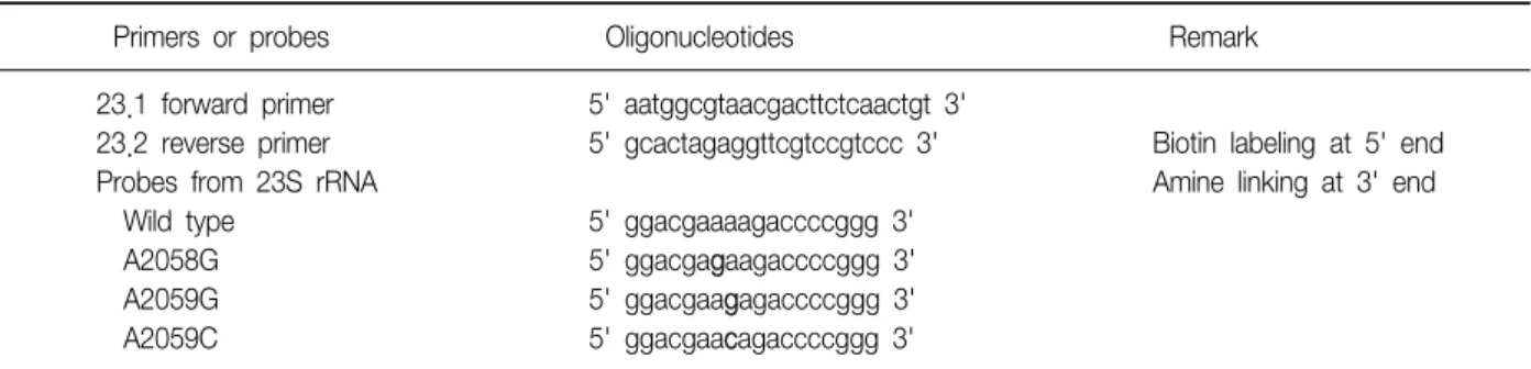
Table 1. Primers and probes for detection of mutation in 23S rRNA gene
Primers or probes Oligonucleotides Remark
23.1 forward primer 5' aatggcgtaacgacttctcaactgt 3'
23.2 reverse primer 5' gcactagaggttcgtccgtccc 3' Biotin labeling at 5' end
Probes from 23S rRNA Amine linking at 3' end
Wild type 5' ggacgaaaagaccccggg 3'
A2058G 5' ggacgagaagaccccggg 3'
A2059G 5' ggacgaagagaccccggg 3'
A2059C 5' ggacgaacagaccccggg 3'
로 동정된 임상 균주 중에서 배양상태가 양호한 220 균주 를 시험에 사용하였다. Reference 균주로는 M. abscessus ATCC 19927와 Staphylococcus aureus ATCC 29213을 사 용하였다.
2. 중합효소연쇄반응
돌연변이 부위인 23S rRNA 유전자의 V domain을 증폭 을 위한 primer들과 중합효소연쇄반응법은 Nash 등의 방 법을 참고하여 실시하였고10, primer들에 대한 염기서열은 Table 1에 나타내었다.
중합효소연쇄반응 용량은 100μl이었고, 구성성분은 10 x Taq polymerase buffer, 10 mM dNTP, 10 pM pri- mer, 2 U Taq polymerase와 10 ul DNA로 구성되었다.
중합효소연쇄반응은 GeneAmp PCR system 9700 (Applied Biosystems, Foster city, CA, USA)으로 실시하 였다. 처음 denaturation 반응은 94oC에서 5분하였고, 이 후 94oC 5분, 60oC 2분, 72oC 2분씩 35회 반복하였으며 마지막 extention 반응으로 72oC 10분을 실시하였다. 이 중합효소연쇄반응 산물의 크기는 대략 720 bp이었다.
3. 역교잡반응법
돌연변이 균주 검출을 위한 probe의 염기서열 및 특징들 은 Table 1에 나타내었다. 역교잡반응은 Honore 등의 방법 을 변형하여 실시하였다14. Biodyne C membrane (Pall corporation, New York, USA) 위에 3' 말단에 amino-기를 첨가한 probe를 공유결합시키고, 활성화한 다음에 중합효 소연쇄반응 산물을 역교잡하여 반응시켰다. Probe와 교잡 된 중합효소연쇄반응 산물은 streptavidin-alkaline phopha- tase를 처리한 후 NBT/BCIP 기질로 발색시켰다.
4. 염기서열분석
모든 임상 균주들의 23S rRNA 유전자의 V domain 지
역 중합효소연쇄반응 산물들을 3730x DNA analyzer (Applied Biosystems, Foster city, CA, USA)를 이용하여 염기서열을 확인하였다.
5. 약제감수성검사법
Clarithromycin 약제에 대한 감수성검사는 NCCLS 검사 법에 의한 broth microdilution 시험법으로 실시하였다15. 50μg/ml 2, 3-Diphenyl-5-(2-thienyl)-tetrazolium chlor- ide (STC: TOKYO KASEI KOGYO CO, LTD., Tokyo, Japan)와 약제 농도 1,024μg/ml부터 1/2배씩 희석하여 0.5μg/ml까지의 clarithromycin 약제가 함유된 Muller Hillton II (Becton Dickinson, Sparks, MD, USA) broth를 microplate 각 well에 100μl씩 분주하였다. Well당 104 cell이 되도록 균액을 접종하여 30oC에서 3∼5일 배양한 후 minimal inhibitory concentration (MIC)을 확인하였 다. 내성 MIC 기준은 ≥8μg/ml으로 하였다15.
결 과 1. 역교잡반응 및 염기서열분석
역교잡반응법을 이용하여 220개의 M. abscessus 임상 균주 중에서 3개의 돌연변이 균주들을 검출하였으며, 중 합효소산물에 대한 염기서열분석을 통하여 이들 균주들 만이 23S rRNA 유전자의 V domain 지역에 점 돌연변이가 형성되어 있음을 확인하였다. 분리된 균주들의 점 돌연변 이 형태는 2058번의 adenine 염기가 guanine 염기로 치환 되었음을 확인하였다(Table 2).
2. Clarithromycin 약제에 대한 약제감수성검사
Broth microdilution 시험법으로 약제감수성검사를 실시 한 결과에서는 220개의 M. abscessus 임상균주 중에서 7개 의 내성 균주들을 검출하였고, 이들의 MIC는 >64μg/ml
Table 2. Clarithromycin MICs and sequencing results for the peptidyltransferase region of the 23S rRNA gene in resistant strains of M. abscessus clinical isolates
Strain Clarithromycin MIC (μg/ml)
23S peptidyltransferase
region Reference strains
M. abscessus ATCC 19927
≤2 −
Staphylococcus aureus ATCC 29213
≤0.25
Mutants
101 >512 A2058G
205 >512 A2058G
249 >256 A2058G
Non-mutants
12 >512 −
173 >512 −
179 >64 −
207 >512 −
- A2058A and A2059A
이었다. 검출된 내성 균주들에는 RHA에 의해 검출된 돌 연변이균들도 포함되었다. Reference 균주의 MIC는 ≤2 μg/ml와 ≤0.25μg/ml이었다(Table 2).
고 찰
항생제 내성에 대한 분자생물학적 기전은 여러 종류가 있으나, 일반적으로 약제 방출, 약제 비활성 혹은 약제 표 적 위치의 변화 등의 특징을 언급할 수 있다.
Macrolide 계열 항생제들의 표적 위치는 리보솜의 50S subunit이며, macrolide 계열 항생제에 대한 내성을 지닌 임상 균주들은 리보솜 50S subunit 내 23S rRNA 유전자의 특정 염기서열이 치환되는 특징을 나타내거나16, Erm methyltransferase에 의하여 23S rRNA 유전자의 특정 염 기부분이 methylation 되어 내성을 나타내는 것으로 보고 되었다17.
Erythromycin과 clarithromycin 같은 macrolide 계열 항생제들은 아마도 리보솜으로부터 peptidly-tRNA를 분 리하여 펩티드 사슬의 형성을 저해함으로써 단백질 합성 초기 단계에서 단백질 형성을 억제하는 역할을 하며18,19, 새로운 리보솜 50S subunits의 조합을 방해하여 세포 내에
서 리보솜의 기능을 점차적으로 소멸시켜 세포 성장을 억 제시키는 기능을 한다20.
비결핵균에 대한 macrolide 계열 항생제 내성에 대한 연구는 M. intracellulare, M. avium, M. chelonae와 M.
abscessus 임상 균주들을 대상으로 23S rRNA 유전자 내의 점 돌연변이에 의한 내성 기전을 보고하였고9-11, 또한 Nash 등은 RGM 균주들 간에 macrolide 계열 항생제 내성 에 관여하는 다양한 Erm 단백질들의 유전자들을 발견하 였다8.
Wallace 등은 clarithromycin으로 치료하여 재발한 환 자들로부터 분리한 M. abscessus 임상 균주들을 대상으로 한 실험에서 이들 균주들이 clarithromycin에 내성을 나타 냈으며, 내성 균주들의 clarithromycin MIC는 ≥16μg/ml 이었고, 23S rRNA 유전자 내에서 염기번호 2058 adenine 이 guanine으로, 염기번호 2059 adenine이 guanine 혹은 cytosine으로 치환된 것을 보고하였다11. 또한 M. ab- scessus 임상 내성 균주와 동일하게 M. intracellulare와 M. avium 임상 내성균주의 경우에도 23S rRNA 유전자의 같은 염기번호 위치에서 다양한 점 돌연변이(A2058G, C, U와 A2059C)가 나타내는 것을 보고하였다9,10.
본 실험에서는 macrolide 계열 항생제가 치료 후 나타 나는 획득내성으로써, 23S rRNA 유전자내에 점 돌연변이 를 일으킨다는 보고를 기초로 하여 역교잡반응법을 개발 하였고, 이 방법을 이용하여 염기번호 2058 adenine이 guanine으로 치환된 형태의 3개의 임상 내성 균주를 확보 하였다. 또한, 약제감수성검사를 이용하여 7개의 임상 내 성 균주를 확보하였는데, 이들 중에는 역교잡반응법에 의 해 분리된 점 돌연변이 균주들도 포함되어 있었다. 그러 나, 염기서열분석 결과에서는 점 돌연변이 균주들의 제외 한 나머지 내성 균주들은 감수성 균주들과 동일한 염기서 열을 나타내고 있었다. 내성으로 나타난 모든 균주들은 MIC가 >64μg/ml으로 고농도 내성을 나타내었다.
본 실험에 사용된 임상 균주들에 대한 환자들의 치료력 은 의뢰 기관의 복잡성과 기록 부재로 인하여 확인할 수가 없었으며, 그러한 이유로 분리한 내성 균주들의 특성이 획득내성인지 자연내성인지에 대해서는 분석할 수는 없 었다. 그러나, M. abscess 균주에서는 점 돌연변이와 다른 종류의 내성 특성을 나타내고 있음을 확인할 수 있었다.
또한 치료 후 분리된 내성 균주들의 특징을 기초로 하여 개발한 역교잡반응법으로 점 돌연변이 균주들에 대해서 는 정확히 분리할 수 있게 되었으나, 실효성에 대해서는 보다 광범위한 검증이 필요하다고 판단되었다.
아직까지 국내의 비결핵균 발생 빈도는 낮은 상황이다.
그러나 점차 증가되는 추세에서 중요시 되고 있는 비결핵 균종들에 대한 내성균들을 확보하여, 치료력과 내성 특성 과의 관계 및 내성 기전들을 분석함으로써 보다 적합하고 신속한 검사법을 개발해야 할 것으로 판단된다.
요 약
연구배경: Mycobacterium abscessus는 빠른 성장성을 지닌 비결핵균중에서 높은 병원성과 약제 내성을 나타내 는 종이며, clarithromycin과 azithromycin 항결핵제가 M.
abscessus에 효과가 있는 유일한 경구용 항결핵제이다.
본 연구에서는 역교잡반응법과 약제감수성검사법을 이용 하여 clarithromycin 약제에 대한 M. abscessus 임상 내성 균주 검출을 시도하였다.
방 법: 역교잡반응법과 약제감수성검사법을 이용하여 220개의 M. abscessus 임상 균주를 대상으로 내성 균주를 분리하였다.
결 과: 약제감수성검사법으로 7개의 임상 내성 균주 들을 검출하였고, 이들 중 3개의 내성 균주는 점 돌연변이 균주로서 역교잡반응법으로도 확인하였다.
결 론: M. abscess 균주에서는 점 돌연변이 및 다른 종류의 내성 특성을 나타내고 있음을 확인할 수 있었다.
참 고 문 헌
1. Brown-Elliott BA, Griffith DE, Wallace RJ Jr. Newly de- scribed or emerging human species of nontuberculous mycobacteria. Infect Dis Clin North Am 2002;16:187- 220.
2. Brown-Elliott BA, Wallace RJ Jr. Clinical and taxonomic status of pathogenic nonpigmented or late-pigmenting rapidly growing mycobacteria. Clin Microbiol Rev 2002;15:716-46.
3. Diagnosis and treatment of disease caused by non- tuberculous mycobacteria. This official statement of the American Thoracic Society was approved by the Board of Directors, March 1997. Medical Section of the American Lung Association. Am J Respir Crit Care Med 1997;156:S1-25.
4. Brown BA, Springer B, Steingrube VA, Wilson RW, Pfyffer GE, Garcia MJ, et al. Mycobacterium wolinskyi sp. nov. and Mycobacterium goodii sp. nov., two new rapidly growing species related to Mycobacterium
smegmatis and associated with human wound in- fections: a cooperative study from the International Working Group on Mycobacterial Taxonomy. Int J Syst Bacteriol 1999;49:1493-511.
5. Buriankova K, Doucet-Populaire F, Dorson O, Gondran A, Ghnassia JC, Weiser J, et al. Molecular basis of in- trinsic macrolide resistance in the Mycobacterium tu- berculosis complex. Antimicrob Agents Chemother 2004;48:143-50.
6. Nash KA. Intrinsic macrolide resistance in Mycobacte- rium smegmatis is conferred by a novel erm gene, erm (38). Antimicrob Agents Chemother 2003;47:3053-60.
7. Nash KA, Zhang Y, Brown-Elliott BA, Wallace RJ Jr.
Molecular basis of intrinsic macrolide resistance in clin- ical isolates of Mycobacterium fortuitum. J Antimicrob Chemother 2005;55:170-7.
8. Nash KA, Andini N, Zhang Y, Brown-Elliott BA, Wallace RJ Jr. Intrinsic macrolide resistance in rapidly growing mycobacteria. Antimicrob Agents Chemother 2006;50:3476-8.
9. Meier A, Kirschner P, Springer B, Steingrube VA, Brown BA, Wallace RJ Jr, et al. Identification of muta- tions in 23S rRNA gene of clarithromycin-resistant Mycobacterium intracellulare. Antimicrob Agents Che- mother 1994;38:381-4.
10. Nash KA, Inderlied CB. Genetic basis of macrolide re- sistance in Mycobacterium avium isolated from patients with disseminated disease. Antimicrob Agents Chemo- ther 1995;39:2625-30.
11. Wallace RJ Jr, Meier A, Brown BA, Zhang Y, Sander P, Onyi GO, et al. Genetic basis for clarithromycin re- sistance among isolates of Mycobacterium chelonae and Mycobacterium abscessus. Antimicrob Agents Chemother 1996;40:1676-81.
12. Coleman K, Athalye M, Clancey A, Davison M, Payne DJ, Perry CR, et al. Bacterial resistance mechanisms as therapeutic targets. J Antimicrob Chemother 1994;33:
1091-116.
13. Fierro JF, Hardisson C, Salas JA. Involvement of cell impermeability in resistance to macrolides in some pro- ducer streptomycetes. J Antibiot (Tokyo) 1988;41:142-4.
14. Honore N, Roche PW, Grosset JH, Cole ST. A method for rapid detection of rifampicin-resistant isolates of Mycobacterium leprae. Lepr Rev 2001;72:441-8.
15. NCCLS. Susceptibility testing of Mycobacteria, Nocar- dia, and other aerobic actinomycetes: approves stan- dard. Wayne, Pennsylvania: NCCLS; 2003.
16. Douthwaite S, Aagaard C. Erythromycin binding is re- duced in ribosomes with conformational alterations in
the 23S rRNA peptidyl transferase loop. J Mol Biol 1993;232:725-31.
17. Goldman RC, Kadam SK. Binding of novel macrolide structures to macrolides-lincosamides-streptogramin B-resistant ribosomes inhibits protein synthesis and bacterial growth. Antimicrob Agents Chemother 1989;
33:1058-66.
18. Andersson S, Kurland CG. Elongating ribosomes in vivo are refractory to erythromycin. Biochimie 1987;69:
901-4.
19. Menninger JR. Mechanism of inhibition of protein syn- thesis by macrolide and lincosamide antibiotics. J Basic Clin Physiol Pharmacol 1995;6:229-50.
20. Champney WS, Tober CL. Superiority of 11,12 carbo- nate macrolide antibiotics as inhibitors of translation and 50S ribosomal subunit formation in Staphylococcus aureus cells. Curr Microbiol 1999;38:342-8.