449
Saccharomyces cerevisiae 의 회분식 배양에서 세라마이드의 생산
김 세 경 ․ 노 용 호 ․ †윤 현 식 인하대학교 생물공학과
(접수 : 2008. 10. 1., 게재승인 : 2008. 10. 22.)
The ceramide contents of Saccharomyces cerevisiae in batch culture
Se Kyung Kim, Yong Ho Noh, and Hyun Shik Yun†
Department of Biological Engineering, Inha University, 253 Yonghyundong Namku, Incheon 402-751, Korea (Received : 2008. 10. 1., Accepted : 2008. 10. 22.)
Ceramide has become a widely used ingredient in cosmetic and pharmaceutical industries, however, only a few yeast strains were investigated for the synthesis of ceramide and the concentration was very low. Ceramide is not only a core intermediate of sphingolipids but also an important modulator of many cellular events including apoptosis, cell cycle arrest, senescence, differentiation, and stress responses. In this study S.cerevisiae was grown in a batch culture and the cellular content of ceramide was measured at different growth phases. The ceramide content was highest at stationary phase and 2.01 mg ceramide/g cell was obtained.
Key Words : Saccharomyces cerevisiae, ceramide, growth phases
서 론
1)
Sphingolipid에 대한 연구는 1874년 Johann Thudichum이 뇌추 출물에서 미지의 지질로서 cerebroside, sphingomyelin을 발견한 때부터 시작되었다(1). 처음에는 뇌, 중추신경계를 중심으로 조직 에서의 구조해석과 sphingolipidosis라는 지질축적증의 병태해명 을 연구의 두 개의 축으로 하여 연구되어 왔다. 약 100년간에 걸친 연구결과 생체막의 구성성분으로서 세포를 구조적으로 유지 하는 것과 생체 내에서의 영양소로서의 역할이 지질의 중요한 기능으로 생각되어 왔다. 1986년에 sphingosine이 in vitro에 서 protein kinase C (PKC)의 강력한 저해제임이 보고되었고(2), sphingolipid에 대한 인식이 단순히 세포막의 구성성분에서 생체 기능을 조절하는 생체내 활성분자로 새롭게 바뀌었다. 지금은 ceramide와 sphingosine으로 대표되는 apoptosis 유도 및 세포사멸 의 신호전달물질로서의 sphingolipid에 대한 연구와 sphingosine 1-phosphate와 sphingosylphosphorylcholine으로 대표되는 세포막 수용체를 가지는 세포간 신호전달물질에 관한 연구가 많이 이루 어지고 있다(3, 4).
†Corresponding Author : Department of Biological Engineering, Inha University, 253 Yonghyundong Namku, Incheon 402-751, Korea
Tel : +82-32-860-7517, Fax : +82-32-876-8751 E-mail : [email protected]
Ceramide 생산의 대표적인 방법은 천연물에서 추출하는 것 이다. 천연으로부터의 추출하는 ceramide의 제조는 3가지 source 에 의한다. 하나는 동물유래의 것으로 주로 소, 돼지 등의 뇌 또는 피부로부터 추출하는 것이다. 그러나 동물기원의 ceramide 는 광우병 및 국제적인 환경단체 등에 의한 동물성원료 사용 금지 운동 등으로 인해 공급이 급격히 줄고 있다(5). 다른 하나 는 대두, 밀, 쌀 등의 식물에서 추출하는 것으로 주로 cerebroside 의 형태이다. Cerebroside는 ceramide의 또 다른 형태로 체내 에서 효소 등에 의해 당 부분이 해리되어 ceramide의 기능을 수행한다. 그러나 cerabrosid는 식물에 존재하는 양이 적어 그 추출량이 극히 적다. 그리고 박테리아나 원핵생물, 곰팡이 등에 존재하는 ceramide를 추출하는 방법이 있다.
천연유래 ceramide의 가격이나 순도 등에서의 문제점으로 인해 이의 해결의 한 방안으로 화학적인 방법이 모색되고 있 다. 그 중 하나가 전합성에 의해 ceramide를 개발하려는 시도 를 일반적으로 serine 또는 단당류에서 출발하여 8-13 단계의 공정을 거치는 것으로 입체화학적 구조 및 공정의 난이도로 인해 많은 연구에도 불구하고 대량 생산에 성공한 예는 아직 없다. 이외에도 생물공학적으로 효소를 이용하여 전구체를 제조한 후 합성에 의해 지방산을 결합시켜 ceramide를 제조 하는 방법이 알려져 있다. Ceramide 제조의 또 다른 방법은 유사 ceramide의 개발이다. 유사 ceramide는 천연 ceramide와 비슷한 구조를 갖고 있어 장벽기능을 발휘하면서도 천연 ceramide가 가격적인 문제로 화장품이나 피부 관련 외용제에 한국생물공학회지 제23권 제5호
Korean J. Biotechnol. Bioeng.
Vol. 23, No. 5, 449-451(2008)
450 Korean J. Biotechnol. Bioeng., Vol. 23, No. 5
효능을 나타낼 정도의 배합이 어려웠던 현실을 극복할 수 있 도록 경제성을 높인 것이다.
다른 미생물과 비교하여 볼 때 yeast는 빠른 생장, 비독성 등 여러가지 재조합 균주로서의 장점을 가지고 있다. Yeast에서의 ceramide 생산은 지금까지 Saccharomyces cerevisiae와 Torulopsis (Candida utilis)에서만 발견되고 있다(6). 본 연구는 Saccharomyces cerevisiae 배양에서 세포 생장에 따라 ceramide의 생산에 미치 는 영향을 알아보는데 그 목적이 있다.
재료 및 방법
효모 균주 및 배양 조건
본 연구에 사용된 균주는 한국 미생물 보존 센터 (KCCM) 에서 분양 받았으며, phytosphingosine-sensitive한 특징을 갖고 있는 Saccharomyces cerevisiae 50515 이다. Saccharomyces cerevisiae 배양을 위한 배지로는 YPD를 사용하였으며 배지 의 조성은 glucose (Samchun pure chemical Co., LTD) 20 g/l, bacto pepton (BD, U.S.A.) 20 g/l, yeast extract (BD, U.S.A.) 10 g/l 이다. 배양은 2 L 삼각 플라스크에 500 ml YPD 배지 를 분주한 후 shaking incubator (Vision Co., Korea)를 이용하여 180 rpm, 30℃에서 배양하였다.
Lipids의 추출
배양이 끝난 균주는 원심분리기 Centrifuge 5403 (Eppendorf Co., Germany)을 사용하여 균체만 회수한 후 동결건조 (Bondiro Ilshin Lab. Co., Korea)하였다. 동결건조 된 균주 1 g에 용매 I (클로로포름:메탄올=1:2, v/v)과 glass bead 2 g을 넣고 강 하게 vortexing 하여 균주를 파쇄 하였다.
파쇄된 균주는 용매 II (클로로포름:메탄올=2:1, v/v) 100 ml 를 첨가하여 30분간 반응시켰다. 반응한 용액은 filtering 하여 60℃에서 evaporation 시켰다. 이와 같은 과정을 4회 더 반복 하였다(7). 이 과정은 샘플내에 있는 polar components와 non-lipid contaminants를 제거하기 위한 과정이다.
Ceramide의 분리 및 정제
추출된 total lipid에서 sphingolipid 부분을 얻기 위해 mild alkaline hydrolysis를 수행하였다(7). 클로로포름을 첨가한 샘플 에 methanol carbon tetrachloride (5:1, v/v) 30 ml와 0.2 M methanolic NaOH 60 ml를 첨가한 후 한 시간동안 반응시켰다.
반응 후 증류수를 첨가하고 1M acetic acid로 중화시켰다. 중화 된 샘플은 4℃에서 12시간동안 방치하여 층분리를 한 후 클로로 포름층만 분리하였다.
HPLC를 이용한 ceramide의 분석
분리된 ceramide는 클로로포름:메탄올 (2:1, v/v)로 10 ml를 맞춘 후 0.2 ㎛ RC filter (Satorius, Germany)로 필터링하여 분석에 사용하였다. 사용된 HPLC는 Acme 9000 HPLC system (Younglin Instrument, Korea)이며 data system은 Autochro 3000 (Younglin Instrument, Korea), detection system은 ELSD Sedex 75 (Sedere, France) 이다. ELSD 조건은 drift temperature 40℃, 분사 가스는 질소 3.5 bar이었다. 분석에 사용된 column은
Spherisorb® 5 mm Silica (Waters, U.S.A.)으로 규격은 4.6×250 mm 이고 이동상은 1.0 ml/min의 클로로포름:메탄올 (96:4, v/v) 이었다(8, 9).
Glucose 분석
배양액은 0.2 ㎛ cellulose acetate filter (Satorius Co., Germany) 로 filtering 하여 RI detector LC-10AD (Shimadzu, Japan)을 장착한 HPLC 10A system (Shimadzu, Japan)으로 분석하였다.
Glucose 분석에 사용된 column은 Aminex HPX-87H (Bio-Rad Laboratories, U.S.A.)이며 0.5M 황산을 0.5 ml/min의 유속으로 흘려주었다.
결과 및 고찰
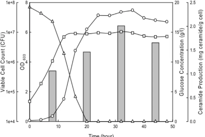
Lipid는 세포내 신호전달체계로서의 기능을 하는 것으로 알려져 왔다. Ceramide나 sphingosine-1-phosphate로 분류되는 sphingolipid는 칼슘 동원, 세포 성장, 세포자살등을 조절하는 강력한 전달체이다. Ceramide 분석은 Ceramide Standard (Avanti U.S.A.)를 기준물질로 하여 ELSD를 장착한 HPLC로 측정하였 으며(Fig. 1) S. cerevisiae의 각기 다른 생장 단계에서 측정하였 다(Fig. 2). 샘플은 exponential phase, early stationary phase, 그리고 균주가 생장이 stationary phase에 도달한 후 12시간, 24시간 뒤에 회수하여 cell 내 ceramide 함량을 측정하였다.
세포 생장이 20시간이 된 후에는 glucose 함량이 완전히 소비 되는 것을 볼 수 있었고, 32시간 뒤에는 viable cell count가 점점 줄어드는 것을 알 수 있었다.
0 2 4 6 8 10 12 14 16 18 20
0 10 20 30 40 50 60 70 80
Intensity (mV)
Time (min)
Figure 1. Chromatogram of ceramide.
(
᠁Ceramide standard, - Cell extract)
Lipid는 세포 membrane에서 에너지 저장과 cell signaling에
사용된다. S. cerevisiae의 ceramide 함량은 stationary phase에
접어든 후 12시간 후에 가장 높은 것으로 나타났다. Ceramide
생산량이 세포 생장에 따르지 않는 것으로 보아 ceramide가
세포막에 사용되기 보다는 signaling molecule로서 더 사용된
다고 사료된다. 세포 생장에서 32시간에 ceramide 생산량은
2.01 mg ceramide/g cell이었다. 이것은 세포 생장의 대수기보다
1.9배, 감속기보다 1.4배 많은 양이다. 세포 생장이 정지기에
들어서면 배지내 glucose의 양은 고갈되고, 세포는 탄소원 결핍
451 Kim, S. K., The ceramide contents of Saccharomyces cerevisiae in batch culture
으로 인한 스트레스에 접어들게 된다. 그 결과 세포 생장 32시간 후의 ceramide의 양이 감소하는 것으로 추정된다.
Time (hour)
0 10 20 30 40 50
Ceramide Production (mg ceramide/g cell)
0.0 0.5 1.0 1.5 2.0 2.5
Glucose Concentration (g/l)
0 5 10 15 20
OD600
0 2 4 6 8
Viable Cell Count (CFU)
1e+4 1e+5 1e+6 1e+7 1e+8
Figure 2. Changes in cell concentration, glucose concentration, and ceramide content in a batch culture of S. cerevisiae.
(-○- OD
600, -□-viable cellcount, -△- glucose concentration,
▓ceramide production)
요 약
Saccharomyces cerevisiae를 배양하여 ceramide를 생산하였다.
각기 다른 growth phase에서의 ceramide 생산량을 비교 실험 하였다. Ceramide는 ELSD가 장착된 HPLC를 통하여 분석하였 으며 stationary phase에 접어든 후 12시간 후에 회수, 추출된 양 (2.01 mg ceramide/g cell)이 최대인 것으로 나타났다.
감 사