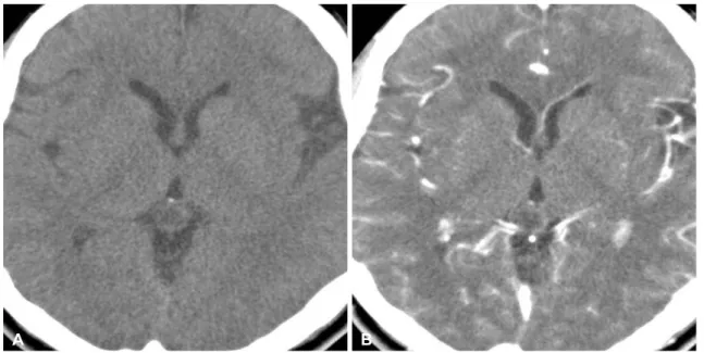
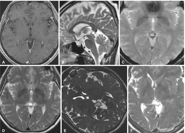
Pineal Cyst Apoplexy: A Rare Complication of Common Entity
5
0
0
전체 글
(2)
(3)
(4)
(5)
수치
관련 문서