Received: Oct 14, 2018 Revised: Jan 28, 2019 Accepted: Feb 5, 2019 Published online Mar 15, 2019 Correspondence to: Jun-Kyu Suh https://orcid.org/0000-0002-1812-9449
National Research Center for Sexual Medicine and Department of Urology, Inha University School of Medicine, 27 Inhang-ro, Jung-gu, Incheon 22332, Korea.
Tel: +82-32-890-3441, Fax: +82-32-890-3097, E-mail: [email protected] Correspondence to: Ji-Kan Ryu https://orcid.org/0000-0003-2125-0212
National Research Center for Sexual Medicine and Department of Urology, Inha University School of Medicine, 27 Inhang-ro, Jung-gu, Incheon 22332, Korea.
Tel: +82-32-890-3505, Fax: +82-32-890-3099, E-mail: [email protected]
*These authors contributed equally to this study as co-first authors.
Copyright © 2020 Korean Society for Sexual Medicine and Andrology
pISSN: 2287-4208 / eISSN: 2287-4690 World J Mens Health 2020 Jan 38(1): 123-131 https://doi.org/10.5534/wjmh.180091
A Simple and Nonenzymatic Method to Isolate Human Corpus Cavernosum Endothelial Cells and Pericytes for the Study of Erectile Dysfunction
Guo Nan Yin1,* , Jiyeon Ock1,* , Min-Ji Choi1 , Kang-Moon Song1 , Kalyan Ghatak1 ,
Nguyen Nhat Minh1 , Mi-Hye Kwon1 , Do-Hwan Seong1 , Hai-Rong Jin2 , Ji-Kan Ryu1 , Jun-Kyu Suh1
1National Research Center for Sexual Medicine and Department of Urology, Inha University School of Medicine, Incheon, Korea,
2Department of Urology, Yantai Yuhuangding Hospital Affiliated to Medical College of Qingdao University, Yantai, China
Purpose: To establish a simple and nonenzymatic technique to isolate endothelial cells (ECs) and pericytes from human cor- pus cavernosum tissue and to evaluate the angiogenic ability of the human cavernous EC or pericytes for the study of high glucose-induced angiopathy.
Materials and Methods: For primary human cavernous EC culture, cavernous tissues were implanted into Matrigel in dishes.
For primary human cavernous pericyte culture, cavernous tissues were settled by gravity into dishes. We performed immuno- cytochemistry and Western blot to determine phenotype and morphologic changes from passage 1 to 5. The primary cultured cells were exposed to a normal-glucose (5 mmol/L) or a high-glucose (30 mmol/L) condition, and then tube formation assay was done.
Results: We successfully isolated high-purity EC and pericytes from human corpus cavernosum tissue. Primary cultured EC showed highly positive staining for von Willebrand factor, and pericyte revealed positive staining for NG2 and platelet-de- rived growth factor receptor-β. Primary cultured EC and pericytes maintained their cellular characteristics up to passage 2 or 3. However, we observed significant changes in their typical phenotype from the passage 4 and morphological characteris- tics from the passage 3. Human cavernous EC or pericytes formed well-organized capillary-like structures in normal-glucose condition, whereas severely impaired tube formation was detected in high-glucose condition.
Conclusions: This study provides a simple and nonenzymatic method for primary culture of human cavernous EC and peri- cytes. Our study will aid us to understand the pathophysiology of diabetic erectile dysfunction, and also be a valuable tool for determining the efficacy of candidate therapeutic targets.
Keywords: Diabetes mellitus; Endothelial cell; Erectile dysfunction; Pericytes
This is an Open Access article distributed under the terms of the Creative Commons Attribution Non-Commercial License (http://creativecommons.org/licenses/by-nc/4.0) which permits unrestricted non-commercial use, distribution, and reproduction in any medium, provided the original work is properly cited.
INTRODUCTION
The penis has a specialized vascular bed and penile erection requires well-coordinated interactions between vascular endothelial cells (ECs), smooth muscle cells, pericytes, and neuronal cells [1-3]. The vascular endo- thelium plays a critical role in the regulation of blood flow and vascular tone [4]. Pericytes surrounding the ECs of microvessels are known to plays a critical role in maintaining vascular homeostasis [5]. We recently documented the differential distribution of EC and pericytes in the erectile tissue of mice and human by using immunohistochemistry [2,3].
Over the past decades, in vitro cell-based models were widely used in the field of vascular biology [6-8] and have given us invaluable insight for understanding physiologic angiogenesis as well as pathophygiology of various vascular diseases. Many different methods for the isolation and cultivation of EC and pericytes from the human umbilical vein, placenta, skeletal muscle, and brain have been reported [9-14]. Although the en- zymatic digestion method is one of the most widely used technique, the enzymes can exert harmful effects on cell surface proteins and negatively affect on cellu- lar viability [15]. Recently, we successfully isolated the EC and pericytes from mouse corpus cavernosum tissue [2,16]. From the clinical perspective of view, however, it is more advantageous to use human cavernous EC and pericytes than use the cells originated from animals.
In the present study, we for the first time established protocol to isolate EC and pericytes from human erectile tissues. Because it is well-known that the passage num- ber of primary cultured cells influences phenotype and morphology of the cells [17], we further determined the changes in phenotypic characteristics and morphologi- cal feature of primary cultured cells according to the passages. Finally, we determined the effects of different glucose concentrations on tube formation by cultivating EC or pericytes under normal- or high-glucose condition.
MATERIALS AND METHODS
1. Human corpus cavernosum tissue and ethics statement
Human corpus cavernosum tissues were obtained from three patients with Peyronie’s disease who have normal erectile function during reconstructive penile surgery: 1 undergoing penoplasty using saphenous vein
graft (age; 55 years), 2 undergoing Nesbit operation (age; 70 and 47 years, respectively). No patient reported diabetes or other cardiovascular diseases. All tissue do- nors provided informed consent, and the experiments were approved by the Ethics Committee and the inter- nal review board of Inha University (No. 2007-730).
2. Isolation and culture of human cavernous endothelial cell
The human cavernous EC were prepared and main- tained as we described previously [16]. Briefly, the corpus cavernosum tissues were cut into two or three pieces and the samples plated on Matrigel-coated (Bec- ton Dickinson, Mountain View, CA, USA) 60-mm cell culture dishes. The Matrigel was polymerized with a 5-minute incubation period at 37°C and 3 mL of complement medium 199 (GIBCO, Carlsbad, CA, USA) supplemented with 20% fetal bovine serum (FBS), 1%
penicillin/streptomycin, 0.5 mg/mL heparin (Sigma-Al- drich, St. Louis, MO, USA), and 5 ng/mL vascular endo- thelial growth factor (R&D Systems Inc., Minneapolis, MN, USA) was added to the dishes. The dishes were incubated at 37°C in a 5% CO2 atmosphere. After the cells were confluent and spread over the bottom of the dish (~3 weeks after the start of culture), only sprout- ing cells were used for subcultivation. The sprouting cells were seeded onto dishes coated with 0.2% gelatin (Sigma-Aldrich).
3. Isolation and culture of human cavernous pericytes
The human cavernous pericytes were prepared and maintained as described previously [2,18]. The fresh adult corpus cavernosum tissues were collected after surgery and transferred into sterile vials containing Hank’s balanced salt solution (GIBCO) and washed twice in phosphate buffer saline. The corpus caver- nosum tissues were cut into several 1-mm pieces and the fragmented pieces settled by gravity into collagen I-coated 35-mm cell culture dishes (Becton Dickin- son). After 30 minutes of incubation at 37°C with 300 µL complement Dulbecco’s modified Eagle Medium (DMEM; GIBCO) supplemented with 10% FBS, 1%
penicillin/streptomycin, and 10 nM human pigment epithelium-derived factor (Sigma-Aldrich), we added an additional 900 µL complement medium and incubated the samples at 37°C in a 5% CO2 atmosphere. The me- dium was changed every 2 days. After the cells were
confluent and spread over the bottom of the dish (~3 weeks after the start of culture), only sprouting cells were used for subcultivation. The sprouting cells were seeded onto dishes coated with 50 µL/mL collagen I (Advanced BioMatrix, San Diego, CA, USA).
4. Characterization of isolated cells
To determine cell type, cells were stained with an- tibody to von Willebrand factor (vWF, an EC marker;
Santa Cruz Biotechnology, Santa Cruz, CA, USA; 1:50), CD34 (an EC marker; Abcam, Cambridge, MA, USA;
1:50), NG2 chondroitin sulfate proteoglycan (a peri- cytes marker; Millipore, San Francisco, CA, USA; 1:50), platelet-derived growth factor receptor-β (PDGFR-β, a pericytes marker; Santa Cruz Biotechnology; 1:50), fibroblast-specific protein 1 (FSP1, a fibroblast marker;
Santa Cruz Biotechnology; 1:50), or DAPI (a nucleus marker; Vector Laboratories Inc., Burlingame, CA, USA). Signals were visualized and digital images were obtained with a confocal microscope (FV1000; Olympus, Tokyo, Japan).
5. In vitro tube formation assay
To evaluate the angiogenic ability of the human cavernous EC or pericytes in normal- or high-glucose condition, the primary cultured human cavernous EC or pericytes were serum-starved (medium 199 supple- mented with 2% FBS and 1% penicillin/streptomycin was used for EC culture, and DMEM supplemented with 2% FBS and 1% penicillin/streptomycin was used for pericyte culture) for 24 hours and were exposed to a normal-glucose (5 mmol/L) or a high-glucose (30 mmol/L) condition for 48 hours as previously described [19-21]. The tube formation assay was performed as previously described [16]. About 50 µL of growth fac- tor-reduced Matrigel (Becton Dickinson) was dispensed into 96-well tissue culture plates at 4°C. After gelling at 37°C for at least 30 minutes, the conditioned hu- man cavernous EC or pericytes were seeded onto the gel at 2×10⁴ cells/well in 200 µL of starvation medium.
The assay was performed in a CO2 incubator and the plates were incubated at 37°C for 24 hours. Images were obtained with a phase-contrast microscope and the numbers of branch points in each well of the plate were counted at a screen magnification of ×40.
6. Western blot
Equal amounts of protein from whole-cell extracts (50
μg/lane) were electrophoresed on 10% sodium dodec- ylsulfate-polyacrylamide gels, transferred to nitrocel- lulose membranes, and probed with antibody to CD34 (an EC marker; Abcam; 1:200), PDGFR-β (a pericytes marker; Santa Cruz Biotechnology; 1:200), or β-actin (a loading control; Abcam; 1:2,000). Results were quanti- fied by densitometry.
7. Statistical analysis
Results are expressed as the mean±standard errors.
We used the Mann–Whitney U-tests or Kruskal–Wal- lis tests for group comparison. We performed statistical analysis with SigmaStat 3.5 software (Systat Software Inc., Richmond, CA, USA). The p-values less than 5%
were considered significant.
RESULTS
1. Localization of endothelial cell and pericytes in the human corpus cavernosum tissue
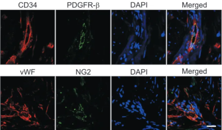
Immunofluorescent double staining of human cav- ernous tissue with antibodies against two different EC markers (CD34 and vWF) or pericytes markers (PDGFR-β and NG2) revealed that EC and pericytes are closely distributed in human erectile tissues (Fig. 1).
2. Isolation of endothelial cell and pericytes from human corpus cavernosum tissue
Representative images of the cells cultivated for 14 days were shown at Fig. 2A and 2C. After cells
CD34 PDGFR- DAPI Merged
vWF NG2 DAPI Merged
Fig. 1. Localization of endothelial cells and pericytes in human cor- pus cavernosum tissue. Immunofluorescent staining of human penile tissue (n=3) performed with antibodies against CD34 or von Willebrand factor (vWF, endothelial cell markers, red) and platelet- derived growth factor receptor-β (PDGFR-β) or NG2 (pericyte mark- ers, green). Scale bar=25 μm. DAPI: 4,6-diamidino-2-phenylindole (a nuclei marker, blue).
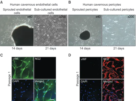
were confluent and spread over the whole bottom (21 days), only sprouting cells were used for subcultiva- tion. However, the primary cultured EC at passage 1 did not show a typical cobble stone appearance, but an elongated spindle-shaped appearance (Fig. 2A). It has been known that primary cultured EC shows different morphology based on the cells of origin [22]. The cells revealed a highly positive staining for an EC marker vWF (green), but did not show positive staining for a pericytes marker NG2 (Fig. 2B). Primary cultured pericyte at passage 1 showed typical multi-directional projections and highly positive staining for a pericytes marker NG2 (red), but did not show positive staining for an EC marker vWF (Fig. 2D).
3. Morphologic and phenotypic changes of human cavernous endothelial cell according to the passages
The phase image of human cavernous EC from pas- sage 1 to passage 5 revealed that EC morphology start-
ed to be changed at passage 3 (Fig. 3A). Fluorescent immunocytochemistry revealed that the cells showed highly positive staining on CD34 and vWF (EC mark- ers), but not on PDGFR-β and NG2 (pericyte markers) up to passage 3. However, more than 60% of human cavernous EC did not express EC markers at passage 5 (Fig. 3B, 3C). Similarly, Western blot analysis revealed a significantly decreased CD34 expression from pas- sage 2 (Fig. 3D, 3E).
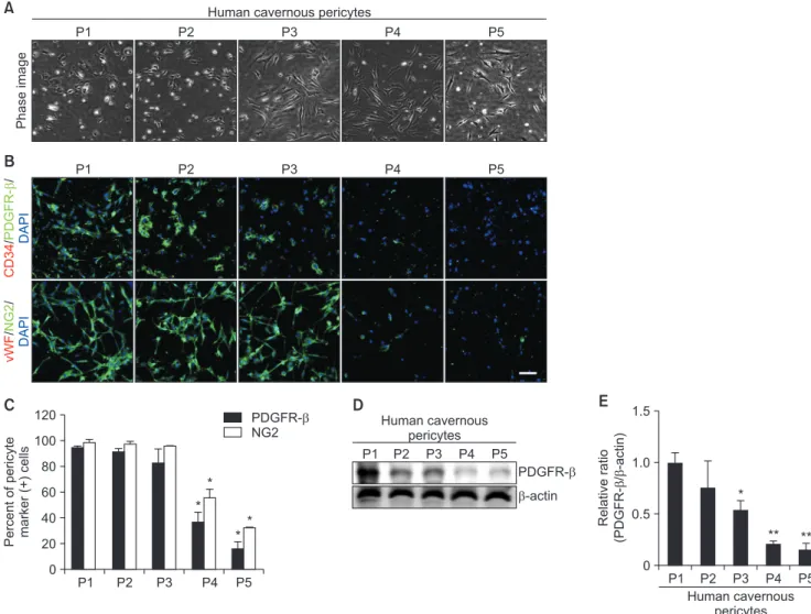
4. Morphologic and phenotypic changes of human cavernous pericyte according to the passages
Similar to the results from human cavernous EC, the phase image of human cavernous pericyte from passage 1 to passage 5 revealed that pericyte morphol- ogy significantly changed from the passage 3 (Fig. 4A).
Fluorescent immunocytochemistry revealed that the cells showed highly positive staining on PDGFR-β and NG2 (pericyte markers), but not on CD34 and vWF (EC
Human cavernous endothelial cells
A B
C D
Sprouted endothelial cells
Sub-cultured endothelial cells
Human cavernous pericytes
Sprouted pericytes Sub-cultured pericytes
14 days 21 days 14 days 21 days
Passage1 Passage1
Fig. 2. Isolation and characterization of human cavernous endothelial cells and pericytes. (A) The human corpus cavernosum tissues were im- planted on a Matrigel-coated 60-mm cell culture dish with endothelial cell culture medium. A representative image of the cells cultivated for 14 days. After cells were confluent and spread over the whole bottom (21 days), only sprouting cells were used for subcultivation. (B) Fluorescent immunocytochemistry of primary human cavernous endothelial cells (passage 1) with antibodies against von Willebrand factor (vWF, endothelial cell marker) and NG2 (a pericyte marker). Nuclei were labeled with the DNA dye DAPI (4,6-diamidino-2-phenylindole). Scale bar=100 μm. (C) The human corpus cavernosum tissues were implanted on a collagen I-coated 35-mm cell culture dishes with pericyte culture medium. A representa- tive image of the cells cultivated for 14 days. After cells were confluent and spread over the whole bottom (21 days), only sprouting cells were used for subcultivation. (D) Fluorescent immunocytochemistry of primary human cavernous pericytes (passage 1) with antibodies against vWF and NG2. Scale bar=100 μm.
markers) up to passage 3. However, more than 60% of human cavernous pericytes did not express pericyte markers at passage 5 (Fig. 4B, 4C). Similarly, Western blot analysis revealed a profound decrease in PDGFR-β expression from passage 3 (Fig. 4D, 4E).
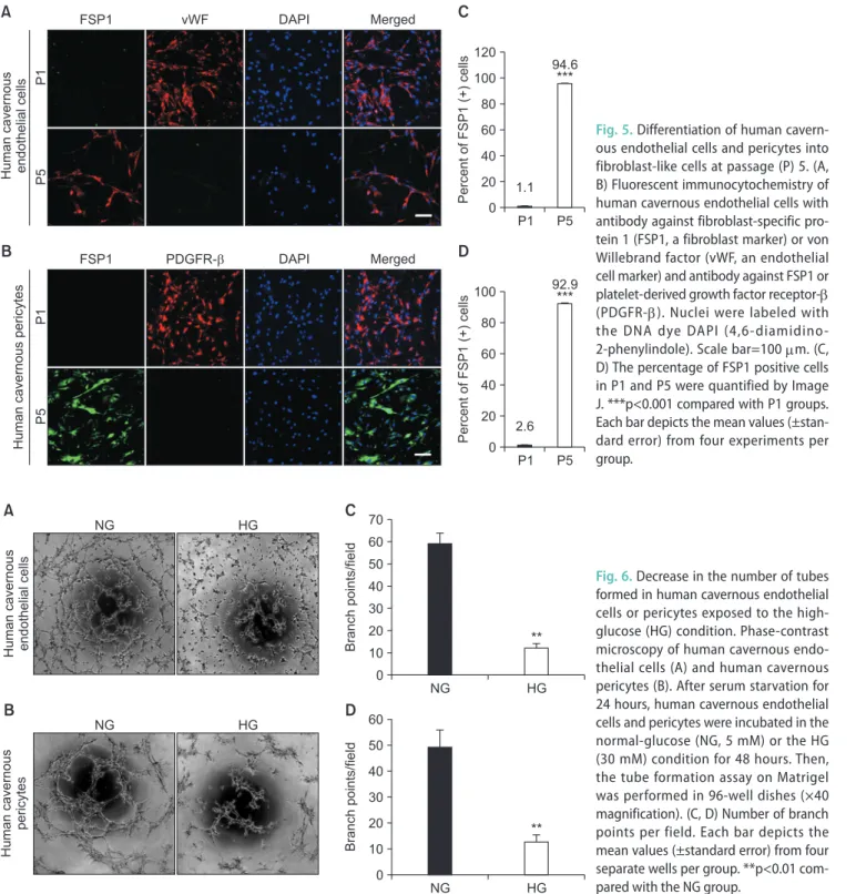
5. Differentiation of human cavernous endothelial cell and pericytes into fibroblast-like cells at passage 5
We determined morphologic and phenotypic char- acteristics of primary cultured cells at passage 5. Both human cavernous EC and pericytes do not express a fibroblast marker FSP1 at passage 1. However, the pri- mary cultured cells showed highly positive staining for
FSP1 (in cavernous EC about 94.6%; in cavernous Peri- cytes about 92.9%) at passage 5 (Fig. 5).
6. Impaired tube formation in human cavernous endothelial cell or pericytes exposed to the high-glucose condition
To test whether primary cultured human cavern- ous EC and pericytes are capable of forming tube-like structures and to determine the effect of high-glucose on tube formation, we performed an angiogenesis as- say on Matrigel in vitro. After 16 hours of incubation on Matrigel, human cavernous EC or pericytes formed well-organized capillary-like structures in normal- glucose condition, whereas the number of tubes was
Phaseimage
B A
CD34//PDGFR- DAPIvWF//NG2 DAPI
Human cavernous ECs
P1 P2 P3 P4 P5
P1 P2 P3 P4 P5
C
PercentofEC marker(+)cells
100 80 60 40 20 0
P1 P2 P3 P4 P5
CD34 vWF
*
*
* *
P1 P2 P3 P4 P5
E
Relativeratio (CD34/-actin)
1.5
1.0
0.5
0
*
*
**
**
D
P1 P2 P3 P4 P5 Human cavernous
endothelial cells
CD34
-actin
Human cavernous endothelial cells Fig. 3. Morphologic and phenotypic changes of human cavernous endothelial cells (ECs) according to the passages (Ps). (A) Phase image of hu- man cavernous ECs from P1 to P5. (B) Fluorescent immunocytochemistry of human cavernous ECs with antibody against CD34 or von Willebrand factor (vWF, EC markers) and antibody against platelet-derived growth factor receptor-β (PDGFR-β) or NG2 (pericyte markers). Nuclei were la- beled with the DNA dye DAPI (4,6-diamidino-2-phenylindole). Scale bar=100 μm. (C) The percentage of CD34- or vWF-positive cells was quanti- fied by Image J. *p<0.05 compared with P1 to P3 groups. (D) Representative Western blot for CD34. (E) Data are presented as the relative density of CD34 to β-actin. The relative ratio measured in the P1 group is arbitrarily presented as 1. *p<0.05 compared with P1 group. **p<0.01 compared with P2 to P3 group. Each bar depicts the mean values (±standard error) from four experiments per group.
significantly decreased when the cells were exposed to high-glucose condition (Fig. 6).
DISCUSSION
The erectile tissue is mainly composed by vascular EC, smooth muscle cells, pericytes and neuronal cells.
The functional and structural derangements of these cellular components play an important role in the pathophysiology of erectile dysfunction (ED) [2,23]. It was reported that the interaction between EC and peri- cytes plays a crucial role in the blood vessel formation and vascular maturation [24]. We recently confirmed
the presence of pericytes in mouse and human corpus cavernosum tissues by using immunohistochemistry, and successfully isolated EC and pericytes from mouse penis [2,16].
To further investigate the cellular mechanisms responsible for physiologic penile erection and patho- physiology of ED, we tried primary culture of EC and pericytes from human corpus cavernosum tissue by using nonenzymatic culture technique. In the pres- ent study, we successfully isolated and cultivated hu- man cavernous EC and pericytes. The morphology and sprouting pattern of the human cavernous EC and pericytes were similar to those cells isolated from
Phaseimage
B A
CD34//PDGFR- DAPIvWF//NG2 DAPI
Human cavernous pericytes
P1 P2 P3 P4 P5
P1 P2 P3 P4 P5
C
Percentofpericyte marker(+)cells
100 80 60 40 20 0
P1 P2 P3 P4 P5
PDGFR- NG2
*
*
*
*
P1 P2 P3 P4 P5
E
Relativeratio (/-actin)PDGFR-
1.5
1.0
0.5
0
*
** **
D
P1 P2 P3 P4 P5 Human cavernous
pericytes
PDGFR-
-actin
Human cavernous pericytes 120
Fig. 4. Morphologic and phenotypic changes of human cavernous pericytes according to the passages (Ps). (A) Phase image of human cavernous pericytes from P1 to P5. (B) Fluorescent immunocytochemistry of human cavernous pericytes with antibody against platelet-derived growth fac- tor receptor-β (PDGFR-β) or NG2 (pericyte markers) and antibody against CD34 or von Willebrand factor (vWF, endothelial cell markers). Nuclei were labeled with the DNA dye DAPI (4,6-diamidino-2-phenylindole). Scale bar=100 μm. (C) The percentage of PDGFR-β or NG2-positive peri- cytes was quantified by Image J. *p<0.05 compared with P1 to P3 groups. (D) Representative Western blot for PDGFR-β. (E) Data are presented as the relative density of PDGFR-β to β-actin. The relative ratio measured in the P1 group is arbitrarily presented as 1. *p<0.05 compared with P1 to P2 groups. **p<0.01 compared with P3 groups. Each bar depicts the mean values (±standard error) from four experiments per group.
mouse corpus cavernosum tissue [2,16]. However, the growth rates of human cavernous EC and pericytes are somewhat lower than those of mouse cavernous EC and pericytes. We previously reported that the mouse cavernous EC and pericytes were confluent and spread over the whole bottom of the dish at 2 weeks [2,16]. However, the human cavernous EC and pericytes were confluent at 3 weeks after cultivation. This find-
ing may result from different metabolic rate and life processes between the mouse and human. The larger the animal, the slower its cellular metabolic rate and life processes. Moreover, the difference in age between mouse (mean age, 8-week-old) and human (mean age, 56-year-old) may also contribute this discrepancy.
The primary cultured EC and pericytes are known to rapidly differentiate and lose their phenotype as
FSP1 vWF DAPI Merged
Humancavernous endothelialcells
A
B
Humancavernouspericytes P1P5
FSP1 PDGFR- DAPI Merged
P1
C
PercentofFSP1(+) cells 100
80 60 40 20 0
P1 P5
***
120
94.6
1.1
D
PercentofFSP1(+)cells 100
80 60 40 20 0
P1 P5
***
92.9
P5 2.6
Fig. 5. Differentiation of human cavern- ous endothelial cells and pericytes into fibroblast-like cells at passage (P) 5. (A, B) Fluorescent immunocytochemistry of human cavernous endothelial cells with antibody against fibroblast-specific pro- tein 1 (FSP1, a fibroblast marker) or von Willebrand factor (vWF, an endothelial cell marker) and antibody against FSP1 or platelet-derived growth fac tor receptor-β (PDGFR-β). Nuclei were labeled with the DNA dye DAPI (4,6-diamidino- 2-phenylindole). Scale bar=100 μm. (C, D) The percentage of FSP1 positive cells in P1 and P5 were quanti fied by Image J. ***p<0.001 compared with P1 groups.
Each bar depicts the mean values (±stan- dard error) from four experiments per group.
C
D
Humancavernous endothelialcells
A
B
Humancavernous pericytes
NG HG
NG HG
Branchpoints/field
0
NG HG
**
70 60 50 40 30 20 10
Branchpoints/field
0
NG HG
**
60 50 40 30 20 10
Fig. 6. Decrease in the number of tubes formed in human cavernous endothelial cells or pericytes exposed to the high- glucose (HG) condition. Phase-contrast microscopy of human cavernous endo- thelial cells (A) and human cavernous pericytes (B). After serum starvation for 24 hours, human cavernous endothelial cells and pericytes were incubated in the normal-glucose (NG, 5 mM) or the HG (30 mM) condition for 48 hours. Then, the tube formation assay on Matrigel was performed in 96-well dishes (×40 magnification). (C, D) Number of branch points per field. Each bar depicts the mean values (±standard error) from four separate wells per group. **p<0.01 com- pared with the NG group.
the passage goes on [25,26], Although some literature have reported that EC and pericytes are stable over several passages [27,28], the stability of primary cul- tured human cavernous EC and pericytes remains a controversial issue. In the present study, we performed immunocytochemistry by using EC and pericyte mark- ers to determine phenotype and morphologic changes from passage 1 to passage 5. Primary cultured EC and pericytes maintained their cellular characteristics up to passage 2. However, there were significant changes in their typical phenotype at passages 4 and 5, and morphologic characteristics from the passage 3 to 5.
More than 60% of the human cavernous EC and peri- cytes did not express their specific cellular markers at passage 5. The cells were elongated and irregular in size, which was similar with fibroblast-like cell. Fur- thermore, the human EC and pericytes showed positive staining for fibroblast-like cell marker FSP1 at passage 5. From these findings, we have reached conclusion that primary cultured human cavernous EC and peri- cytes should be used at earlier passages (up to passage 2).
To further examine whether the primary cultured human cavernous EC and pericytes are capable of forming tube-like structures under different glucose concentration, we performed tube formation assay by cultivating the cells under normal- or high-glucose condition. Similar to the results from the our previous study in mice [29], the number of tube was significant- ly lower in human cavernous EC and pericytes exposed to high-glucose condition than in the cells exposed to the normal-glucose condition.
To the best of our knowledge, this is the first study to provide protocol to isolate human cavernous EC and pericytes. Although our in vitro model may not com- pletely represent the complexity of in vivo diabetic ED, this model will be a valuable tool to understand the role of the each cellular component in physiologic pe- nile erection and pathophysiology of diabetic ED.
CONCLUSIONS
We established a simple and nonenzymatic method to isolate human cavernous EC and pericytes. Human cav- ernous EC or pericytes function study will be a valuable tool for determining the efficacy of candidate therapeu- tics targeting angiogenesis and may open a new avenue to develop novel therapeutic modalities for ED.
ACKNOWLEDGEMENTS
This work was supported by the National Research Foundation of Korea (NRF) grant (Guo Nan Yin, 2018R1C1B6003829] and Ji-Kan Ryu [2016R1A2B2010087]) and by a Medical Research Center Grant (Ji-Kan Ryu, 2014R1A5A2009392) funded by the Korean government (Ministry of Science, ICT and Future Planning).
Disclosure
The authors have no potential conflicts of interest to disclose.
Author Contribution
Research conception & design: Yin GN, Ock J, Ryu JK, Suh JK. Performing the experiments: Yin GN, Ock J, Choi MJ, Song KM. Data acquisition: Ghatak K, Minh NN. Data analysis and interpretation: Yin GN, Ock J, Choi MJ. Statistical analysis:
Kwon MH, Jin HR. Drafting of the manuscript: Yin GN, Ock J, Ryu JK. Critical revision of the manuscript: Seong DH, Suh JK. Receiving grants: Yin GN, Ryu JK. Approval of final manu- script: all authors.
Data Sharing Statement
The data analyzed for this study have been deposited in HARVARD Dataverse and are available at https://doi.
org/10.7910/DVN/Q8KIQD.
REFERENCES
1. Andersson KE. Mechanisms of penile erection and basis for pharmacological treatment of erectile dysfunction. Pharma- col Rev 2011;63:811-59.
2. Yin GN, Das ND, Choi MJ, Song KM, Kwon MH, Ock J, et al. The pericyte as a cellular regulator of penile erection and a novel therapeutic target for erectile dysfunction. Sci Rep 2015;5:10891.
3. Yin GN, Park SH, Choi MJ, Limanjaya A, Ghatak K, Minh NN, et al. Penile neurovascular structure revisited: immuno- histochemical studies with three-dimensional reconstruction.
Andrology 2017;5:964-70.
4. Verma S, Buchanan MR, Anderson TJ. Endothelial function testing as a biomarker of vascular disease. Circulation 2003;
108:2054-9.
5. Díaz-Flores L, Gutiérrez R, Varela H, Rancel N, Valladares F.
Microvascular pericytes: a review of their morphological and
functional characteristics. Histol Histopathol 1991;6:269-86.
6. Bauer AL, Jackson TL, Jiang Y. A cell-based model exhibiting branching and anastomosis during tumor-induced angiogen- esis. Biophys J 2007;92:3105-21.
7. Bookholt FD, Monsuur HN, Gibbs S, Vermolen FJ. Math- ematical modelling of angiogenesis using continuous cell- based models. Biomech Model Mechanobiol 2016;15:1577- 600.
8. Finkenzeller G, Torio-Padron N, Momeni A, Mehlhorn AT, Stark GB. In vitro angiogenesis properties of endothelial pro- genitor cells: a promising tool for vascularization of ex vivo engineered tissues. Tissue Eng 2007;13:1413-20.
9. Baudin B, Bruneel A, Bosselut N, Vaubourdolle M. A protocol for isolation and culture of human umbilical vein endothelial cells. Nat Protoc 2007;2:481-5.
10. Bryan BA, D’Amore PA. Pericyte isolation and use in endo- thelial/pericyte coculture models. Methods Enzymol 2008;
443:315-31.
11. Maier CL, Shepherd BR, Yi T, Pober JS. Explant outgrowth, propagation and characterization of human pericytes. Micro- circulation 2010;17:367-80.
12. Mogensen C, Bergner B, Wallner S, Ritter A, d’Avis S, Nini- chuk V, et al. Isolation and functional characterization of pericytes derived from hamster skeletal muscle. Acta Physiol (Oxf) 2011;201:413-26.
13. Rops AL, van der Vlag J, Jacobs CW, Dijkman HB, Lensen JF, Wijnhoven TJ, et al. Isolation and characterization of con- ditionally immortalized mouse glomerular endothelial cell lines. Kidney Int 2004;66:2193-201.
14. Tigges U, Welser-Alves JV, Boroujerdi A, Milner R. A novel and simple method for culturing pericytes from mouse brain.
Microvasc Res 2012;84:74-80.
15. Bernas MJ, Cardoso FL, Daley SK, Weinand ME, Campos AR, Ferreira AJG, et al. Establishment of primary cultures of human brain microvascular endothelial cells to provide an in vitro cellular model of the blood-brain barrier. Nat Protoc 2010;5:1265-72.
16. Yin GN, Ryu JK, Kwon MH, Shin SH, Jin HR, Song KM, et al.
Matrigel-based sprouting endothelial cell culture system from mouse corpus cavernosum is potentially useful for the study of endothelial and erectile dysfunction related to high-glucose exposure. J Sex Med 2012;9:1760-72.
17. Bala K, Ambwani K, Gohil NK. Effect of different mitogens and serum concentration on HUVEC morphology and char-
acteristics: implication on use of higher passage cells. Tissue Cell 2011;43:216-22.
18. Neng L, Zhang W, Hassan A, Zemla M, Kachelmeier A, Frid- berger A, et al. Isolation and culture of endothelial cells, peri- cytes and perivascular resident macrophage-like melanocytes from the young mouse ear. Nat Protoc 2013;8:709-20.
19. Allen DA, Harwood S, Varagunam M, Raftery MJ, Yaqoob MM. High glucose-induced oxidative stress causes apoptosis in proximal tubular epithelial cells and is mediated by mul- tiple caspases. Faseb J 2003;17:908-10.
20. Rajesh M, Mukhopadhyay P, Bátkai S, Haskó G, Liaudet L, Drel VR, et al. Cannabidiol attenuates high glucose-induced endothelial cell inflammatory response and barrier disrup- tion. Am J Physiol Heart Circ Physiol 2007;293:H610-9.
21. Yu T, Sheu SS, Robotham JL, Yoon Y. Mitochondrial fission mediates high glucose-induced cell death through elevated production of reactive oxygen species. Cardiovasc Res 2008;
79:341-51.
22. Langley RR, Ramirez KM, Tsan RZ, Van Arsdall M, Nilsson MB, Fidler IJ. Tissue-specific microvascular endothelial cell lines from H-2K(b)-tsA58 mice for studies of angiogenesis and metastasis. Cancer Res 2003;63:2971-6.
23. Moon KH, Park SY, Kim YW. Obesity and erectile dysfunc- tion: from bench to clinical implication. World J Mens Health 2019;37:138-47.
24. Armulik A, Genové G, Betsholtz C. Pericytes: developmental, physiological, and pathological perspectives, problems, and promises. Dev Cell 2011;21:193-215.
25. Armulik A, Abramsson A, Betsholtz C. Endothelial/pericyte interactions. Circ Res 2005;97:512-23.
26. Dellavalle A, Sampaolesi M, Tonlorenzi R, Tagliafico E, Sac- chetti B, Perani L, et al. Pericytes of human skeletal muscle are myogenic precursors distinct from satellite cells. Nat Cell Biol 2007;9:255-67.
27. Capetandes A, Gerritsen ME. Simplified methods for consis- tent and selective culture of bovine retinal endothelial cells and pericytes. Invest Ophthalmol Vis Sci 1990;31:1738-44.
28. Helmbold P, Nayak RC, Marsch WC, Herman IM. Isolation and in vitro characterization of human dermal microvascular pericytes. Microvasc Res 2001;61:160-5.
29. Yin GN, Park SH, Song KM, Limanjaya A, Ghatak K, Minh NN, et al. Establishment of in vitro model of erectile dysfunc- tion for the study of high-glucose-induced angiopathy and neuropathy. Andrology 2017;5:327-35.