PGHN
Invited Review
Received:December 26, 2012, Revised:December 27, 2012, Accepted:December 28, 2012
Corresponding author: Jeong Kee Seo, Division of Pediatric Gastroenterology, Hepatology and Nutrition, Department of Pediatrics, Seoul National University Children's Hospital, 101, Daehak-ro, Jongno-gu, Seoul 110-769, Korea. Tel: +82-2-2072-3468, Fax: +82-2-743-3455, E-mail:
jkseo@snu.ac.kr
Copyright ⓒ 2012 by The Korean Society of Pediatric Gastroenterology, Hepatology and Nutrition
This is an openaccess article distributed under the terms of the Creative Commons Attribution NonCommercial License (http://creativecommons.org/licenses/by-nc/3.0/) which permits unrestricted noncommercial use, distribution, and reproduction in any medium, provided the original work is properly cited.
Diagnosis of Wilson Disease in Young Children: Molecular Genetic Testing and a Paradigm Shift from the Laboratory Diagnosis
Jeong Kee Seo
Division of Pediatric Gastroenterology, Hepatology and Nutrition, Department of Pediatrics, College of Medicine, Seoul National University, Seoul, Korea
Wilson disease (WD) is an autosomal recessive disorder of copper metabolism that results in accumulation of copper primarily in the liver, brain and cornea. Mutations in the WD gene, ATP7B, cause failure of copper excretion from hepatocyte into bile and a defective synthesis of ceruloplasmin. More than 500 mutations are now recognized, scat- tered throughout the ATP7B gene. Since WD has protean clinical presentations, awareness of WD in clinical practice is important for the early diagnosis and prevention of accumulated copper toxicity. Molecular genetic testing is playing an increasingly important role in the diagnosis of WD in uncertain cases and family screening. Siblings should be screened for WD once an index case has been diagnosed. Discrimination of heterozygotes from asymptomatic pa- tients is essential to avoid inappropriate lifelong therapy for heterozygotes. Genetic testing, either by haplotype analy- sis or by mutation analysis, is the only definite solution for differentiating heterozygote carriers from affected asympto- matic patients. Routine genetic testing, because of the multitude of documented mutations, has been thought to be impractical until recently. However, genetic testing is now being more actively applied to the diagnosis of WD, partic- ularly in young children in whom conventional biochemical diagnosis has much limitation and only genetic testing is able to confirm WD. Because advancement of modern biochemical technology now allows more rapid, easier, and less expensive mutation detection, direct DNA sequencing could be actively considered as the primary mode of diag- nostic investigation rather than a supplementary test to the conventional biochemical tests. This review will focus on the recent advancement of molecular genetics and genetic diagnosis of WD in very young children on the basis of research data of the Seoul National University Children’s Hospital and recent literature. (Pediatr Gastroenterol Hepatol Nutr 2012; 15: 197∼209)
Key Words: Wilson disease, ATP7B, Genetic diagnosis, Mutation, Child
Fig. 1. (A) Severe cirrhotic liver in a 13 year old girl with Wilson disease who under- went the first liver transplan- tation in Korea in 1988 [11], (B) Micro and macro-nodular cirrhosis of the removed liver after liver transplantation at Seoul National University Children's Hospital.
Table 1. Clinical Manifestations of 516 Korean Patients with Wilson Disease
Total No.
(N=516)
Children (N=264)
Adults (N=252) Hepatic symptoms
Neurologic symptoms Psychiatric symptoms Hemolytic anemia Musculoskeletal symptoms Renal disease
439 (85) 181 (35) 47 (9) 28 (5) 21 (4) 11 (2)
231 (89) 54 (20) 17 (6) 17 (6) 10 (4) 6 (2)
208 (83) 127 (50) 30 (11) 11 (4) 11 (4) 5 (2) Values are presented as number (%).
INTRODUCTION
Wilson disease (WD) is an autosomal recessive disorder of copper metabolism that results in accu- mulation of copper in the liver, brain, cornea, kid- ney, and other tissues.
WD occurs at a frequency of approximately 1 in 30,000-50,000 worldwide. In a recent nationwide survey for WD in Korea, the estimated prevalence rate of WD in the pediatric population is one in 37,000 [1,2].
Since the description of WD in 1912 by Samuel Alexander Kinnier Wilson as a “familial syndrome of progressive lenticular degeneration in the brain associated with cirrhosis of the liver” [3], there was a major breakthrough in WD research in 1993 when the WD gene ATP7B was first cloned [4-10].
The protein product of ATP7B gene is a copper transporting adenosine triphosphatase (ATPase), which is mainly expressed in hepatocytes. Muta- tions in ATP7B cause the functional loss as a cop- per transporter and result in impairment of hep- atic biliary copper excretion and also copper in- corporation into apo-ceruloplasmin. Accumulation of copper in the body ultimately leads to liver dis- ease, neurologic symptoms, and Kayser-Fleischer corneal rings which have been described as
"classical symptom triad" of WD (Fig. 1) [11].
In young children, the onset of manifestations is primarily hepatic. Neurologic and psychiatric onset is usually observed in older children and adolescents [12,13].
Asymptomatic elevation of aminotransferases can be observed in children less than 3 years. The first manifestations of WD can be observed in adults greater than 60 years of age.
Children with WD typically present with liver disease including chronic hepatitis with asympto- matic elevations of aminotransferases, cirrhosis and occasionally fulminant hepatic failure (Table 1 and 2) [1]. Neurologic symptoms of WD including tremor, dysphagia, dystonia, rigidity, dysarthria, and choreoathetosis usually occur later in child- hood and in adults. The prevalence of neurologic symptoms increases with age. About half of the adult patients with WD present with neurologic symptoms (Table 1) [1].
Early diagnosis and appropriate management can cure WD. If left untreated, however, WD can progress to fatal hepatic failure or severe neuro- logic deterioration and death.
Because the misdiagnosis and delay in treat-
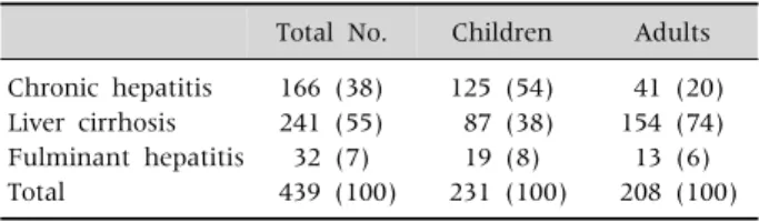
Table 2. Presentation Modes of Liver Diseases in Korean Children and Adults with Wilson Disease
Total No. Children Adults Chronic hepatitis
Liver cirrhosis Fulminant hepatitis Total
166 (38) 241 (55) 32 (7) 439 (100)
125 (54) 87 (38) 19 (8) 231 (100)
41 (20) 154 (74) 13 (6) 208 (100) Values are presented as number (%).
Fig. 2. Three types of defects in copper dependent traffic- king pathway among various types of missense mutations [15,17,18]. CTR1: copper-tran- sporter 1, ER: endoplasmic reticulum, TGN: trans-Golgi network.
ment lead to fatal deterioration, applying appro- priate diagnostic tests early in life is most important. However, the diagnosis of WD is very difficult particularly in young children who fre- quently show atypical or insufficient findings of biochemical and clinical tests for WD.
It has recently been documented that molecular genetic testing detects WD earlier and more se- curely in very young children who frequently do not meet the diagnostic criteria of laboratory tests for WD.
In the past, genetic testing was once considered impractical because of the great numbers of WD causing mutations reported in literature and most of these mutations beging very rare.
In recent years, direct genetic diagnosis including full DNA sequencing has become much easier and is more rapid than before. As the cost of molecular genetic testing has been decreasing, genetic diag-
nosis is replacing the copper related biochemical tests and is more actively used particularly in very young children as one of the initial diagnostic workup tests for WD.
In this article, the author reviewed recent ad- vancements in the diagnosis of WD in young chil- dren and suggested an algorithm for the diagnosis of WD on the basis of the research experience at Seoul National University Children’s Hospital and recently published literature.
INTRA-CELLULAR COPPER TRAFFI- CKING PATHWAY
Dietary copper is absorbed from the intestinal epi- thelium into the blood. And copper in the blood, which is bound to albumin, is delivered to the liver.
In the liver, copper enters the hepatocyte through the copper-transporter 1 (CTR1), and is transported into hepatocytes. Then a specific copper chaperone ATOX1 carries copper to the ATP7B protein (copper- transporting ATPase) located in the trans-Golgi net- work (TGN). Copper is then transported by ATP7B from the TGN into the apical membrane-trafficked vesicles, and copper is then excreted into bile (Fig.
2 and 3) [14-18].
The relocation ATP7B depends on copper con- centrations. When copper concentrations are low
Fig. 3. The failure of Cu de- pendent trafficking pathway and functional defects of copper transport to apoenzyme in representative missense muta- tions [15,17,18]. ER: endoplas- mic reticulum, TGN: trans-Golgi network.
or in the basal state, ATP7B is located in the TGN, where they deliver copper to the apo-ceruloplas- min. When copper concentrations are elevated, ATP7B moves out of the TGN and is relocated to cytoplasmic vesicles. These copper containing vesi- cles move periphery to the plasma membrane where the accumulated copper is released into the bile canaliculus. After releasing copper, vesicles are recycled and ATP7B returns back to the TGN.
In WD, ATP7B and copper cannot leave the TGN because of a conformational change including misfolding in ATP7B variants. The functional loss of ATP7B as a copper transporter results in copper accumulation in the hepatocyte and copper tox- icity related symptoms occur.
The mutated ATP7B is nonfunctional as a cop- per transporter. In patients with ATP7B muta- tions, severe impairments of both copper release into the bile and copper incorporation into cer- uloplasmin result in high hepatic copper contents, very low levels of copper bound serum cer- uloplasmin, and low biliary copper.
There seems to be a difference in the failure of copper trafficking pathway among various muta- tions at the molecular level (Fig. 3) [15-20].
Mutations of ATP7B may cause three types of lo- calization defects: 1) a normal steady state, con- stitutive localization within the TGN, but no re- sponse to copper [17,19]; 2) mislocalization at the
endoplasmic reticulum (ER); 3) constitutive local- ization at the cell periphery (Fig. 2).
The mislocalization at the ER seems to be the most common type of failure in copper trafficking pathway. It is found in R778L and H1069 muta- tions [17]. ER mislocalization of ATP7B is often due to misfolding and associated with proteasomal degradation [21-23].
And in patients with other type of mutations, copper and ATP7B binding complex are con- stitutively localized at the cell periphery and cop- per is not excreted into bile.
However, clinical correlation with these molec- ular level differences among various mutations is not clearly documented at the present.
Not all mutations in ATP7B disrupt both the copper transport into bile and the delivery to cer- uloplasmin (Fig. 3) [15-20]. Most of the mutation failuretions including R778L and H1069Q have both of these two defects. Some mutations such as G943S and M769V seem to have only one defect. In G943S, the failure of copper trafficking pathway to bile canalicuil was found but intact cu- proenzyme biosynthesis was shown in the com- plementation assay. These types of mutation could explain the normal serum ceruloplasmin level in some patients with WD. In one patient who had compound heterozygote mutations with G943S, serum ceruloplasmin was reported to be normal
Table 3.Human Mutation Database (2012)
Mutation types Number (%)
Nucleotide substitutions (missense/nonsense) Nucleotide substitutions (splicing)
Nucleotide substitutions (regulatory) Small deletions
Small insertions Small indels Gross deletions
Gross insertions/duplications Complex rearrangements Repeat variations Total
410 (63) 57 (9)
7 110 (17) 49 (8)
6 11 0 1 0 651 (100)
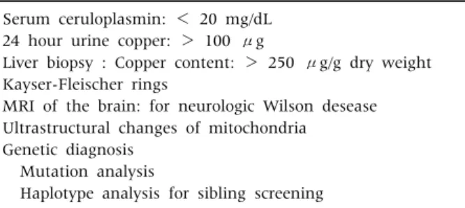
Table 4. Diagnosis of Wilson Disease Serum ceruloplasmin: < 20 mg/dL 24 hour urine copper: > 100 μg
Liver biopsy : Copper content: > 250 μg/g dry weight Kayser-Fleischer rings
MRI of the brain: for neurologic Wilson desease Ultrastructural changes of mitochondria
Genetic diagnosis Mutation analysis
Haplotype analysis for sibling screening
[17]. Further studies are needed to confirm the correlation of the localization defects in the cell with laboratory findings or clinical severity ob- served in patients with WD.
MUTATIONS OF ATP7B
The ATP7B is a large gene which has 21 exons varying from 77 to 1,234 bp [4]. ATP7B, a protein product of the gene, is copper-transporting P type ATPase. The ATP7B gene is expressed mainly in the liver and kidney. The highest expression is shown in hepatocytes. The copper delivery to the apo-ceruloplasmin and copper excretion into bile canaliculi from hepatocytes are essential functions of the ATP7B. Mutations cause conformational changes such as misfolding in the ATP7B which result in loss of function as a copper transporter.
Since the discovery of WD gene, more than 500 different mutations have been documented so far, and novel mutations are continuously being reported. Single nucleotide substitutions-missense or nonsense mutations are the most common mutations. Small deletions, small insertions and splicing site mutations are the next common mu- tation types (Table 3) [10].
Mutations differ among ethnic groups. Arg778Leu mutation is located on exon 8 in the trans-mem- brane domain 4. It is the most common mutation in East Asian countries. This mutation has seldom been reported in European populations, so far. An
allele frequency of Arg778Leu in Korean children with WD is 37-41% [2,24-27]. The A874V, L1083F, and N1270S are the next common mutations in Korea, and the total allele frequency of these 4 mu- tations is 64% [2]. A higher frequency of Arg778Leu was also reported in Taiwanese [28,29], Chinese [30], and Japanese [31] patients.
His1069Gln mutation, which is located on exon 14 close to the ATP binding domain, is the most common mutation in western countries. This mu- tation accounts for about one third of WD muta- tions in European and American populations, with the highest allele frequency of 73% reported in polish patients [32].
Patients with WD usually have two different mutations rather than two identical mutations.
Since most patients with WD are compound heter- ozygotes and most mutations are very rare, geno- type/phenotype correlation studies are difficult to perform.
DIAGNOSIS OF WD AND LIMITATIONS OF LABORATORY TESTS IN YOUNG CHILDREN
Clinical suspicion of WD is the most important first step for the early diagnosis. WD should be con- sidered in the differential diagnosis of children with unknown chronic hepatitis, unusual neurologic or psychiatric symptoms, and hemolytic anemia.
The diagnosis of WD often depends on the de- tection of low serum ceruloplasmin levels, increased 24 hour urinary copper excretion, Kayser-Fleisher rings in the descemet membrane of the cornea, and
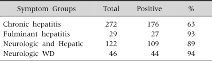
Table 6. Kayser-Fleisher Rings in 469 Korean Patients with Wilson Disease (WD)
Symptom Groups Total Positive % Chronic hepatitis
Fulminant hepatitis Neurologic and Hepatic Neurologic WD
272 29 122 46
176 27 109 44
63 93 89 94
Fig. 4. Structural abnorma- lities of mitochondriae in the hepatocytes from the article of Sternlieb (Hepatology 1992;
16:728-32) [33].
Table 5.Diagnostic Investigations for Wilson Disease Diagnostic Lab. Patients No.
Ceruloplasmin (mg/dL) Under 7
7-20 Over 20
24-hour urinary copper excretion (μg) Under 40
40-100 Over 100 Kayser-Fleischer ring Present
Absent
335 (68) 136 (28) 21 (4)
17 (4) 46 (10) 406 (86)
336 (73) 126 (27) Values are presented as number (%).
increased hepatic copper contents, (Table 4). None of the laboratory parameters alone allows a definite diagnosis of WD. A combination of any two of these four laboratory findings strongly supports for a di- agnosis of WD.
In 550 pediatric and adult patients detected on a nation-wide survey of WD in Korea, low serum ceruloplasmin (<20 mg/dL), high 24-hour urine copper (>100 μg), and Kayser-Fleischer rings were found in 96%, 86%, and 73% of the patients (Table 5) [1]. High hepatic copper content (>250 μg/g of dry weight liver) were found in 88% of the tested patients.
The Kayser-Fleischer ring, which is copper depos- its in the periphery of the cornea, is very useful particularly for the diagnosis of patients with neu- rologic symptoms (Table 6). In the neurologic form of WD, the absence of Kayser-Fleischer rings is ex- tremely rare. If slit lamp examinations reveal Kayser- Fleischer rings in children presenting with typical neurologic manifestations, a definite diag- nosis of WD can be made without further bio- chemical tests.
In clinical practice, less invasive investigations including measurements of serum ceruloplasmin levels, urine collections for copper content, and slit lamp examinations for Kayser-Fleischer rings are performed more frequently at the initial stages of WD diagnosis than hepatic copper assays which need invasive liver biopsy. None of the bio- chemical tests are highly sensitive or specific.
Structural abnormalities of the mitochondria
Fig. 5. High signal intensity in the putamen on T2 weighted magnetic resonance image (A) and reversal of the lesion after the- rapy in children with neurologic WD (B). From a case of Seoul National University Children's Hospital [35].
Table 7. 24 Hour Urine Copper Excretion (μg) in 114 Children
with Wilson Disease (p=0.0035)
Over 100 (diagnostic)
100-40 (equivocal)
Under 40 (normal) Under 6 years
(n=66) Over 6 years (n=88) Total
19/26 (73)
84/88 (96)
103/114 (90)
5/26 (19)
3/88 (3)
8/114 (7)
2/26 (8)
1/88 (1)
3/114 (3) Values are presented as number (%). At Seoul National University Children’s Hospital.
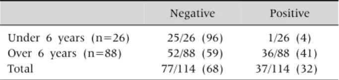
Table 8. Presence of a Kayser-Fleischer Ring by Slit Lamp Examinations in 114 Korean Children
Negative Positive Under 6 years (n=26)
Over 6 years (n=88) Total
25/26 (96) 52/88 (59) 77/114 (68)
1/26 (4) 36/88 (41) 37/114 (32) Values are presented as number (%). At Seoul National Univer- sity Children’s Hospital.
within hepatocytes are regarded as an important and characteristic feature of WD (Fig. 4) [33]. One of the most striking changes is the deformity of the mitochondrial cristae. Cystic dilatation of the cris- tae results from separation of the outer membrane from the inner membrane of the cristae. The size and shape of the mitochondria are very pleomor- phic. Some mitochondria may contain vacuoles and dense inclusion bodies in the cytoplasm.
If cholestatic liver disease is not present, these mitochondrial changes could be considered patho- gnomonic findings of WD. Although the ultra-struc- tural abnormalities of mitochondriae are very char- acteristic and specific, diagnostic sensitivity is low.
Mitochondrial changes are not prominent in most of the children with WD who are diagnosed in early childhood. Children with WD rarely show all these characteristic mitochondrial changes.
Magnetic resonance imaging (MRI) inves- tigations of the brain should be a part of the eval- uation in all children with neurologic WD, and should be considered prior to treatment. Liver MRI including multiple hypointense nodules on T2–
weighted images, correlates with the severity of hepatic dysfunction, and also demonstrates rever- sible changes after clinical improvement [34].
In children with the neurologic form of WD, the most frequently identified abnormality on MRI is bilateral symmetrical high signal intensity in the putamen on T2-weighted images. This lesion sub-
sides after chelating therapy (Fig. 5) [35].
LIMITATIONS OF DIAGNOSTIC LABO- RATORY TESTS FOR WD IN VERY YOUNG CHILDREN
Intermediate or equivocal values in the 24 hour urinary copper are more common in young chil- dren (Table 7).
In patients over 6 years of age, all but 4 children (96%) showed increased 24 hour urinary copper above 100 μg. In contrast, only 73% of children under the age of 6 showed increased 24 hour uri- nary copper above 100 μg. In addition, 7 of 26 children (27%) did not meet the diagnostic cut off value of urinary copper above 100 μg.
Young children rarely present with Kayser-Fleischer ring. A Kayser-Fleischer ring was detected with a slit lamp examination in 41% of children older than 6 years of age. In contrast, only one child out of 26 chil- dren under 6 years old showed a Kayser-Fleischer ring (Table 8).
In order to increase the diagnostic accuracy of bi-
Table 9. Scoring System Developed at the Eighth International Meeting on Wilson Disease, Leipzig, 2003 (Ferenci et al. [36]) Kayser-Fleischer rings Liver copper (in absence of cholestasis)
Present 2 >250 μg/g 2
Absent 0 50-250 μg/g 1
Neurologic symptoms (or typical brain MRI) Normal (<50 μg/g) −1
Present 2 Rhodamine-positive hepatocyte on biopsy* 1
Absent 0 Urinary copper (in absence of acute hepatitis)
Serum ceruloplasmin Normal 0
Normal (>0.2 g/L) 0 1-2× ULN 1
0.1-0.2 g/L 1 >2× ULN 2
<0.1 g/L 2 Normal, but > 5× ULN after D-penicillamine 2
Coombs-negative hemolytic anemia Mutation analysis
Present 1 Two chromosome mutations 4
Absent 0 One chromosome mutation 1
No mutations detected 0
Total score Evaluation
≥4 Diagnosis highly likely
2-3 Diagnosis probable, more tests needed
≤1 Diagnosis very unlikely
*If no quantitative liver copper analysis available. MRI: magnetic resonance imaging, ULN: upper limit of normal.
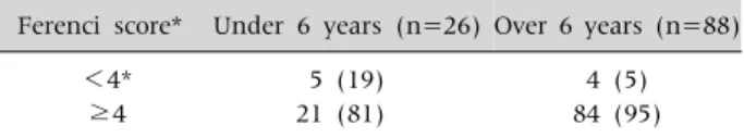
Table 10.Age Difference in the Validity of Ferenci Score in 114 Children with WD
Ferenci score* Under 6 years (n=26) Over 6 years (n=88) <4*
≥4
5 (19) 21 (81)
4 (5) 84 (95) Values are presented as number (%). At Seoul National University Children’s Hospital. *Diagnosis of Wilson disease is established.
ochemical and clinical investigations, Ferenci diag- nostic scoring system (Table 9) was developed at the Eighth International Meeting on WD, in 2001 [36].
The Ferenci scoring system has limitations for the diagnosis of WD, particularly in very young children. We evaluated the validity of the Ferenci scoring system using biochemical and clinical pa- rameters, without applying the mutation analysis results, in 114 children with WD. The Ferenci scor- ing system has excellent diagnostic value and can detect WD in about 95% of the children older than 6 years of age. However, the diagnosis of WD is absent in about 19% of young children under 6 years of age when the scoring system is applied to this very young age group (Table 10).
GENETIC TESTING AND AN ALGO- RITHM FOR THE DIAGNOSIS OF WD IN INFANTS AND PRESCHOOL CHILDREN
Although majority of patients diagnosed with WD are in the age range between 5 and 35 years old, very young children with WD are being re- ported more frequently in recent literature. Severe impairments in ATP7B including large deletion, in- sertion, nonsense mutations and splice site muta- tions can lead to an earlier onset of WD.
Thomas et al. [14] reported severe liver disease in a 3 year old boy with 54 amino acids missing in ATP7B immediately after the first discovery of WD gene. Early childhood cases of WD were re- ported in a 13 month old child with hyper- transaminasemia, in a 2 year old child with chronic hepatitis, in a 3 year old child with cirrhosis and in a 5 year old child with acute liver failure [37-40].
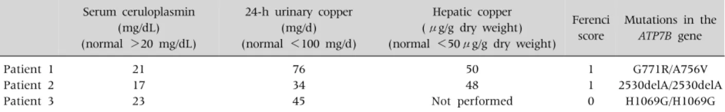
Caprai et al. [41] reported 3 children who failed to be diagnosed with WD solely on the basis of biochemical diagnostic criteria. These children showed near normal serum ceruloplasmin levels.
The 24-hour urinary copper excretions were not high, and hepatic copper contents were below 50 μg/g dry weight, Ferenci scores of these patients
Table 11. Copper Metabolism Parameters of the 3 Patients Described in Details Serum ceruloplasmin
(mg/dL) (normal >20 mg/dL)
24-h urinary copper (mg/d) (normal <100 mg/d)
Hepatic copper (μg/g dry weight) (normal <50μg/g dry weight)
Ferenci score
Mutations in the ATP7B gene Patient 1
Patient 2 Patient 3
21 17 23
76 34 45
50 48 Not performed
1 1 0
G771R/A756V 2530delA/2530delA
H1069G/H1069G
Table 12. Clinical and Biochemical Features of a 9 Month Old Korean Infant with Wilson Disease Age (month)
AST/ALT (IU/L) Copper in urine (μg/d) Ceruloplasmin (mg/dL)
9 102/122
22 113/144
<7
23 90/105
14.9
<7
24 171/194
15.2
26 185/285
62.2
<7
27 262/401
30 111/162
421.4
<7
Mutations G1186S/4006delA
Kayser-Fleischer ring
Copper in the liver (μg/g dry wt)
Negative
748 AST: aspartate aminotransferase, ALT: alanine aminotransferase. At Seoul National University Children's Hospital.
(Table 11), were low and the diagnosis of WD was very unlikely. Nevertheless, all 3 children showed two mutations in the ATP7B gene. Only the molec- ular genetic testing was able to confirm WD in these patients.
In our unit, we reported the youngest case of WD in a 9 month-old male infant who visited hospitals because of persistently elevated aminotransferases [42]. His ceruloplasmin level was very low (<7 mg/dL). The 24 hour urine copper concentrations were not high, and Kayser-Fleischer ring was absent.
The diagnosis of WD was established through genetic testing in this patient, who was confirmed to be a compound heterozygote of G1186S and 4006delA (Table 12).
Molecular genetic testing is playing an increas- ingly important role in the diagnosis of WD in un- certain cases and family screening. About 20% of heterozygote carriers have low serum ceruloplasmin levels, borderline normal urinary copper concen- trations, and moderate elevations of hepatic copper (50-250 μg/g dry weight). Molecular genetic testing is a reliable tool for distinguishing securely healthy heterozygote carriers from affected presymptomatic patients. It could avoid inappropriate lifelong ther- apy in heterozygotes.
For the screening of siblings, molecular genetic testing either by mutation analysis or haplotype analysis is the only definite solution [43-45].
When the 2 mutations are identified in an index patient, mutation analysis is most efficient in fam- ily screening strategy. The cost necessary for checking the presence of already identified 2 mu- tations is less expensive; about 10% of the cost for a complete DNA sequencing in the index patient.
If the mutations of the index patient are un- known, haplotype analysis with closely linked mi- crosatellite markers is useful for family screening.
Because a large number of mutations have been recognized in the ATP7B gene, mutation screening was considered impractical. However, in recent years, direct genetic diagnosis including complete DNA sequencing has become much easier and more rapid than in the past. Therefore, for un- certain cases, genetic diagnosis is now becoming the most specific and sensitive diagnostic test, and is being included among the initial diagnostic workup tests, although the cost of genetic testing is still high to be used extensively in all patients.
Currently reported mutation detection rate is very high.
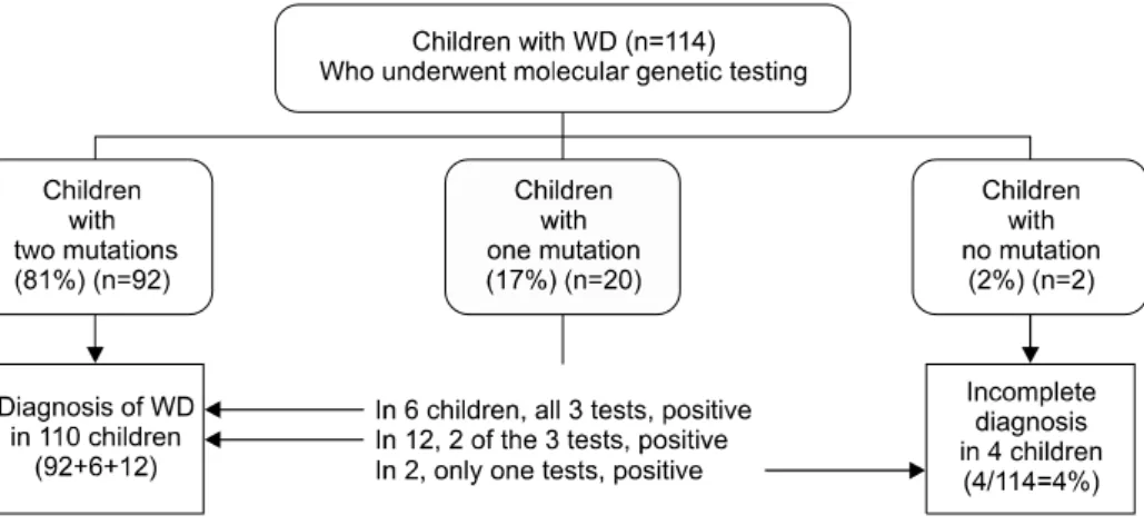
In 114 children with unexplained hepatitis at
Fig. 6. Genetic diagnosis and copper metabolism associated 3 tests in 114 children with Wilson disease (WD) at Seoul National University Children’s Hospital (Low ceruloplasmin+
high urine copper+Kayser-Fl- eischer ring).
our children’s hospital, complete gene sequencing of the coding regions of all the exons and flanking regions of the intron was confirmed. In 81% of children, two mutations were found and a definite diagnosis of WD was confirmed. In 17%, one mu- tation was found and no second mutation was de- tected after a complete DNA sequencing. No mu- tations were found in only 2 children, who had a definite clinical diagnosis of WD after observations of their response to therapy and long-term clinical follow-ups (Fig. 6).
If 2 mutations are found, and one of them was not previously reported, it should be determined whether this mutation is a WD causing novel mu- tation or a normal variant in the patient’s popula- tion. A couple of methods are available to de- termine the functional activity and to predict con- sequences of the protein structural changes of newly identified variants [17,46,47].
On-line prediction programs such as SIFT (Sor- ting Intolerant from Tolerant), PolyPhen (Polymor- phism Phenotyping), and Align-GVGD (Align- Grantham Variation Grantham Deviation) are avail- able for the evaluation of newly identified un- classified variants. Conservation, structure, bio- chemical properties, and models of the 3-dimen- sional protein structure of variants are evaluated by the computational predictive algorithms and compared with original residue at the affected site.
The functional activity of newly identified ATP7B variants can be evaluated in a yeast com-
plementation assay utilizing ATP7B homolog Ccc2p in a strain of Saccharomyces cerevisiae [46,47].
This functional assay helps to distinguish normal from disease causing mutations. With immuno- fluorescence microscopic examinations, failure of the copper dependent trafficking pathway of the ATP7B variants can be also detected.
The functional assays in the yeast mutant Ccc2p model confirmed 508 WD causing variants includ- ing 267 missense mutations in 644 variants which were identified by diagnostic sequencing of ATP7B (http://www.wilsondisease.med.ualberta.ca/data- base.asp).
An allele frequency study in the normal pop- ulation for an unknown variant is useful. At least 100 alleles with 50 persons should be included.
When one mutation is detected in a child and the mutation has been previously reported, this child is either a heterozygote carrier coincidentally having unknown hepatitis rather than WD, or a WD patient in whom the second mutation is not identified. In this group of the children with one mutation, it was not difficult to confirm the diag- nosis of WD; two of the 3 tests including cer- uloplasmin measurement, urinary copper assay, and Kayser- Fleischer ring examination were pos- itive in 60% of this group of children and all 3 tests were positive in 30% of this group of children (Fig.
6). In 10% of children who were found to have one known mutation, one test was positive and other 2 tests were negative.
Fig. 7. Diagnostic approach for Wilson disease (WD) in infants and preschool children – paradigm shift in the era of molecular genetics (at Seoul National University Children’s Hospital).
A long term clinical follow up and evaluation of the treatment response usually confirms the diag- nosis of WD in such cases. Therefore, in almost all of the children with unexplained hepatitis, a defi- nite diagnosis of WD could be established with the direct DNA sequencing of ATP7B performed in the initial investigation. Children who have no muta- tion are extremely rare, and the diagnosis of WD in these patients could be established with long term follow-ups and treatment responses evaluations.
Even in patients who have one known mutation and one positive laboratory test, WD is highly likely.
Liver biopsy for hepatic copper and mitochondrial structural abnormalities or brain MRI may be rec- ommended to aid in establishing the diagnosis of WD in this group of patients. Long-term evaluation of the response to treatment also can be very helpful in some patients who have no mutations.
Further investigations including Multiplex Liga- tion-dependent Probe Amplification (MLPA) can be attempted to search for large gene defects such as whole exon deletions which are not easily de- tected by direct DNA sequencing method. MLPA assay is a recently developed highly sensitive new method for detecting copy number variations in genomic DNA sequences. MLPA can detect large deletions and duplications on whole exon levels, which are not found by direct sequencing. The MLPA assay in combination with a complete DNA sequencing is found to increase the diagnostic ac- curacy in some pediatric genetic diseases such as
Peutz–Jeghers syndrome [48] or Alagille syndrome, which are known to have large gene defects.
However, large gene defects in ATP7B are ex- tremely rare in patients with WD, therefore MLPA assay may not be efficient.
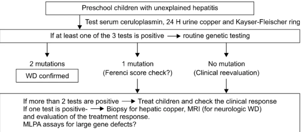
The diagnostic algorism of WD in our children’s hospital for young children is shown in Fig. 7.
For infants and preschool children with un- explained hepatitis, we test ceruloplasmin levels, 24-hour urinary copper concentrations and Kayser- Fleischer ring with slit lamp examinations. If at least one of these less invasive tests is positive, rou- tine genetic testing is recommended to all children as one of the initial investigational examinations.
When ATP7B mutations are detected, we extend mutation analyses to all family members for genet- ic screening.
If 2 mutations are found, we treat the patient with a definite diagnosis of WD. If one mutation is found and more than 2 laboratory tests are pos- itive, we regard the patient as having a tentative diagnosis of WD and we evaluate the treatment re- sponse during follow-ups. If one mutation is found and only one test is positive, we recommend fur- ther studies such as liver biopsy for hepatic copper, or brain MRI. And we evaluate the treatment response.
When a detected mutation is not previously re- ported, allele frequency study in our population is performed to check whether it is a normal variant.
If genetic testing is not performed, many of the
very young children may not be diagnosed as hav- ing WD because these patients usually have no Kayser-Fleischer rings, equivocal levels of bio- chemical tests, and low Ferenci scores.
Sequencing of ATP7B could be considered first as an initial test, particularly in young children, for a definitive diagnosis of WD and for secure family screening.
REFERENCES
1. Seo JK, Kim YS, Hahn CJ, Baik SK. A nationwide sur- vey for prevalence and clinical characteristics of Wilson disease in Korea. Korean J Hepatol 2004;10(Suppl):
5-15.
2. Seo JK. Wilson disease: an update. Korean J Hepatol 2006;12:333-63.
3. Wilson SAK. Progressive lenticular degeneration: a familial nervous disease associated with cirrhosis of the liver. Brain 1912;34:295-507.
4. Bull PC, Thomas GR, Rommens JM, Forbes JR, Cox DW. The Wilson disease gene is a putative copper trans- porting P-type ATPase similar to the Menkes gene. Nat Genet 1993;5:327-37.
5. Chelly J, Monaco AP. Cloning the Wilson disease gene.
Nat Genet 1993;5:317-8.
6. Petrukhin K, Fischer SG, Pirastu M, Tanzi RE, Chernov I, Devoto M, et al. Mapping, cloning and genet- ic characterization of the region containing the Wilson disease gene. Nat Genet 1993;5:338-43.
7. Tanzi RE, Petrukhin K, Chernov I, Pellequer JL, Wasco W, Ross B, et al. The Wilson disease gene is a copper transporting ATPase with homology to the Menkes dis- ease gene. Nat Genet 1993;5:344-50.
8. Yamaguchi Y, Heiny ME, Gitlin JD. Isolation and char- acterization of a human liver cDNA as a candidate gene for Wilson disease. Biochem Biophys Res Commun 1993;197:271-7.
9. Petrukhin K, Lutsenko S, Chernov I, Ross BM, Kaplan JH, Gilliam TC. Characterization of the Wilson disease gene encoding a P-type copper transporting ATPase: ge- nomic organization, alternative splicing, and struc- ture/function predictions. Hum Mol Genet 1994;3:
1647-56.
10. Cox DW, Moore SD. Copper transporting P-type ATPases and human disease. J Bioenerg Biomembr 2002;34:333-8.
11. Kim ST, Park YH, Lee KU, Kum SJ, Youn YK, Seo JK, et al. An experience of first liver transplantation in
Korea. J Korean Transplant Soc 1988;2:27.
12. Moon JS, Ko JS, Seo JK. Long-term clinical follow-up of Korean children with Wilson disease; twenty years' experience. J Korean Pediatr Soc 2001;44:127-38.
13. Seo JK, Moon HR. Hepatitis in childhood as a manifes- tation of treatable Wilson’s disease. Kor J Gastroenter- ol 1983;15:55-64.
14. Thomas GR, Forbes JR, Roberts EA, Walshe JM, Cox DW. The Wilson disease gene: spectrum of mutations and their consequences. Nat Genet 1995;9:210-7.
15. Lutsenko S, Barnes NL, Bartee MY, Dmitriev OY.
Function and regulation of human copper-transporting ATPases. Physiol Rev 2007;87:1011-46.
16. Schaefer M, Hopkins RG, Failla ML, Gitlin JD.
Hepatocyte-specific localization and copper-dependent trafficking of the Wilson's disease protein in the liver.
Am J Physiol 1999;276:G639-46.
17. Forbes JR, Cox DW. Copper-dependent trafficking of Wilson disease mutant ATP7B proteins. Hum Mol Genet 2000;9:1927-35.
18. de Bie P, Muller P, Wijmenga C, Klomp LW. Molecular pathogenesis of Wilson and Menkes disease: correla- tion of mutations with molecular defects and disease phenotypes. J Med Genet 2007;44:673-88.
19. Ambrosini L, Mercer JF. Defective copper-induced traf- ficking and localization of the Menkes protein in pa- tients with mild and copper-treated classical Menkes disease. Hum Mol Genet 1999;8:1547-55.
20. Cater MA, La Fontaine S, Shield K, Deal Y, Mercer JF.
ATP7B mediates vesicular sequestration of copper: in- sight into biliary copper excretion. Gastroenterology 2006;130:493-506.
21. Meusser B, Hirsch C, Jarosch E, Sommer T. ERAD: the long road to destruction. Nat Cell Biol 2005;7:766-72.
22. Forbes JR, Hsi G, Cox DW. Role of the copper-binding domain in the copper transport function of ATP7B, the P-type ATPase defective in Wilson disease. J Biol Chem 1999;274:12408-13.
23. DiDonato M, Hsu HF, Narindrasorasak S, Que L Jr, Sarkar B. Copper-induced conformational changes in the N-terminal domain of the Wilson disease cop- per-transporting ATPase. Biochemistry 2000;39:
1890-6.
24. Seo JK. Molecular genetic testing and diagnosis of Wilson disease. Korean J Pediatr Gastroenterol Nutr 2008;11(Suppl):72S-82S.
25. Seo JK, Kim JW. Mutation analysis of Wilson disease Gene: Arg778Leu mutation in Korean Children.
Korean J Pediatr Gastroenterol Nutr 1999;2:164-8.
26. Yoo HW. Identification of novel mutations and the three most common mutations in the human ATP7B gene of
Korean patients with Wilson disease. Genet Med 2002;4(6 Suppl):43S-8S.
27. Bae SH, Kim JW, Seo JK. Haplotype analysis and possi- ble founder effect at the R778L mutation of the ATP7B gene in Korean patients with Wilson disease. Korean J Hepatol 2009;15:309-19.
28. Tsai CH, Tsai FJ, Wu JY, Chang JG, Lee CC, Lin SP, et al. Mutation analysis of Wilson disease in Taiwan and description of six new mutations. Hum Mutat 1998;12:370-6.
29. Wan L, Tsai CH, Tsai Y, Hsu CM, Lee CC, Tsai FJ.
Mutation analysis of Taiwanese Wilson disease patients. Biochem Biophys Res Commun 2006;345:
734-8.
30. Xu Y, Fan Y, Yu L, Jiang Y, Yang R, Han Y, et al.
Identification of a mutation hotspot in exon 8 of Wilson's disease gene by cycle sequencing. Zhonghua Yi Xue Yi Chuan Xue Za Zhi 1998;15:284-7.
31. Nanji MS, Nguyen VT, Kawasoe JH, Inui K, Endo F, Nakajima T, et al. Haplotype and mutation analysis in Japanese patients with Wilson disease. Am J Hum Genet 1997;60:1423-9.
32. Czlonkowska A, Rodo M, Gajda J, Ploos van Amstel HK, Juyn J, Houwen RH. Very high frequency of the His1069Gln mutation in Polish Wilson disease patients. J Neurol 1997;244:591-2.
33. Sternlieb I. Fraternal concordance of types of abnormal hepatocellular mitochondria in Wilson's disease.
Hepatology 1992;16:728-32.
34. Cheon JE, Kim IO, Seo JK, Ko JS, Lee JM, Shin CI, et al. Clinical application of liver MR imaging in Wilson's disease. Korean J Radiol 2010;11:665-72.
35. Kim TJ, Kim IO, Kim WS, Cheon JE, Moon SG, Kwon JW, et al. MR imaging of the brain in Wilson disease of childhood: findings before and after treatment with clinical correlation. AJNR Am J Neuroradiol 2006;27:
1373-8.
36. Ferenci P, Caca K, Loudianos G, Mieli-Vergani G, Tanner S, Sternlieb I, et al. Diagnosis and phenotypic classification of Wilson disease. Liver Int 2003;23:
139-42.
37. Gow PJ, Smallwood RA, Angus PW, Smith AL, Wall AJ,
Sewell RB. Diagnosis of Wilson's disease: an experience over three decades. Gut 2000;46:415-9.
38. Iorio R, D'Ambrosi M, Mazzarella G, Varrella F, Vecchione R, Vegnente A. Early occurrence of hyper- transaminasemia in a 13-month-old child with Wilson disease. J Pediatr Gastroenterol Nutr 2003;36:637-8.
39. Beyersdorff A, Findeisen A. Morbus Wilson: case report of a two-year-old child as first manifestation. Scand J Gastroenterol 2006;41:496-7.
40. Wilson DC, Phillips MJ, Cox DW, Roberts EA. Severe hepatic Wilson's disease in preschool-aged children. J Pediatr 2000;137:719-22.
41. Caprai S, Loudianos G, Massei F, Gori L, Lovicu M, Maggiore G. Direct diagnosis of Wilson disease by mo- lecular genetics. J Pediatr 2006;148:138-40.
42. Kim JH, Kim JH, Seo KS, Ko JS, Chang JY, Yang HR, et al. Genetically confirmed Wilson disease in a 9 month-old boy with elevation of aminotransferase.
World J Gastroenterol 2012. [being in press]
43. Stewart EA, White A, Tomfohrde J, Osborne-Lawrence S, Prestridge L, Bonne-Tamir B, et al. Polymorphic mi- crosatellites and Wilson disease (WD). Am J Hum Genet 1993;53:864-73.
44. Kim JW, Kim SI, Seo JK. A genetic linkage study of Wilson disease in Korean families. J Korean Pediatr Soc 1993;36:1596-612.
45. Thomas GR, Roberts EA, Walshe JM, Cox DW.
Haplotypes and mutations in Wilson disease. Am J Hum Genet 1995;56:1315-9.
46. Luoma LM, Deeb TM, Macintyre G, Cox DW.
Functional analysis of mutations in the ATP loop of the Wilson disease copper transporter, ATP7B. Hum Mutat 2010;31:569-77.
47. Forbes JR, Cox DW. Functional characterization of mis- sense mutations in ATP7B: Wilson disease mutation or normal variant? Am J Hum Genet 1998;63:1663-74.
48. Yang HR, Ko JS, Seo JK. Germlinemutation analysis of STK11 gene using direct sequencing and multiplex ligation-dependent probe amplification assay in Korean children with Peutz-Jeghers syndrome. Dig Dis Sci 2010;55:3458-65.
![Fig. 1. (A) Severe cirrhotic liver in a 13 year old girl with Wilson disease who under-went the first liver transplan-tation in Korea in 1988 [11], (B) Micro and macro-nodular cirrhosis of the removed liver after liver transplantation at Seoul Nation](https://thumb-ap.123doks.com/thumbv2/123dokinfo/5411719.222508/2.892.458.801.681.825/severe-cirrhotic-wilson-disease-transplan-nodular-cirrhosis-transplantation.webp)
![Fig. 3. The failure of Cu de- de-pendent trafficking pathway and functional defects of copper transport to apoenzyme in representative missense muta-tions [15,17,18]](https://thumb-ap.123doks.com/thumbv2/123dokinfo/5411719.222508/4.892.95.617.182.446/failure-pendent-trafficking-functional-transport-apoenzyme-representative-missense.webp)