Copyright © 2015 Korean Neurotraumatology Society 135
Introduction
Skull defect is caused by trauma, tumor, congenital defor- mity and post operation defect in neurosurgery field. Recon- struction of skull defect is need for cerebral protection, biome- chanical stability and improvement of cosmetic deformity.8) Multiple materials are used for skull reconstruction. Nowa- days, bone graft substitutes are used increasingly as alterna- tive to autograft and allograft because of their shortcom- ings.2,11)
GeneX® (Biocomposites Ltd., Staffordshire, UK) is intro- duced as one of bone graft substitutes. Some studies report- ed about complications related to GeneX® using for bony re- construction on various parts.1,7) However, complications of GeneX® using for skull reconstruction is not reported yet.
GeneX® was used recently to patients who had post-opera- tive skull defect for reconstruction in our hospital. We ob- served some complications related to GeneX®.
Case Report
Case 1
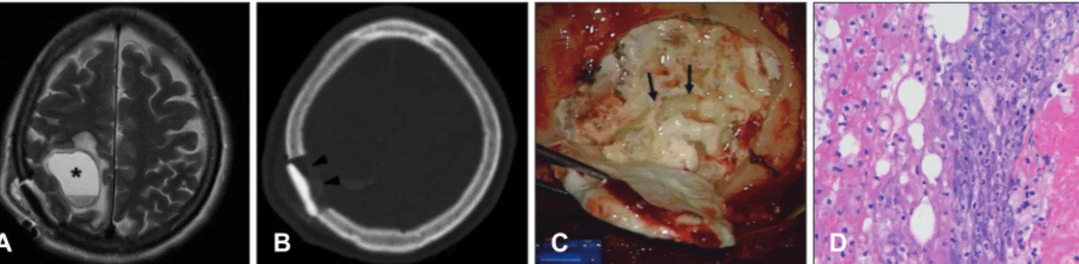
A 62-year-old female had skull mass on right parietal bone. She was underwent operation for removal of skull mass and reconstruction of defect on skull with 10 cc of GeneX®. Nine days after the operation, she complained of neglecting on left side of the body and dizziness. Brain mag- netic resonance image (MRI) and brain computed topogra- phy (CT) scan were checked for evaluation of the neurologic deficit. There was round cystic lesion in right parietal paren- chyme under the operation site in brain MRI (Figure 1A), and brain CT scan shows loss of the reconstructed material along the inner side (Figure 1B). Infectious signs such as fe- ver and myalgia were not appeared, and blood levels of white blood cell, erythrocyte sedimentation rate, and C-reactive peptide belonged normal limits. She was underwent opera- tion for removal of the reconstructed material and the cyst with irrigation. In operative field, turbid fluid collection was observed around reconstructive material, and the cyst was seen under the defect site, and it was enclosed in a whitish membrane and collected turbid fluid with many tiny whitish particles inside (Figure 1C). Reconstruction of the defect was done by using mini-plate and contourable mesh plate.
Removed materials from the lesion were referred for histo- logic examination, and it was identified to acute inflam- mation (Figure 1D).
Lytic Complications after Skull Reconstruction Using GeneX
®Jin-Hack Park, MD, Sang-Jun Suh, MD, Yoon-Soo Lee, MD, Jeong-Ho Lee, MD, Kee-Young Ryu, MD, and Dong-Gee Kang, MD
Department of Neurosurgery, Daegu Fatima Hospital, Daegu, Korea
Multiple methods and materials are available for bone defect reconstruction. Bone graft substitute is one of the materials used for reconstruction of bone defect and have been widely used recently. This report describes some cases about com- plications related to GeneX® which is introduced as mixture of calcium sulfate and β-tricalcium phosphate at manufacturer’s official web site. It informed of 3 patients who suffered wound inflammation, serous cyst after using GeneX® for reconstruct- ing skull defect.
(Korean J Neurotrauma 2015;11(2):135-138) KEY WORDS: Skull defect ㆍBone substitutes ㆍPostoperative complications.
Received: February 25, 2015 / Revised: June 16, 2015 Accepted: June 19, 2015
Address for correspondence: Sang-Jun Suh, MD
Department of Neurosurgery, Daegu Fatima Hospital, 99 Ayang- ro, Dong-gu, Daegu 41199, Korea
Tel: +82-53-940-7339, Fax: +82-53-954-7417 E-mail: [email protected]
cc This is an Open Access article distributed under the terms of Cre- ative Attributions Non-Commercial License (http://creativecommons.
org/licenses/by-nc/3.0/) which permits unrestricted noncommercial use, distribution, and reproduction in any medium, provided the original work is properly cited.
CASE REPORT
Korean J Neurotrauma 2015;11(2):135-138
pISSN 2234-8999 / eISSN 2288-2243 http://dx.doi.org/10.13004/kjnt.2015.11.2.135
136 Korean J Neurotrauma 2015;11(2):135-138 Lytic Complications by GeneX®
??? 단축제목 보내주십시오 ???
Case 2
A 70-year-old woman with a history of breast ductal cell carcinoma and lung metastasis visited due to scalp mass. In physical examination, somewhat hard mass was checked under subcutaneous tissue. Heterogeneous enhanced mass with osteolysis around was found on left frontal bone in en- hanced brain CT scan. She was underwent operation for re- moval of the mass, and 10 cc of GeneX® was used for skull reconstruction. The mass was identified to metastasis on left frontal bone. One month later, she complained about swell- ing and local pain on previously operative site (Figure 2A).
There was no definite systemic or local infectious sign in laboratory test. There was cystic lesion instead of recon- structed material on follow up brain CT scan (Figure 2B).
She was underwent operation of wound revision and irriga- tion. In operative field, there was cyst which contained many whitish particles assumed to GeneX® on skull recon- struction site (Figure 2C). Newly reconstruction was done with using contourable mesh plate. After operation, her dis- comfort was resolved. In histologic examination, it was iden- tified to chronic inflammation (Figure 2D).
Case 3
A 49-year-old woman had skull defect on left frontal bone after undergoing surgery of craniotomy and aneurysmal clipping for spontaneous subarachnoid hemorrhage due to
ruptured anterior communicating artery aneurysm. She felt cosmetic complex about depression along the inferior mar- gin of temporal line of frontal bone and protrusion at the in- ferior portion of temporal fossa. Skull reconstruction was done in such a way that 5 cc of GeneX® filled along the pre- viously craniotomy gap and fat graft was done around the depression site. Two months later, there was developed swelling, fluid collection, and local pain on operative site.
Fluid aspiration was performed on swelling site. The aspi- rated fluid looked turbid brown, and whitish particles gener- ated from GeneX® were observed in the fluid. There was no definite systemic or focal infectious sign. She suffered from delayed wound healing. After daily wound dressing was done for a month, her local inflammation was healed. De- pression of the inferior margin of temporal line of frontal bone and protrusion at the inferior portion of temporal fossa were remained.
Discussion
Many materials have been developed for reconstruction of bony defect. It is divided largely into autologous bone, allol- ogous bone and bone graft substitutes. Autograft is recog- nized to be gold standard, because it provides osteoconduc- tive, osteoinductive, osteogenic properties.2) But, autologous bone has complications with donor site morbidity, such as FIGURE 1. A: Brain MRI shows round cyst on right parietal lobe (asterisk). B: Brain computed tomography shows loss of the recon- structed material along the inner side (black arrowheads). C: Photograph shows turbid fluid with multiple tiny particles (black ar- rows). D: Inflammatory cells are mainly composed of neutrophil (hematoxylin-eosin, original magnification, ×400).
A B C D
FIGURE 2. A: Swelling on previous operation site is seen (dotted line). B: Follow up brain computed tomography shows cystic le- sion instead of reconstructed material (white arrowheads). C: Photograph shows cyst which is enclosed in a whitish membrane and collected turbid fluid with multiple tiny particles inside (black arrowheads). D: Lymphocytes are scattered (hematoxylin-eosin, origi- nal magnification, ×400).
A B C D
Jin-Hack Park, et al.
(2명 경우 Sun-Ah Choi and Hae Ja Kim) Sun-Ah Choi?, et al.
http://www.kjnt.org 137 donor site pain, infection.11) Allologous bone is another con-
siderable choice, but allograft has risk for transferring viral disease, nonunion and immunologic response.2) Because of those shortcomings, bone graft substitutes are used as alter- native, and new products are developing even now.
Bone graft substitutes have variety in composition, mech- anism of action. GeneX® is introduced as “absorbable, os- teoconductive, synthetic bone graft material with a nega- tively charged surface to accelerate bone growth in trauma, spine and non-unions” at manufacturer’s official web site (www.biocomposites.com). It composed of calcium sulfate and β-tricalcium phosphate (βTCP) in a same ratio.10) βTCP have porous with vary in size, and it is similar with trabecu- lar. When bone tissue attached to βTCP surface, osteoid grows and is mineralized, and it turns to bone tissue, and it resorbs at finally.2) Precise mechanism that calcium sulfate acts as osteoconductive has not been discovered.9) Some ar- ticles reported that calcium sulfate stimulate new bone for- mation and dissolve rapidly. According to the official web site, GeneX® has advantage that is negatively charged sur- face to enhance cell attachment and proliferation compared with untreated calcium phosphate. It is injected to fill the bone defect site, and sets within 15 minutes at normal body temperature. Yang et al.10) presented their animal research which filled with GeneX® to vertebral body defect site of sheep, and they represented their experimental results that GeneX® facilitate osteogenesis more rapidly compared with not using GeneX® group.
Although GeneX® has effective to bone reconstruction, complications relate to GeneX® also reported. Two papers de- scribed complications which occurred after the use of GeneX®. Saadoun et al.7) reported 3 patients who suffered complica- tions related of GeneX® after using for spinal fusion. They also supported their experience by representing mouse ex- perimental results that GeneX® caused significant muscle and adjacent tissue necrosis at injected site. Friesenbichler et al.1) reviewed 31 patients retrospectively who underwent bone graft with GeneX® after bone tumor removal in orthopedic
department, 5 of the 31 patients had developed complica- tions related to GeneX®. Their patients suffered from the complications which commonly involved sterile inflamma- tion, delayed wound healing, local pain and inflammatory cystic formation. They commonly reported marked local aseptic inflammation and inflammatory cystic formation after using GeneX®.1,7) In our institutes, GeneX® was used for skull reconstruction to 6 patients in the last 2 years (Table 1).
Half of them exhibited complications related to GeneX® such as wound swelling or inflammatory cyst containing the fluid and whitish particles assumed GeneX®. All of compli- cated patients suffered from self-limited local inflammation, not systemic infection. It was considered foreign-body reac- tion, because the lesions were localized to GeneX® graft sites and all of the patients were healed with removing of the material without using antibiotics.
Both βTCP and calcium sulfate are believed to safe to man. But, some articles reported complications of using cal- cium sulfate. Most frequent complication is persistent serous drainage from wounds.3) But, there was no definite patho- physiologic explanation about mechanisms which led to in- flammatory reaction. Some hypotheses about mechanism of the inflammation are suggested. One of them is that accu- mulation of calcium-rich fluid which is derived from too early graft resorption induced inflammation.5,6) Another one is that response to osmotic effect caused by not dissolved cal- cium phosphate.4,5) Based on our experiences, it is postulated that fluid collection on dead space by early resorption of GeneX® particle before bone regeneration may cause in- flammation to adjacent tissue. Also, other materials such as blood or cerebrospinal fluid can pool into the dead space, and it may inhibit bone regeneration and cause the local inflam- mation.
Conclusion
Bone graft substitute is one of the options for skull recon- struction. It is developing now and new products are intro- TABLE 1. Information of patients
Case Sex/age Diagnosis Dosage of GeneX® (cc) Complication
1 M/73 Benign fibrous osseous on skull 10 None
2 F/49 Skull defect due to craniotomy for ruptured
ACoA aneurysm 10 Soft tissue inflammation
3 F/70 Metastatic skull tumor 5 Soft tissue inflammation
4 M/46 Skull defect due to craniotomy for ruptured
ACoA aneurysm 5 None
5 F/67 Skull defect due to craniotomy for ruptured
right MCA aneurysm 5 None
6 F/62 Benign fibrous osseous on skull 10 Inflammatory cyst
ACoA: anterior communicating artery, MCA: middle cerebral artery, M: male, F: female
138 Korean J Neurotrauma 2015;11(2):135-138 Lytic Complications by GeneX®
??? 단축제목 보내주십시오 ???
duced. GeneX® is launched recently, so data about efficacy and problem of the products are short. It is easy to use for bone void filler, but early resorption of GeneX® before bone regeneration can induce serous cyst, local inflammation. So, attention is required in case of using GeneX® to large bone defect which needs long time to bone formation. Especially, more attention is need for using GeneX® adjacent to nervous system such as skull can cause neurologic deficit by the lo- cal inflammation and the cyst. In addition, broad study is required about safety and complication of GeneX®.
■ The authors have no financial conflicts of interest.
REFERENCES
1) Friesenbichler J, Maurer-Ertl W, Sadoghi P, Pirker-Fruehauf U, Bodo K, Leithner A. Adverse reactions of artificial bone graft sub- stitutes: lessons learned from using tricalcium phosphate geneX®. Clin Orthop Relat Res 472:976-982, 2014
2) Giannoudis PV, Dinopoulos H, Tsiridis E. Bone substitutes: an update. Injury 36 Suppl 3:S20-S27, 2005
3) Johnson LJ, Clayer M. Aqueous calcium sulphate as bone graft for voids following open curettage of bone tumours. ANZ J Surg 83:564-570, 2013
4) Kelly CM, Wilkins RM, Gitelis S, Hartjen C, Watson JT, Kim PT.
The use of a surgical grade calcium sulfate as a bone graft substitute:
results of a multicenter trial. Clin Orthop Relat Res (382):42-50, 5) Lee GH, Khoury JG, Bell JE, Buckwalter JA. Adverse reactions to 2001 OsteoSet bone graft substitute, the incidence in a consecutive series.
Iowa Orthop J 22:35-38, 2002
6) Robinson D, Alk D, Sandbank J, Farber R, Halperin N. Inflamma- tory reactions associated with a calcium sulfate bone substitute.
Ann Transplant 4:91-97, 1999
7) Saadoun S, Macdonald C, Bell BA, Papadopoulos MC. Dangers of bone graft substitutes: lessons from using GeneX. J Neurol Neu- rosurg Psychiatry 82:e3, 2011
8) Spetzger U, Vougioukas V, Schipper J. Materials and techniques for osseous skull reconstruction. Minim Invasive Ther Allied Technol 19:110-121, 2010
9) Thomas MV, Puleo DA. Calcium sulfate: Properties and clinical applications. J Biomed Mater Res B Appl Biomater 88:597-610, 10) Yang HL, Zhu XS, Chen L, Chen CM, Mangham DC, Coulton 2009 LA, et al. Bone healing response to a synthetic calcium sulfate/
β-tricalcium phosphate graft material in a sheep vertebral body defect model. J Biomed Mater Res B Appl Biomater 100:1911-1921, 2012 11) Younger EM, Chapman MW. Morbidity at bone graft donor sites.
J Orthop Trauma 3:192-195, 1989