Degos-Like Lesions Associated with SLE
Vol. 29, No. 2, 2017 215
Received June 14, 2016, Revised July 14, 2016, Accepted for publication July 20, 2016
Corresponding author: Kee Suck Suh, Department of Dermatology, Kosin University College of Medicine, 262 Gamcheon-ro, Seo-gu, Busan 49267, Korea. Tel: 82-51-990-6145, Fax: 82-51-990-3041, E-mail: ksderm98@
unitel.co.kr
This is an Open Access article distributed under the terms of the Creative Commons Attribution Non-Commercial License (http://creativecommons.
org/licenses/by-nc/4.0) which permits unrestricted non-commercial use, distribution, and reproduction in any medium, provided the original work is properly cited.
Copyright © The Korean Dermatological Association and The Korean Society for Investigative Dermatology
pISSN 1013-9087ㆍeISSN 2005-3894
Ann Dermatol Vol. 29, No. 2, 2017 https://doi.org/10.5021/ad.2017.29.2.215
CASE REPORT
Degos-Like Lesions Associated with Systemic Lupus Erythematosus
Min Soo Jang, Jong Bin Park, Myeong Hyeon Yang, Ji Yun Jang, Joon Hee Kim, Kang Hoon Lee, Geun Tae Kim1, Hyun Hwangbo, Kee Suck Suh
Departments of Dermatology and 1Internal Medicine, Kosin University College of Medicine, Busan, Korea
Degos disease, also referred to as malignant atrophic pap- ulosis, was first described in 1941 by Köhlmeier and was in- dependently described by Degos in 1942. Degos disease is characterized by diffuse, papular skin eruptions with porce- lain-white centers and slightly raised erythematous te- langiectatic rims associated with bowel infarction. Although the etiology of Degos disease is unknown, autoimmune dis- eases, coagulation disorders, and vasculitis have all been considered as underlying pathogenic mechanisms. Approx- imately 15% of Degos disease have a benign course limited to the skin and no history of gastrointestinal or central nerv- ous system (CNS) involvement. A 29-year-old female with history of systemic lupus erythematosus (SLE) presented with a 2-year history of asymptomatic lesions on the dorsum of all fingers and both knees. The patient had only skin lesions and no gastrointestinal or CNS vasculitis symptoms. Her skin le- sions were umbilicated, atrophic porcelain-white lesions with a rim of erythema. On the basis of clinical, histologic, and laboratory findings, a diagnosis of Degos-like lesions as- sociated with SLE was made. The patient had been treated for SLE for 7 years. Her treatment regimen was maintained over a 2 month follow-up period, and the skin lesions improved slightly with no development of new lesions. (Ann Dermatol
29(2) 215∼218, 2017) -Keywords-
Degos disease, Degos-like lesions, Systemic lupus eryth- ematosus
INTRODUCTION
Degos disease is a rare systemic vaso-occlusive disorder.
Degos-like lesions associated with systemic lupus eryth- ematosus (SLE) are a type of vasculopathy. Almost all Degos-like lesions have the clinical pathognomonic ap- pearance of porcelain-white, atrophic papules with pe- ripheral telangiectatic erythema1. Such lesions can be found in at least two distinctive clinical settings: (1) apparent idi- opathic disease, either classic Degos disease or its benign variant; and (2) cutaneous manifestations in several con- nective tissue diseases such as SLE, antiphospholipid syn- drome, dermatomyositis, and systemic sclerosis2. The course, treatment, and prognosis of these diseases has sub- stantially varied. To date, one case of systemic Degos dis- ease with SLE has been reported in the Korean literature3. Herein, we report the first case of cutaneous Degos-like le- sions without systemic involvement associated with SLE in Korea.
CASE REPORT
A 29-year-old woman presented to our clinic with a two-year history of asymptomatic atrophic white lesions.
Two years ago, the lesions began as pink dome-shaped papules on her dorsal fingers and knees bilaterally. These papules gradually turned into umbilicated, atrophic porce- lain-white lesions with a rim of erythema. Seven years
MS Jang, et al
216 Ann Dermatol
Fig. 1. (A∼C) Multiple erythematous papules with porcelain-white scars surrounded by an erythematous rim were seen on dorsum of all fingers and both knees.
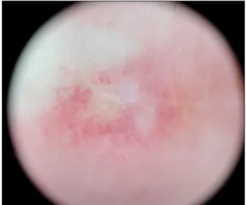
Fig. 2. On dermoscopy, a central, whitish, structureless area surrounded by an erythematous rim is seen (×10).
ago, the patient was diagnosed with SLE and was being treated in a rheumatology clinic. On physical examina- tion, atrophic, porcelain-white scars surrounded by an er- ythematous rim were seen on the dorsum of all fingers and both knees (Fig. 1). The patient also had a malar rash and arthritis, but did not have symptoms of gastrointestinal tract involvement or central nervous system (CNS) vasculi- tis, such as blurred vision and hemiparesis. Her neuro- logical exam was unremarkable. On dermoscopy, a cen- tral whitish, structureless area surrounded by an eryth- ematous rim of short vessels was seen (Fig. 2). Laboratory testing was positive for anti-nuclear antibody (1:1,280), anti-ds-DNA antibody, anti-smith antibody, anti-Ro anti- body, and anti-La antibody, but negative for anticardio- lipin immunoglobulin (Ig)G, anticardiolipin IgM, and lu- pus anticoagulant. Histologic findings showed hyper- keratosis, epidermal atrophy, and dermal sclerosis in the central portion. It also showed lymphocytic infiltration around vessels, fibrinoid necrosis of the vessel wall, and thrombus deposition in the lumen (Fig. 3). On the basis of these clinical, dermoscopic, laboratory, and histologic findings, the diagnosis of Degos-like lesions associated with SLE was made. The patient was being treated with
hydroxychloroquine (300 mg/day), prednisolone (8 mg/day), and cyclosporine (50 mg/day) for seven years, and bera- prost (20 μg/day) and pentoxifylline (400 mg/day) for 2 years for SLE. Because the patient’s skin lesions improved
Degos-Like Lesions Associated with SLE
Vol. 29, No. 2, 2017 217 Fig. 3. (A) Histologic findings showed hyperkeratosis, epidermal atrophy, and dermal sclerosis in the central portion (H&E, ×40).
(B) Lymphocytic infiltration around vessels, fibrinoid necrosis of the vessel wall, and thrombus deposition in the lumen were seen (H&E, ×400).
gradually during the two-month follow-up period, we con- tinued her treatment as before without further inter- vention, and no new lesions developed.
DISCUSSION
Degos disease, also known as malignant atrophic pap- ulosis, is a rare systemic vaso-occlusive disorder first de- scribed by Köhlmeier1 in 1941 as erythematous papules with a central porcelain-white atrophic lesion. A combina- tion of coagulopathy, endothelial cell damage, auto- immune disease, and vasculitis have been proposed as un- derlying pathogenic mechanisms, but none of these have been confirmed4.
Degos disease is classified into two types by associated clinical symptoms and underlying disease2. First, classic Degos disease has characteristic skin lesions and affects other organs with multiple limited infarcts. Skin lesions are usually the first manifestation of the disease, although its poor prognosis is the result of gastrointestinal and CNS involvement5. With systemic involvement, the reported mean survival time is approximately 2 years, but it varies from less than 1 year to more than 12 years6. Conversely, approximately 15% of Degos disease cases are a benign form often limited to the skin without gastrointestinal or CNS involvement. These cases generally exhibit long-term survival. Second, Degos-like lesions can occur in patients with connective tissue diseases such as SLE, antiphos- pholipid antibody syndrome, dermatomyositis, and sys- temic sclerosis7,8. Because of the broad overlap in clinical features and histopathological findings indistinguishable from those of cutaneous lupus erythematosus in skin le-
sions of Degos disease, Ball et al.8 did not conceive of Degos disease as a specific disease, but as a distinctive pattern expressive of different pathologic processes.
SLE is a chronic multiorgan auto-immune inflammatory disease and skin lesions are the second most frequent clin- ical manifestation. Nonspecific disease-related skin lesions are frequently seen, usually in the active phase of the dis- ease9. Vasculopathy presenting as an acute or subacute manifestation of lupus can occur as Degos-like lesions and be etiologically implicated in the pathogenesis of Degos disease8. In our patient, there was no evidence of abnor- mality in platelet aggregation or blood fibrinolytic activity.
She was found to have positive anti-nuclear antibody (1:1,280), anti-Ro antibody, anti-La antibody, anti-ds-DNA antibody, and anti-smith antibody titers at the time of diagnosis. Considering that the presence of vasculitis in patients with SLE is associated with anti-La antibody10, we assumed that our patient also had vasculitis, which can cause Degos-like skin lesions. By considering her clinical and laboratory findings, we suggested that her cutaneous Degos-like lesions were associated with SLE.
The skin lesions of Degos disease consist of three different evolution stages: (1) erythematous papules of onset; (2) er- ythematous papules with purple or necrotic centers with or without central crust; and (3) porcelain-white scars sur- rounded by an erythematous rim2. Dermoscopic findings consist of three different patterns, each corresponding to a different evolution stage. Dermoscopically, in the early stages, papules are characterized by a combination of a reddish-to-purple background and purpuric dots. At the in- termediate stage, papules display a targetoid pattern with necrotic centers surrounded by erythematous halos.
MS Jang, et al
218 Ann Dermatol
Finally, healed lesions have a whitish, structureless center surrounded by a rim of short, slightly curved vessels11. Harvell et al.12 described sequential changes in the histol- ogy of Degos lesions. Early lesions manifest a superficial and deep perivascular lymphocytic infiltrate, interstitial mucin, and vacuolar interface dermatitis. Fully developed lesions typically show prominent lymphocytic vasculitis, intramural fibrin deposition, central epidermal atrophy, and a developing zone of papillary dermal sclerosis and mucin deposition. Late stage lesions often demonstrate a classic inverted wedge-shaped zone of sclerosis with an atrophic epidermis and a sparse lymphocytic infiltrate in the upper half of the dermis. Given features of the three different stages, the cutaneous lesions in our patient were late stage, as all lesions corresponded with the clinical, dermoscopic, and histologic findings of that stage.
Currently, there is no consistently effective therapy for Degos disease, although Kim et al.13 reported that Degos-like lesions associated with Behçet’s disease improved after treatment with aspirin and dipyridamole. Heparin has been successfully used in the acute phase, although other fibrinolytic agents have been ineffective. Immunosuppression with corticosteroids or other agents may worsen skin le- sions and complications of Degos disease. Other therapies without proven benefits include antibiotics, arsenic, chlor- oquine, azathioprine, methotrexate, phenylbutazone, and interferon5,14. Our patient was treated with hydroxychlo- roquine, prednisolone, and cyclosporine for seven years, and with beraprost and pentoxifylline for two years for SLE. Her treatment was continued without modification, and her skin lesions slightly improved.
Considering all clinical, histological, and laboratory find- ings, we suggest that the cutaneous lesions in our patient were a reaction pattern. We suspect that her skin lesions were caused by peripheral arterial occlusion resulting from vasculitis associated with SLE. On the basis of our ex- perience with this case, when pathognomonic Degos-like lesions that manifest with porcelain white scars sur- rounded by an erythematous rim occur, definitive diag- nosis should be made after a careful clinical history and examinations that include physical, histological, and labo- ratory evaluation for SLE and other connective tissue diseases. Herein, we report a case of Degos-like lesions associated with SLE.
CONFLICTS OF INTEREST
The authors have nothing to disclose.
REFERENCES
1. Köhlmeier W. Multiple hautnekrosen bei thromboangiitis obliterans. Arch Dermatol Syphilol 1941;181:792-793.
2. Lipsker D. Malignant atrophic papulosis (Degos disease). In:
Goldsmith LA, Katz SI, Gilchrest BA, Paller AS, Leffell DJ, Wolff K, editors. Fitzpatrick’s dermatology in general me- dicine. 8th ed. New York: McGraw Hill, 2012:2072-2076.
3. Lee JH, Ryu HJ, Park YB, Choi BY, Lee EY, Lee YJ, et al. A case of systemic lupus erythematosus with Degos' disease. J Korean Rheum Assoc 2007;14:256-262.
4. Magro CM, Poe JC, Kim C, Shapiro L, Nuovo G, Crow MK, et al. Degos disease: a C5b-9/interferon-α-mediated endo- theliopathy syndrome. Am J Clin Pathol 2011;135:599-610.
5. Theodoridis A, Makrantonaki E, Zouboulis CC. Malignant atrophic papulosis (Köhlmeier-Degos disease)-a review.
Orphanet J Rare Dis 2013;8:10.
6. Subbiah P, Wijdicks E, Muenter M, Carter J, Connolly S.
Skin lesion with a fatal neurologic outcome (Degos' disease).
Neurology 1996;46:636-640.
7. Tsao H, Busam K, Barnhill RL, Haynes HA. Lesions resembling malignant atrophic papulosis in a patient with dermatomyositis. J Am Acad Dermatol 1997;36:317-319.
8. Ball E, Newburger A, Ackerman AB. Degos' disease: a distinctive pattern of disease, chiefly of lupus erythematosus, and not a specific disease per se. Am J Dermatopathol 2003;25:308-320.
9. Cardinali C, Caproni M, Bernacchi E, Amato L, Fabbri P.
The spectrum of cutaneous manifestations in lupus erythe- matosus--the Italian experience. Lupus 2000;9:417-423.
10. Ramos-Casals M, Nardi N, Lagrutta M, Brito-Zerón P, Bové A, Delgado G, et al. Vasculitis in systemic lupus erythe- matosus: prevalence and clinical characteristics in 670 patients. Medicine (Baltimore) 2006;85:95-104.
11. Anker JP, Kaminska-Winciorek G, Lallas A, Nicoletti S, Januszewski K, Mazzei ME, et al. The dermoscopic variability of Degos disease at different stages of progression. Der- matol Pract Concept 2014;4:59-61.
12. Harvell JD, Williford PL, White WL. Benign cutaneous Degos' disease: a case report with emphasis on histopathology as papules chronologically evolve. Am J Dermatopathol 2001;23:116-123.
13. Kim YJ, Yun SJ, Lee SC, Lee JB. Degos disease associated with Behçet's disease. Ann Dermatol 2015;27:235-236.
14. Melnik B, Hariry H, Vakilzadeh F, Gropp C, Sitzer G.
Malignant atrophic papulosis (Köhlmeier-Degos disease).
Failure to respond to interferon alpha-2a, pentoxifylline and aspirin. Hautarzt 2002;53:618-621.