202
Copyrights © 2015 The Korean Society of RadiologyINTRODUCTION
Tuberculosis (TB) remains a major health problem in Korea since its incidence has not decreased significantly in recent de- cades. The estimated incidence rate of TB in Korea was 87.6/
100000 population in 2012. The incidence of extrapulmonary TB has increased along with the growing number of immuno- compromised patients (1). Cervical lymphadenitis is the most common presentation of extrapulmonary TB. However, extra- pulmonary TB lacks specific clinical manifestations and can mimic many diseases; thus, the diagnosis may be intricate (2).
TB abscess also shows variable findings in imaging studies;
hence, it is often difficult to radiographically distinguish it from the other neoplasms (3).
We report a rare case of a huge retroperitoneal TB abscess
mimicking a retroperitoneal cystic mass. The mass was diag- nosed as TB abscess by histopathologic examination after surgi- cal excision. No active pulmonary or spinal TB involvement was identified. The patient was successfully treated with anti-TB medications.
CASE REPORT
A 55-year-old man presented with a palpable mass in the right lower abdominal region which was first noticed 2 months ago. The patient had pulmonary TB 26 years ago, and he had successfully completed the treatment. He was otherwise healthy and did not have any other symptoms or abnormal physical findings. Laboratory results were within their normal range.
His chest X-ray showed several calcified nodules and fibrosis
Case Report
pISSN 1738-2637 / eISSN 2288-2928 J Korean Soc Radiol 2015;72(3):202-205 http://dx.doi.org/10.3348/jksr.2015.72.3.202
Received September 12, 2014; Accepted October 29, 2014Corresponding author: Seunghwan Cha, MD Department of Radiology, Yonsei University Wonju College of Medicine, Wonju Severance Christian Hospital, 20 Ilsan-ro, Wonju 220-701, Korea.
Tel. 82-33-741-1462 Fax. 82-33-732-8281 E-mail: peace22@yonsei.ac.kr
This is an Open Access article distributed under the terms of the Creative Commons Attribution Non-Commercial License (http://creativecommons.org/licenses/by-nc/3.0) which permits unrestricted non-commercial use, distri- bution, and reproduction in any medium, provided the original work is properly cited.
Large cystic masses originating from the retroperitoneal space are rare, and cystic tumors are often considered preferentially in the differential diagnosis. However, it is difficult to make a correct diagnosis. A 55-year-old man presented with a palpa- ble abdominal mass. A computed tomography (CT) scan detected a mass mimicking a large cystic tumor in the retroperitoneal space anterior to the psoas muscle. The mass had an enhanced outer margin, an irregular inner margin, and several sur- rounding necrotic lymph nodes. However, histopathologic examination followed by an exploratory laparotomy confirmed that the mass was consistent with a tubercu- lous (TB) abscess. A retroperitoneal TB abscess without spinal or active pulmonary TB is very rare. To the best of our knowledge, there are no published reports of a retroperitoneal TB abscess confirmed by both CT scan and surgical pathology in the Korean literature. We report a rare case of a huge retroperitoneal TB abscess that can mimic a cystic tumor.
Index terms Tuberculous Abscess Retroperitoneal Cystic Mass Retroperitoneal Tuberculosis
A Large Tuberculous Abscess Mimicking a Retroperitoneal Cystic Mass: A Case Report
1후복막 낭성 종양으로 오인될 수 있는 거대 결핵 농양: 증례 보고1
Kwang Suk Kim, MD
1, Seunghwan Cha, MD
1, Jihyun Ahn, MD
1, Miyeon Cho, MD
2Departments of 1Radiology, 2Pathology, Yonsei University Wonju College of Medicine, Wonju Severance Christian Hospital, Wonju, Korea
Kwang Suk Kim, et al
203
jksronline.org J Korean Soc Radiol 2015;72(3):202-205
pathologic examination by hematoxylin and eosin staining re- vealed granulomatous inflammation and caseous necrosis consis- tent with TB abscess (Fig. 1F); however, acid-fast staining and polymerase chain reaction test for Mycobacterium tuberculosis were negative. Immunohistochemical staining for D2-40, CD34, CD31, and calretinin was negative. There was no endothelial lining along the inner wall of the lesion, which ruled out the possibility of a pre-existing mesothelial or lymphangitic cyst.
Anti-TB therapy with isoniazid, rifampicin, pyrazinamide, and ethambutol was administered for 6 months. After the comple- tion of treatment, the patient did not develop any further symp- toms during the follow-up.
DISCUSSION
Retroperitoneal abscesses are a uncommon clinical problem and they have a high mortality rate. They can occur as complica- in the right upper lung field, indicating old healed TB, but it
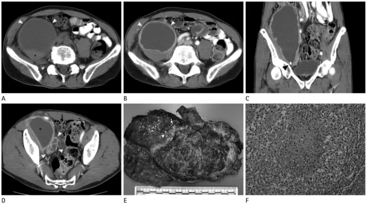
showed no interval changes when compared with the previous X-ray image which was taken 6 years ago. An abdominal comput- ed tomography (CT) scan detected an oval-shaped cystic mass (8.3 × 6.5 × 20.9 cm) in the right lower retroperitoneal space. The mass had a well-defined outer margin, an irregular inner margin, wall enhancement, and hemorrhage. This mass compressed the right psoas muscle but it was relatively well demarcated from the muscle (Fig. 1A, B). There were focal wall calcifications within the mass (Fig. 1C). Several necrotic lymph nodes were observed around the mass in the right obturator space, and there was no evidence of ascites (Fig. 1D). Based on the radiographic findings, we considered cystic tumors that occur in the retroperitoneal space such as complicated cystic lymphangioma, mucinous cyst- adenocarcinoma, neurogenic tumors with cystic degeneration, or other undifferentiated sarcomas. Total surgical excision of the mass and adjacent lymph nodes was performed (Fig. 1E). Histo-
Fig. 1. Oval-shaped cystic mass in the right lower retroperitoneal space.
A. Precontrast axial image shows a large retroperitoneal mass (white arrowheads) compressing the psoas muscle (asterisk).
B. Postcontrast axial image shows that the cystic mass has a well-defined outer margin, an irregular inner margin, and wall enhancement (white arrowheads).
C. Coronal image shows focal calcifications (black arrowhead) within the mass.
D. Multiple lymph nodes with necrotic changes (white arrowheads) are observed around the large mass (asterisk).
E. Gross specimen of the excised mass.
F. Histological examination shows granulomatous inflammation with central caseous necrosis consistent with tuberculosis (hematoxylin and eo- sin stain, × 200).
E B
D A
F C
A Large Tuberculous Abscess Mimicking a Retroperitoneal Cystic Mass
204
J Korean Soc Radiol 2015;72(3):202-205 jksronline.orgIn conclusion, this is a rare case of TB abscess presenting as a large retroperitoneal cystic mass without active pulmonary or vertebral involvement. The diagnosis was based on CT imaging and histopathology. To the best of our knowledge, there are no published reports of retroperitoneal TB abscess which was con- firmed by both CT scan and surgical pathology in the Korean literature. As TB remains endemic in Korea and with the con- tinued increase in multidrug-resistant TB infections, it is impor- tant to know various imaging features of extrapulmonary TB in- cluding TB abscess. In this respect, this rare case provides an uncommon yet important differential diagnosis of a retroperito- neal cystic lesion.
REFERENCES
1. Kwon YS, Koh WJ. Diagnosis of pulmonary tuberculosis and nontuberculous mycobacterial lung disease in Korea.
Tuberc Respir Dis (Seoul) 2014;77:1-5
2. Golden MP, Vikram HR. Extrapulmonary tuberculosis: an overview. Am Fam Physician 2005;72:1761-1768
3. Barbalinardo RJ, Hamilton GB, Eliot GR, Lazaro EJ, Haycock C. Tuberculous retroperitoneal lymphadenopathy mimick- ing metastatic pancreatic carcinoma. J Natl Med Assoc 1986;78:385-387
4. Karoui S, Bibani N, Ouaz A, Serghini M, Chebbi F, Nouira K, et al. Retroperitoneal abscess: a rare localization of tuber- cular infection. Gastroenterol Res Pract 2010;2010:475130 5. Epstein BM, Mann JH. CT of abdominal tuberculosis. AJR
Am J Roentgenol 1982;139:861-866
6. Yang DM, Jung DH, Kim H, Kang JH, Kim SH, Kim JH, et al.
Retroperitoneal cystic masses: CT, clinical, and pathologic findings and literature review. Radiographics 2004;24:
1353-1365
7. Shin YS, Kim HJ, Kim MK. Juxta-adrenal malignant schwan- noma with lymph node metastases. Can Urol Assoc J 2013;
7:E657-E659
8. Fong Y, Coit DG, Woodruff JM, Brennan MF. Lymph node metastasis from soft tissue sarcoma in adults. Analysis of data from a prospective database of 1772 sarcoma pa- tients. Ann Surg 1993;217:72-77
9. Chuang HJ, Liu JY, Chang FW, Chou CC, Loh CH. Vertebral tuberculosis presenting as a large retroperitoneal cyst. Tai- tions of other diseases such as perforated appendicitis, diverticu-
litis, perforated colonic or duodenal cancer, Crohn’s disease of the bowel, pancreatitis, or trauma. They are often caused by poly- microbial infections, and the common pathogens are Escherichia coli, Klebsiella pneumoniae, Enterococcus spp., and Staphylo- coccus aureus. However, abdominal TB abscesses are uncom- mon in immunocompetent patients. Pathogenesis of such cases can be through hematogenous or lymphatic dissemination from active pulmonary TB or a direct extension from an adjacent or- gan (4).
Extrapulmonary TB abscess can present with variable radiolog- ic features. It is difficult to differentiate it from lymphoma, malig- nant tumors, or various inflammatory conditions. Furthermore, the differential diagnosis becomes more complex when extrapul- monary TB presents as a soft tissue mass, showing involvement of the peritoneum, or an abscess. Epstein and Mann (5) discussed that it is important to differentiate other neoplasms from TB ab- scess. Therefore, the mass in our case should also be differentiated from cystic tumors occurring in the retroperitoneum.
Retroperitoneal tumors with a cystic component can be cystic lymphangiomas, mucinous cystic tumors, or neurogenic tumors with cystic degeneration. Also, malignant tumors can resemble a cystic mass if they have extensive necrosis. Unlike in this case, typical cystic lymphangiomas and mucinous cystic tumors have a thin, smooth wall and may have septa within the lesion. It is known that among neurogenic tumors, schwannomas can dis- play cystic degeneration as well as focal areas of punctate calcifi- cation. However, they can be differentiated from TB abscess be- cause schwannomas are seldom accompanied by necrotic lymph nodes. Among malignant tumors, retroperitoneal leiomyosarco- mas tend to develop massive cystic degeneration. They can show central necrosis more commonly than the other sarcomas, whereas calcifications are not typically present. Furthermore, sarcoma accompanied by lymphadenopathy is quite rare (6-8).
Chuang et al. (9) reported a case of vertebral TB presenting as a large retroperitoneal cyst which has radiographic similarities with our case. There are also several reports of an iliopsoas ab- scess that originated from spinal TB (10). If there were findings suspicious for spinal TB or active pulmonary TB, TB abscess would have been highly suggested on CT imaging in our case.
However, there was no evidence of vertebral TB or active pul- monary TB in our patient.
Kwang Suk Kim, et al
205
jksronline.org J Korean Soc Radiol 2015;72(3):202-205
es. Postgrad Med J 2004;80:459-462 wan J Obstet Gynecol 2008;47:247-249
10. Mallick IH, Thoufeeq MH, Rajendran TP. Iliopsoas abscess-
후복막 낭성 종양으로 오인될 수 있는 거대 결핵 농양: 증례 보고1
김광석
1· 차승환
1· 안지현
1· 조미연
2후복막에서 기원한 커다란 낭성 병변은 흔하지 않다. 또한 이런 경우에 대개 낭성 종양을 먼저 생각하기 마련이고 진단이 쉽지 않다. 본원에 복부 종괴감으로 내원한 환자가 있어 컴퓨터단층촬영을 시행하였다. 거대 낭성 종양으로 오인될 수 있 는 병변이 요근 앞쪽 후복막에 있었다. 이 병변의 변연은 조영증강이 되고 불규칙한 내부 변연을 보였으며 주변으로 괴사된 림프절이 동반되어 있었다. 하지만 이 낭성 병변은 진단적 개복술 후 결핵 농양으로 진단되었다. 이 농양은 척추 결핵과 활 동성 폐결핵 없이 나타난 보기 드문 증례로, 국내에서 컴퓨터단층촬영 사진과 수술적으로 진단된 증례가 문헌으로 보고된 적은 없다. 그래서 낭성 종양으로 오인될 수 있는 드문 커다란 결핵 농양 1예를 보고한다.
연세대학교 원주의과대학 원주세브란스기독병원 1영상의학과, 2병리과