INTRODUCTION
Endoscopic retrograde cholangiopancreatography (ERCP) is commonly performed to diagnose and treat hepato-pancreato- biliary diseases. But it has a higher potential for procedure-relat- ed complications compared to other endoscopic procedures in the upper gastrointestinal tract (1). Even in the best hands, seri- ous complications from therapeutic ERCP occur in 2.5–8% of cases, with mortality ranging from 0.5–1.1% (2). Guidewire cannulation is widely used for bile duct cannulation during ERCP. Although complications of ERCP due to guidewire-relat- ed injury are rare and can be managed conservatively, they can be life-threatening (3). We report on a case of massive hemobilia due to hepatic arteriobiliary fistula caused by guidewire-associ-
ated injury during ERCP that was successfully treated with he- patic arterial embolization. To the best of our knowledge, this is the first report of a hepatic arteriobiliary fistula caused by guide- wire-associated injury during ERCP.
CASE REPORT
A 67-year-old woman presented with acute upper abdominal pain, nausea, and vomiting. She denied any fever, chills, recent gastrointestinal bleeding, or taking anticoagulant medication.
Biochemistry tests revealed cholestatic liver function and pan- creatitis. Laboratory tests data were as follows: total bilirubin, 3.0 mg/dL (0.1–1.2 mg/dL); direct bilirubin, 2.1 mg/dL (0.1–0.4 mg/dL); aspartate aminotransferase, 360 U/L (15–40 U/L); ala-
Case Report
pISSN 1738-2637 / eISSN 2288-2928 J Korean Soc Radiol 2015;72(5):348-351 http://dx.doi.org/10.3348/jksr.2015.72.5.348
Received September 5, 2014 Accepted January 8, 2015
Corresponding author: Jae Cheol Hwang, MD Department of Radiology, Ulsan University Hospital, University of Ulsan College of Medicine,
877 Bangeojinsunhwando-ro, Dong-gu, Ulsan 682-714, Korea.
Tel. 82-52-250-8913 Fax. 82-52-230-1155 E-mail: stent@paran.com
This is an Open Access article distributed under the terms of the Creative Commons Attribution Non-Commercial License (http://creativecommons.org/licenses/by-nc/3.0) which permits unrestricted non-commercial use, distri- bution, and reproduction in any medium, provided the original work is properly cited.
Although endoscopic retrograde cholangiopancreatography (ERCP) is an effective modality for diagnosis and treatment of biliary and pancreatic diseases, the risk for procedure-related complications is high. Hemorrhage is one of major complica- tions of ERCP. Most ERCP-associated bleeding is primarily a complication related to sphincterotomy rather than diagnostic ERCP. We are reporting a case of massive hemobilia due to hepatic arteriobiliary fistula caused by guidewire-associated injury during ERCP, which was successfully treated with transarterial embolization of the hepatic artery.
Index terms
Endoscopic Retrograde Cholangiopancreatography Complication
Hepatic Arteriobiliary Fistula Hemobilia
Embolization
Massive Hemobilia due to Hepatic Arteriobiliary Fistula during Endoscopic Retrograde Cholangiopancreatography: An Extremely Rare Guidewire-Related Complication
1내시경적 역행성 담췌관 조영술 도중 간동맥-담도 누공에 의한 대량 혈담즙증: 희귀한 유도철사 관련 합병증1
Jeong Gu Nam, MD
1, Young Woo Seo, MD
1, Jae Cheol Hwang, MD
1, Young Cheol Weon, MD
1, Byeong Seong Kang, MD
1, Sung Jo Bang, MD
2, Min Seo Bang, MD
1Departments of 1Radiology, 2Internal Medicine, Ulsan University Hospital, University of Ulsan College of Medicine, Ulsan, Korea
Copyrights © 2015 The Korean Society of Radiology
348
Jeong Gu Nam, et al
349
jksronline.org J Korean Soc Radiol 2015;72(5):348-351
denly complained of sharp upper abdominal pain and was ex- tremely irritability during ERCP. Duodenoscopy revealed fresh blood gushing from the papilla. The patient was immediately moved to the angiographic suites for an angiography. Hepatic arteriography revealed active contrast extravasation into the bile duct from the anterior superior branch of the right hepatic ar- tery (Fig. 1). Fluoroscopy following arteriography showed irreg- ular, cast-like filling defects that occupied the majority of the bile duct (Fig. 2). Subsequently, selective coil embolization of the hepatic artery branch was successfully performed (Fig. 3). Per- cutaneous transhepatic biliary drainage (PTBD) was performed simultaneously. Although the patient remained hemodynami- cally stable, follow-up laboratory tests on the same day revealed nine aminotransferase, 314 U/L (0–40 U/L); alkaline phospha-
tase, 315 U/L (25–100 U/L); γ-glutamyltransferase, 182 U/L (0–
50 U/L); amylase, 529 U/L (20–104 U/L); and lipase, 1723 U/L (5.6–51.3 U/L). Coagulation was within normal limits. Abdomi- nal computed tomography (CT) revealed diffuse dilatation of the common bile duct with left intrahepatic duct stones and a normal pancreatic appearance.
The next day, she underwent ERCP under a diagnosis of cho- ledocholithiasis with biliary pancreatitis. Duodenoscopy re- vealed no mass in the ampulla of Vater and confirmed that there was no bleeding from the papilla. To cannulate the bile duct, a 0.025-inch guidewire was advanced during the sphincterotomy under endoscopic and fluoroscopic guidance. The patient sud-
Fig. 1. Selective hepatic arteriography showed an arteriobiliary fistula (arrow) with active contrast extravasation into the bile duct (arrow- heads).
Fig. 3. Common hepatic arteriography after selective coil emboliza- tion showed coils (arrow) that were placed at the hepatic artery branch and no longer contrast extravasation.
Fig. 2. Fluoroscopy following hepatic arteriography revealed irregular cast-like filling defects (arrows) in the bile duct due to massive hemo- bilia.
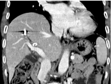
Fig. 4. Coronal reformatted contrast-enhanced CT scan demonstrated no evidence of subcapsular hepatic hematoma or perihepatic free air, suggesting liver perforation.
Massive Hemobilia due to Hepatic Arteriobiliary Fistula during Endoscopic Retrograde Cholangiopancreatography
350
J Korean Soc Radiol 2015;72(5):348-351 jksronline.orgpy, or hepatobiliary surgery (9). The possible explanation for an arteriobiliary fistula include direct injury to the arteries of the bile duct, tumor necrosis, mycotic pseudoaneurysm of hepatic artery, and endothelial toxicity due to chemotherapeutic medi- cation. In the present case, the hepatic arteriobiliary fistula re- sulted from a direct injury to the hepatic artery, which led to massive hemobilia requiring urgent treatment.
Although complications of ERCP that are caused by guide- wire-related injury are rare and can be managed conservatively, they may be life-threatening and require arteriographic emboli- zation or surgery. In the literature, subcapsular hematomas that have been the most commonly reported guidewire-associated complication of ERCP, which usually developed a few hours af- ter ERCP and was successfully managed conservatively or by a simple drainage procedure (4). Massive hemobilia from hepatic arteriobiliary fistula should be considered a serious complica- tion that requires more aggressive and urgent therapeutic inter- ventions. Transcatheter arterial embolization is a safe and effec- tive life-saving treatment for massive hemobilia and should be considered a first-line therapy (10). The primary goal of therapy is to decrease pulsatile blood pressure distal to the artificial oc- clusion. The risk of hepatic necrosis is minimal with superselec- tive embolization. In the present case, selective angiography of the hepatic arteries was performed immediately after ERCP and selective coil embolization of the hepatic artery was successfully performed.
In conclusion, massive hemobilia in the present case was most likely due to an arteriobiliary fistula caused by a guidewire-related injury during ERCP and was successfully treated with selective coil embolization of the hepatic artery. A hepatic arteriobiliary fis- tula caused by guidewire-associated injury during ERCP is an ex- tremely rare complication, but should be part of the differential diagnosis when there is bleeding at the site of a sphincterotomy.
More attention should be paid to the possibility of guidewire-re- lated complications, and if an arteriobiliary fistula is suspected, immediate hepatic angiography is required to detect and even- tually embolize the damaged artery.
REFERENCES
1. Aliperti G. Complications related to diagnostic and thera- peutic endoscopic retrograde cholangiopancreatography.
decreased hemoglobin levels from 11.4 to 9.2 g/dL. The patient had no further bleeding.
Follow-up CT was performed 2 weeks after coil embolization, which showed no evidence of a subcapsular hematoma or peri- hepatic free air, which suggests liver perforation (Fig. 4). During the 1-month follow-up period, the patient’s liver function and pancreatic enzymes were normalized, and the intrahepatic duct stones were cholangioscopically removed.
DISCUSSION
There are a few published reports of guidewire-associated in- jury during ERCP that resulted in subcapsular hepatic hemato- ma (4), subcapsular biloma (5), liver parenchyma perforation (6), and portobiliary fistula (7). Most of these reports explain that these complications are the result of accidental puncture of the intrahepatic bile duct by the guidewire. To the best of our knowledge, this is the first report of a hepatic arteriobiliary fistu- la caused by guidewire-associated injury during ERCP.
Post ERCP-associated hemorrhage is primarily a complica- tion related to the sphincterotomy rather than the diagnostic ERCP. Bleeding following a sphincterotomy has been variably reported in < 1% to 10% of the patients. Most sphincterotomy site bleedings are managed successfully by supportive conserva- tive measures with or without an endoscopic hemostatic proce- dure. In only a small percentage of cases, refractory bleeding oc- curs and requires angiographic embolization or surgery (8). In addition, while bleeding at the site of a sphincterotomy usually presents as duodenal bleeding, hemobilia due to arteriobiliary fistula presents as massive hemorrhage in the intrahepatic bile duct, which increases intraductal pressure and may aggravate biliary complications such as obstructive jaundice, cholangitis, and sepsis. Therefore, if there is sudden fresh bleeding from the bile duct, as in this case, a diagnosis of hepatic arteriobiliary fis- tula should be considered to differentiate from sphincterotomy site bleeding.
Although hemobilia is an uncommon cause of upper gastro- intestinal bleeding, its incidence is increasing due to more fre- quent use of local diagnostic and therapeutic interventions. As a result, hemobilia, which used to occur as a consequence of blunt or penetrating liver trauma in the past, is now mostly iatrogenic following liver biopsy, PTBD, hepatic intraarterial chemothera-
Jeong Gu Nam, et al
351
jksronline.org J Korean Soc Radiol 2015;72(5):348-351
Endoscopic Retrograde Cholangiopancreatography. Case Rep Gastroenterol 2011;5:487-491
7. Kawakami H, Kuwatani M, Kudo T, Ehira N, Yamato H, Asaka M. Portobiliary fistula: unusual complication of wire-guided cannulation during endoscopic retrograde cholangiopan- creatography. Endoscopy 2011;43 Suppl 2 UCTN:E98-E99 8. Ferreira LE, Baron TH. Post-sphincterotomy bleeding: who,
what, when, and how. Am J Gastroenterol 2007;102:2850- 2858
9. Green MH, Duell RM, Johnson CD, Jamieson NV. Haemobi- lia. Br J Surg 2001;88:773-786
10. Srivastava DN, Sharma S, Pal S, Thulkar S, Seith A, Bandhu S, et al. Transcatheter arterial embolization in the man- agement of hemobilia. Abdom Imaging 2006;31:439-448 Gastrointest Endosc Clin N Am 1996;6:379-407
2. Hart R, Classen M. Complications of diagnostic gastroin- testinal endoscopy. Endoscopy 1990;22:229-233
3. Enns R, Eloubeidi MA, Mergener K, Jowell PS, Branch MS, Pappas TM, et al. ERCP-related perforations: risk factors and management. Endoscopy 2002;34:293-298
4. Del Pozo D, Moral I, Poves E, Sanz C, Martín M. Subcapsu- lar hepatic hematoma following ERCP: case report and re- view. Endoscopy 2011;43 Suppl 2 UCTN:E164-E165 5. Dupas JL, Mancheron H, Sevenet F, Delamarre J, Delcense-
rie R, Capron JP. Hepatic subcapsular biloma. An unusual complication of endoscopic retrograde cholangiopancrea- tography. Gastroenterology 1988;94(5 Pt 1):1225-1227 6. Kayashima H, Ikegami T, Kasagi Y, Hidaka G, Yamazaki K,
Sadanaga N, et al. Liver Parenchyma Perforation following
내시경적 역행성 담췌관 조영술 도중 간동맥-담도 누공에 의한 대량 혈담즙증: 희귀한 유도철사 관련 합병증1
남정구
1· 서영우
1· 황재철
1· 원영철
1· 강병성
1· 방성조
2· 방민서
1내시경적 역행성 담췌관 조영술은 담췌관 질환의 진단 및 치료에 효과적인 검사법이다. 하지만 시술 관련 합병증의 발생 가능성이 있으며, 출혈은 이러한 합병증 중 하나이다. 대개 내시경적 역행성 담췌관 조영술 관련 출혈은 팽대부 괄약근 절 개술에 의해 발생한다. 저자들은 내시경적 역행성 담췌관 조영술 도중 발생한 간동맥-담도 누공에 의해 대량 혈담즙증이 생긴 환자에서 선택적 간동맥 색전술을 통해 성공적으로 치료한 증례를 보고하고자 한다.
울산대학교 의과대학 울산대학교병원 1영상의학과, 2내과