Received: June 28, 2018 Revised: July 4, 2018 Accepted: July 4, 2018
Correspondence to
The Korean Society of Clinical Neurophysiology
Daeil Building 1111, 12 Insadong-gil, Jongno-gu, Seoul 03163, Korea Tel: +82-2-2291-2290 Fax: +82-2-737-6531 E-mail: kscn@kscn.or.kr
* These authors contributed equally to this work.
Basic concepts of needle electromyography
Jee-Eun Kim1*, Jin Myoung Seok2*, Suk-Won Ahn3, Byung-Nam Yoon4, Young-Min Lim5, Kwang-Kuk Kim5, Ki-Han Kwon6, Kee Duk Park7, Bum Chun Suh8; and on behalf of the Korean Society of Clinical Neurophysiology Education Committee
1Department of Neurology, Seoul Medical Center, Seoul, Korea
2Department of Neurology, Soonchunhyang University Cheonan Hospital, Soonchunhyang University College of Medicine, Cheonan, Korea
3Department of Neurology, Chung-Ang University Hospital, Chung-Ang University College of Medicine, Seoul, Korea
4Department of Neurology, Seoul Paik Hospital, Inje University College of Medicine, Seoul, Korea
5Department of Neurology, Asan Medical Center, University of Ulsan College of Medicine, Seoul, Korea
6Department of Neurology, Hallym University Dongtan Sacred Heart Hospital, Hwaseong, Korea
7Department of Neurology, Mokdong Hospital, Ewha Womans University School of Medicine, Seoul, Korea
8Department of Neurology, Kangbuk Samsung Hospital, Sungkyunkwan University School of Medicine, Seoul, Korea
Clinical evaluations, nerve conduction studies, and electromyography play major comple- mentary roles in electrophysiologic diagnoses. Electromyography can be used to assess pathologic changes and localize lesions occurring in locations ranging from motor units to anterior-horn cells. Successfully performing electromyography requires knowledge of the anatomy, physiology, and pathology of the peripheral nervous system as well as sufficient skill and interpretation ability. Electromyography techniques include acquiring data from visual/
auditory signals and performing needle positioning, semiquantitation, and interpretation.
Here we introduce the basic concepts of electromyography to guide clinicians in performing electromyography appropriately.
Key words: Electromyography; Electrodiagnosis; Needles; Neuromuscular diseases
Ann Clin Neurophysiol 2019;21(1):7-15 https://doi.org/10.14253/acn.2019.21.1.7
SpeCial arTiCle
The Korean SocieTy of clinical neurophySiology
Received: December 21, 2017 Revised: January 3, 2018 Accepted: January 3, 2018
Correspondence to
The Korean Society of Clinical Neurophysiology
Daeil Building 1111, 12 Insadong-gil, Jongno-gu, Seoul 03163, Korea Tel: +82-2-2291-2290 Fax: +82-2-737-6531 E-mail: kscn@kscn.or.kr
Basic requirements for visual evoked potentials
Hung Youl Seok1, Eun-Mi Lee2, Kee Duk Park3, Dae-Won Seo4; and on behalf of the Korean Society of Clinical Neurophysiology Education Committee
1Department of Neurology, Keimyung University School of Medicine, Daegu, Korea
2Department of Neurology, Ulsan University Hospital, University of Ulsan College of Medicine, Ulsan, Korea
3Department of Neurology, Mokdong Hospital, Ewha Womans University College of Medicine, Seoul, Korea
4Department of Neurology, Samsung Medical Center, Sungkyunkwan University School of Medicine, Seoul, Korea
Visual evoked potentials (VEPs) are frequently used to assess the anterior and posterior visual pathways. In particular, the use of VEPs have been increasing in various fields such as evalua- tion of the optic nerves in patients with multiple sclerosis. The performance of VEP test can be affected by various factors such as stimulus type and subject condition, and its interpretation is also difficult. However, there have been no guidelines for performing and interpreting VEPs in Korea. Therefore, we aimed to provide comprehensive information regarding basic require- ment and interpretation for VEPs.
Key words: Visual evoked potential; Optic nerve; Visual pathway
Ann Clin Neurophysiol 2018;20(1):12-17 https://doi.org/10.14253/acn.2018.20.1.12
THE KOREAN SOCIETY OF CLINICAL NEUROPHYSIOLOGY
INTRODUCTION
A visual evoked potential (VEP) measures an electrophysiological response of the visual pathway to a patterned or unpatterned visual stimulus. It is a reliable, sensitive, and non- invasive technique that can measure impairment of visual pathways.1-4 While stimulation with a relatively low frequency (up to 4/s) generates transient VEPs, stimulation with a high frequency (over 10/s) generates responses corresponding to relatively simple waves in accordance to the stimulation. These are called steady-state VEPs.5 Responses induced by a patterned stimulus are called patterned VEPs (PVEPs), while those induced by an un- patterned stimulus are called flash VEPs (FVEPs).1-4 In this overview we describe compre- hensive information regarding basic requirement and interpretation for VEPs.
SPECIAL ARTICLE
ORCID Jee Eun Kim
http://orcid.org/0000-0002-3811-3479 Jin Myoung Seok
http://orcid.org/0000-0002-1484-2968 Suk-Won Ahn
http://orcid.org/0000-0002-9979-4589 Byung-Nam Yoon
http://orcid.org/0000-0003-0946-0276 Young-Min Lim
http://orcid.org/0000-0001-5074-812X Kwang-Kuk Kim
http://orcid.org/0000-0002-9641-5870 Ki-Han Kwon
http://orcid.org/0000-0001-6399-2236 Kee Duk Park
http://orcid.org/0000-0003-1628-6683 Bum Chun Suh
http://orcid.org/0000-0002-3947-5615
IntroductIon
An electrodiagnostic study (EDX) involves two main proce- dures: a nerve conduction study (NCS) and electromyogra- phy (EMG). Except for a few exceptional circumstances, such as when examining the acute stage of nerve injury, EMG should be followed by NCS in order to achieve an accurate diagnosis.1 EMG measures the electrical activity of muscle fibers either individually or collectively, and can be used to characterize pathognomic changes in muscles, nerves, roots, and anterior-horn cells. EMG results can offer additional in- formation to that available from an NCS that will assist in lo- calizing or distinguishing disorders. Needle EMG is crucial for diagnosing radiculopathy, myopathy, plexopathy, and mo- tor neuron disease. An accurate diagnosis requires the ex- amining physician (or a technologist being supervised by a physician) to have received adequate training and obtained a comprehensive knowledge of neurologic and musculo- skeletal diseases. Here we present the basic concepts and techniques of needle EMG and suggest some interpretation methods that can be applied in real clinical settings.
Standard technIque for data acquISItIon
Equipment
Electrical activity is recorded using surface or needle elec- trodes, with needle electrodes being used more frequently in clinical settings, and so here we focus on EMG studies per- formed with needle electrodes. The following equipment is essential for performing a needle EMG study: EMG machine, needle, cables, gloves, and ground electrodes. The ground electrode is attached to the tested limbs to ensure electrical safety and minimize electrical noise. The EMG needle is con- nected to the EMG machine via a cable, and the examiner needs to wear disposable gloves to protect himself/herself from blood-mediated diseases. The EMG needle should not be reused.
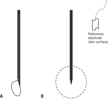
A concentric or monopolar needle can be used, each of which has pros and cons. In a concentric needle, a thin wire passing through the center of the shaft acts as the recording electrode, and the insertion tube is the reference electrode.
Because the tip of the concentric needle is cut obliquely, the
recording area of a concentric needle is shaped like a tear- drop (Fig. 1).2 In contrast, a monopolar needle is coated with Teflon and its tip (without this coating) acts as the recording (active) electrode.2 A reference electrode is not included in a monopolar needle, and so a surface electrode needs to be attached to nearby skin. The recording tip of a monopolar needle has a pin shape and its recording area has a spherical pattern around the tip.
These differences between concentric and monopolar needles result in both the amplitude and duration of mea- sured motor-unit action potentials (MUAPs) being slightly smaller for the former. A monopolar needle is cheaper and less painful for subjects because it is thinner than a concen- tric needle, but it requires a separate reference electrode, and the associated electrode impedance mismatch is likely to increase the electrical noise of an EMG study involving a monopolar needle.2
Preparing patients for EMG studies
Successfully performing an EMG study is strongly depen- dent on good cooperation from the patient. Before starting
A
fig. 1. Types and recording area of electromyography needles. (A) A concentric needle and its recording field (side view). A wire that acts as the active electrode runs through the center of the needle shaft (white), and the shaft acts as the reference electrode. (B) A monopolar needle and its recording field (side view). The needle tip without a Teflon coat- ing acts as the active electrode, and an additional reference electrode needs to be attached to nearby skin.
B
EMG, the physician should inform the patient about the pro- cedure, including the possibility of pain induced by needle movements. Relaxing the muscles during needle insertion can reduce the pain. The pain can be aggravated after skin penetration when the needle is close to the nerve or posi- tioned in the end-plate zone, and the patient should be en- couraged to inform the examiner about any such discomfort that occurs during the investigation. The physician should check for any medical condition before the study that may increase the likelihood of complications, such as the pres- ence of cardiac pacemakers or having anticoagulation or bleeding disorders.3
Muscle selection and needle insertion
The muscle for testing can be selected by reviewing the clinical history and the results of the neurologic examination and preceding NCS of the patient. The muscles to be exam- ined and the order in which they are examined should be determined before the EMG study, and modified during it if need be. If the patient is suspected to have radiculopathy, the symptomatic limb and paraspinal muscles are chosen for evaluation. Proximal muscles, especially in weak limbs, are selected for evaluating myopathy. Motor neuron disease should be assessed by examining multiple distal and proxi- mal muscles in order to detect any widespread denervation.
Superficial palpable muscles that are not close to major vessels, viscera, or nerve trunks are generally selected. The most-informative or less-painful muscles should be tested first, because a significant proportion of EMG investigations are not completed due to the patient not tolerating the as- sociated pain.
After the initial planning for muscle selection, the needle piercing point is cleaned with alcohol and then dried. The insertion points for the muscle are guided based on ana- tomical landmarks, and the patient is asked to contract the muscle to confirm the position. After telling the patient that stinging pain can develop within a few seconds and making him/her relax, the physician quickly penetrates the skin so as to position the needle in the tested muscle. The patient is asked to slightly activate the muscle in order to check that the needle is in the correct position.
Performing EMG
When performing EMG on individual muscles, four types
of independent activity need to be analyzed sequentially:
1) insertional activities, 2) spontaneous activities at rest, 3) voluntary activities during low-to-moderate muscle contraction, and 4) interference pattern during maximal contraction. Sometimes the interference-pattern analysis can be replaced by isolated MUAP and recruitment analysis during low-to-moderate contractions.4 The needle should be moved by 0.5-1 mm along a straight line and paused for more than 1 s to look for abnormal spontaneous activities.2 Typically 5 to 30 recordings are required to traverse the muscle fiber, depending on its diameter.4 This needling pro- cedure is repeated at two to four different angles (e.g., four quadrants) at the same puncture site without removing the needle from the skin in order to minimize patient discom- fort.
The sweep speed of the oscilloscope is generally set at 5-10 ms/division, with its sensitivity set at 50 μv/division for evaluating the insertional and spontaneous activities.2,4 When analyzing voluntary activities, the sensitivity should be 200 μv/division and the sweep speed 5-10 ms/division.2,4 The EDX physician can also use slower sweep speeds of 50- 100 ms/division to calculate the firing pattern or rate during recruitment analysis.4 A bandpass filter from ≈30 Hz to 10,000 Hz (even higher) is normally used.4
Insertional activity
Rapidly inserting an electrode into a muscle will normally induce temporary electrical activity that lasts slightly longer than the electrode insertion time, which is due to both the direct physical stimulation and the denervation of muscle fibers. This so-called insertional activity may appear in the form of a high-frequency positive sharp wave, fibrillation, or complex repetitive discharge (CRD). Insertion activity normally does not last longer than 300 ms, and such activity lasting for longer than 300 ms after the needle movement has stopped is abnormal, which can be observed in cases of denervation, myopathy, or inflammation.2 Moreover, inser- tional activity is reduced in muscle with fibrotic/fatty chang- es.2 Five to ten short advances of the needle are appropriate for evaluating insertional activity.
Spontaneous activity
Healthy resting muscles are normally electrically silent, with the only exception being the activity that occurs in the end-
plate zone at the neuromuscular junction (NMJ).2 The spon- taneous activity is optimally checked in fully relaxed muscles, since otherwise the physician may misinterpret MUAPs as abnormal spontaneous potentials. To achieve muscle re- laxation, the physician can divert the patient’s attention by talking, instructing the patient to contract the antagonist muscle, positioning the tested muscle in the neutral position, or providing verbal encouragement to relax the muscle.4 Normal spontaneous activity
End-plate noise/spikes
Electrical activity associated with the physiologic release of acetylcholine quanta in the end-plate zone can be recorded in a resting muscle. These miniature end-plate potentials (also called end-plate noise) have short durations (less than 3 ms) and low amplitudes (< 10 μV), and are monophasic negative waves that are typically heard as seashell sounds.5
An electrode positioned at the muscle end plate can stim- ulate the axon terminal and produce action potentials with the following characteristics: an irregular and high firing rate (≤ 50 Hz), biphasic small amplitudes (100-200 μV), short periods (3-4 ms), and an initial negative (upward) deflection.2,4 These potentials are called as end-plate spikes, and in an EMG ma- chine they sound like splashes of oil on a hot frying pan.
Abnormal spontaneous activity
Activities from muscle fibers: positive sharp waves, fibrillation and CRD, and myotonic discharges
When a nerve innervating a muscle is damaged (corre- sponding to denervation), the muscle fiber will become su- persensitive and result in spontaneous discharge potentials being recorded as fibrillation or positive sharp waves. These two activities have the same electropathologic meanings, but their morphologic differences come from the relation- ship between the origin of the waveform in the endomysi- um and the recording site.2 Fibrillation is characterized by small-amplitude (20-200 μV) and short-duration (1-5 ms) bi- or triphasic spikes with an initial positive deflection and a highly regular firing pattern (0.5-15 Hz).4 The sound gener- ated by this potential is similar to the sound of rain on a tin rooftop. Positive sharp waves also have a regular firing pat- tern (usually 0.5-10 Hz, but can be up to 30 Hz) and start with
an acutely angled positive curve, followed by a negative wave with a longer period (10-30 ms) and larger amplitude than a fibrillation potential.4 A semiquantitative method is used to score fibrillation and positive sharp waves, with rare spontaneous potentials scored as 1+ and a screen filled with spontaneous potentials as 4+.2 Fibrillation and positive waves typically appear 7-10 days after nerve transection, but the onset is delayed when muscles further from the nerve damage are examined.6 These two types of potential can be detected in any denervated muscles and can be seen in di- verse diseases such as neuropathies, myopathies, and lower motor neuron diseases.
CRD is another type of abnormal spontaneous potential that can occur by the activation of groups of neighboring muscle fibers. One muscle fiber acting as a motor will ac- tivate other muscle fibers via electrical synapses. Fatigued muscle fibers result in the termination of discharges, but the wave appears again with a new motor unit discharging. CRD characteristically starts and ends abruptly, and its firing pat- tern is highly regular (1-100 Hz).4 CRD consists of stereotypic polyspikes (three to five) with amplitudes of 50-500 μV and durations less than 50 ms,4 and sounds like a machine gun.
Myotonic discharges are another type of spontaneous activity originating from muscle fibers, and they consist of brief fibrillation and positive waveforms with a waxing and waning firing rate and amplitude. Myotonic discharges have a regular firing pattern, but the firing rate explosively varies in the range of 40-100 Hz.4 These fluctuations have the high- ly distinct electrical sound of a dive bomber. Myotonic dis- charges are not only seen in myopathies with myotonia (e.g., myotonic dystrophy and congenital paramyotonia), but also in other diseases such as hyperkalemic periodic paralysis, polymyositis, toxic myopathy, and a few axonal disorders.4
Activities associated with motor units: fasciculation, myokymic discharges, and neuromyotonia
Fasciculation is a single spontaneous randomly discharging potential from a motor unit. Its shape and size can vary (either normal or abnormal MUAPs) depending on its originating motor unit.2 Notably, fasciculation involves firing at a very low rate (1-2 Hz) and in an irregular manner that cannot be reproduced by voluntary contractions, since the minimum firing rate of MUAPs during voluntary contractions is 4 Hz.2 Fasciculation during EMG frequently sounds like popcorn
popping. Although it can occur in healthy conditions (e.g., benign fasciculation syndrome), it is more frequently ob- served under pathologic conditions such as a chronic neuro- genic disease (e.g., motor neuron disease, peripheral axonal neuropathies, and radiculopathies) and also in metabolic conditions such as hyperthyroidism.2
Myokymic discharges are spontaneous, grouped poten- tials from the same motor unit bursting in a rhythmic or semirhythmic pattern. Each bursting group comprises 2-10 MUAPs with either a normal or abnormal appearance, and the firing rate of the burst groups is usually 40-60 Hz, with an electrically silent state between the discharges (lasting 0.1- 10 s).4 These electrical features make myokymic discharges sound like people marching.4 Typical diseases that can show myokymic discharges include radiation-induced nerve in- jury, chronic nerve compression, Guillain-Barré syndrome, hypocalcemia, brainstem tumors, and multiple sclerosis.2
Neuromyotonia involves spontaneous, high-frequency (100-300 Hz), repetitive, decremental MUAPs resulting from hyperexcitability of motor axons.4 Neuromyotonia manifests as a pinging sound in EDX, and is seen in hyperexcitable nerve syndromes such as Isaac’s syndrome and other chron- ic neuropathic diseases such as spinal muscular atrophy or hereditary motor neuropathies.4
MUAP analysis during contractions of low-to-moderate in- tensity
After first analyzing the electrical activity in the resting state, an EDX diagnosis involves analyzing MUAPs in order to dif- ferentiate between normal and abnormal conditions and to identify whether an abnormal finding is due to neuropathic or myopathic disorders. Analyzing MUAPs also facilitates the understanding of the time course and severity of the injury. The patient is asked to evenly contract the tested muscle at a low-to-moderate intensity, which results in only a few (typically one to four) MUAPs appearing on the screen of the EMG machine. Only MUAPs that are close to the recording needle with short rise times (< 500 μs) and a sharp appearance should be included in the analysis.2 The needle position must be changed if necessary to ensure that actual MUAPs are recorded. The morphology (amplitude, duration, and number of phases) and stability of each MUAP and their recruitment patterns during voluntary contraction are analyzed either qualitatively or quantitatively, with qual-
itative estimations being more common in clinical settings.
Qualitative analysis involves the physician assessing more than 20 MUAPs at different positions in order to verify that the MUAPs selected for the analysis are representative of the entire muscular system.2 Qualitatively analyzed data are compared to age-matched values for the same muscle in order to detect any abnormalities.2
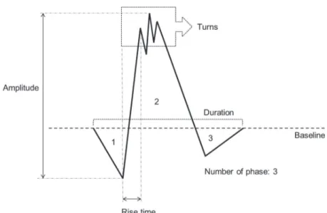
Morphology analysis of MUAP
The following characteristics of MUAPs are analyzed in detail to identify if the data are indicative of a normal motor unit or a specific disease pattern (Fig. 2):
1) Amplitude 2) Duration 3) Rise time
4) Number of phases/turns/satellite potentials 5) Stability
Each of these aspects can be influenced by physiologic (age, selected muscle, temperature, and force), technical (needle type and position relative to the muscle fiber, and filter settings), and pathologic factors.3
Amplitude and duration
The amplitude is calculated as the maximum amplitude between the positive and negative peaks of the main spikes.
Because the tissue between the needle and muscle fibers acts as a high-frequency filter, only 2-12 muscle fibers that are very close to the needle tip will affect the amplitude.2 The duration, which is measured as the time from the initial deflection until the signal returns to the baseline, reflects the activity of muscle fibers within 2.5 mm of the electrode tip.2 Nevertheless, the status of the motor units is represent- ed more by the duration than by the amplitude, primarily because muscle fibers contribute more to the period of the MUAP than to the spike amplitude, and also because move- ment of the electrode affects the duration less than the am- plitude.4 The amplitudes of MUAPs vary widely between in- dividual subjects, but mostly they are within the range from 100 μV to 2 mV when using a concentric needle.2 The MUAP duration is normally 5-15 ms in most limbs, and it typically increases for old age, low temperatures, and distal limbs.2 The pitch and volume of the electrical sound are generally associated with the duration and amplitude, respectively, of the MUAP.
Rise time
The rise time is defined as the time period between the larg- est positive and negative amplitudes (major spike), and it re- flects how far the muscle fiber is from the recording needle tip, it can be used to select the MUAPs to be analyzed. Only signals from nearby fibers in motor units (with rise times <
500 μs) should be analyzed when assessing the morpholo- gy.2
Number of phases/turns/satellite potentials
The number of phases is defined as the number of area made by division of MUAPs waves with baseline. This can also be quantified as the number of times that the signal crosses the baseline of the MUAP plus 1.2 An MUAP with more than four phases is defined as a polyphasic potential, and reflects desynchronized activation of muscle fibers within a motor unit.2 Normally fewer than 15% of MUAPs in muscles are polyphasic, although this proportion can be up to 25% in healthy deltoid muscles.2 An abnormal condi- tion such as neuropathic or myopathic disease is suspected when more than 15% of MUAPs are polyphasic (> 25% in deltoid muscles).2
Changes in the directions of the MUAP that do not pass through the baseline but produce a thorny shape are called turns (or serrations).4 Turns have the same physiologic meaning as polyphasic potentials, with the presence of more than five turns considered to be abnormal.5 A satellite potential is a time-linked potential that follows the main po-
tential slowly, and occurs frequently in the presence of early reinnervation.2
Stability
A healthy MUAP shows a uniform morphology during repet- itive firings. The development of any variability in the config- uration (number of phases) or amplitude at a fixed position suggests impairment of neuromuscular transmission.2 Not only changes to the NMJ such as due to myasthenia gravis or Lambert-Eaton syndrome, but also any denervation con- dition such as neuropathic/myopathic disorders can make MUAPs unstable.2
Firing-pattern analysis (recruitment and interference-pattern analy- sis)
MUAPs during voluntary contractions show a semirhythmic pattern that appears orderly but with some variation during the interpotential interval.4 MUAPs can be differentiated from spontaneous abnormal activities such as fibrillation/
positive sharp waves and myotonic discharges based on fir- ing patterns; for example, a fibrillation/positive sharp wave is regular, whereas a myotonic discharge shows a waxing and waning amplitude.4
Recruitment
Changes in the number of motor units in muscles can be indirectly estimated with EMG by employing voluntary con- tractions. Recruitment involves the relationship between the
fig. 2. Parameters for the morphology evaluation of motor-unit action potentials.
firing rate and the number of different MUAPs required to produce a certain contraction. To exert a larger force, the fir- ing rate of the initial motor units initially increases, and then additional motor units start to be activated and participate in producing the contraction. In the normal state, motor units will start firing at 5-8 Hz and start to recruit other MUAPs even during low-intensity contractions (8-10 Hz).2
The recruitment frequency is one parameter for quanti- fying recruitment, and corresponds to the firing rate when the next motor unit is recruited. Thinking backwards, the re- cruitment ratio between the maximum firing rates of initially activated motor units to the number of different shapes of MUAP firing can be calculated.4 This ratio is normally about 5:1, which means that four or more units should be firing at 20 Hz.4 Notably, the frequency and ratio of recruitment should be measured during firing rates of at least 15 Hz, because in several pathologic condition selective higher threshold, large size of motor unit are lost.5
Recruitment can be classified into normal, reduced, and early (rapid) recruitment, and poor activation. Reduced re- cruitment can be seen in primary neuropathic disease with axonal loss or conduction block and end-stage myopathy.5 Poor activation shows a low firing rate but a normal recruit- ment pattern. This can be due to poor cooperation by pa- tients or other upper motor neuron disease, and should be differentiated from true reduced-recruitment conditions.2 Early recruitment refers to the presence of a large number of MUAPs when generating a minimal force. Early recruitment happens when several muscle fibers eliminate in motor unit and motor unit became smaller and generate less force, as in myopathies and neuromuscular junction disorder with conduction block.5
Interference pattern: MUAP analysis during maximal con- traction
Greater activation results in overlapping of individual MUAPs to produce an interference pattern. A healthy complete in- terference pattern exhibits the rapid movement of MUAPs and increased amplitude, eventually showing as a thick line with an amplitude of 2-4 mV when maximal contraction is reached.2 The greatest applied force will result in the con- traction of many motor units, which will make it difficult to distinguish individual MUAPs. However, in a neuropathic disorder there will be a reduction in the firing rate of motor
units during maximal contraction and the interference pat- tern will look like a picket fence with each abnormal MUAP being discernible even when the patient contracts maximal- ly.2 Maximal contractions by a myopathy patient will pro- duce the normal number of MUAPs, but they will have small amplitudes, be of short duration, and overlap.2
Many physicians prefer to check recruitment only based on the interference pattern. However, analyzing the interfer- ence pattern during strong voluntary contractions is painful, and it is difficult to judge the relationship between the firing rate and the number of MUAPs when there are overlapping electrical signals. In most cases, performing recruitment analysis during only moderate effort is sufficient.4
Interpretation of EMG findings
Basic principles for interpreting needle EMG findings Interpreting the results of needle EMG is a crucial aspect of electrophysiologic studies. Performing accurate diagnoses of neuromuscular disorders using needle EMG requires electromyographers to receive sufficient training and have a comprehensive knowledge of neuromuscular anatomy, physiology, and diseases.1,7-9 This means that a considerable amount of time is needed to achieve adequate skill and knowledge in performing the needle EMG procedure and interpreting the findings, and continuous efforts should be made to master this.
Planning of needle EMG and interpreting the results based on the clinical context and the results of NCS
Needle EMG and NCS are very sensitive tests, during which examiners can encounter subtle or minimal abnormalities caused by subclinical diseases even in the absence of clini- cal symptoms and signs. Moreover, numerous physiologic and technical factors can affect the results of a needle EMG study, and so the electromyographer should always inter- pret any detected needle EMG abnormalities in the context of their clinical relevance.
Examiners must attempt to obtain adequate data for a diagno- sis and minimize patient discomfort
Needle EMG is uncomfortable, and sometimes even painful.
Patients may tolerate only a very small number of needling interventions on muscles, and especially young children fre-
quently cannot complete an entire examination.10 Planning which muscles to sample in needle EMG is crucial, with the most-important muscles for a diagnosis being selected first, and routine needle EMG examinations of many muscles being avoided. Moreover, the electromyographer should be prepared to modify the initial EMG plan based on the anal- ysis results obtained in real time; this approach will allow an accurate diagnosis to be obtained from an examination of the minimum number of muscles.
Avoiding overestimation of clinically insignificant or uncorrelat- ed abnormalities in needle EMG
The results of NCS and abnormal findings of needle EMG sometimes do not fit or are not correlated with the clinical diagnosis, which results in the electromyographer not being able to make a definitive diagnosis based on needle EMG.
The clinical assessment should be repeated in this situation.
Moreover, electromyographers need to understand the lim- itations of a needle EMG study, and always try to avoid over- estimating clinically uncorrelated or insignificant findings of a needle EMG study, since they could eventually lead to unnecessary or even harmful treatments.8
Common patterns of needle EMG for correlations between clinical and electrophysiologic findings
A needle EMG study can identify the precise location of a lesion, which is not possible clinically. Moreover, specific pat- terns of spontaneous activities and MUAP characteristics can provide useful information about the underlying pathologic process.2,5,8
Normal results
If the results of needle EMG studies are normal, the electro- myographer should reassess the clinical diagnosis of the patient—the patient could indeed be clinically normal, but the possibility of the clinical symptoms being caused by dis- orders of the central nervous system should be considered.
The EMG findings can be normal in the presence of hyper- acute axonal loss. The processing time of reinnervation with changes in MUAP can result in the only abnormal finding of needle EMG in an acute setting being a decreased recruit- ment of MUAPs, and this might not be detected during the test if the lesion is not severe. The results of needle EMG can also be normal if the pathology underlying the neuropathy
is mainly demyelinating, in which case NCS findings are important for the diagnosis. A conduction block in demye- linating neuropathies can result in decreased recruitment of MUAPs. NMJ disorders can present with normal findings in a needle EMG study depending on their severity: a mild NMJ disorder can result in only slight variations of the firing of muscle fibers, and both the morphology and recruitment of the MUAPs can be normal.
Neuropathic pattern
A pattern of acute or subacute axonal neuropathy can be observed after several days and weeks after the onset, which shows denervation potentials including fibrillation and positive sharp waves in needle EMG, while the morpholo- gy of MUAP remains normal. The occurrence of Wallerian degeneration and reinnervation can make abnormal spon- taneous activity more obvious, with the following morphol- ogy changes appearing in MUAPs: long durations, high amplitudes, and/or polyphasic with decreased recruitment.
Denervation potentials will disappear in chronic axonal loss.
MUAP morphologic changes often persist for a long time or even indefinitely. Pure demyelinating neuropathy will pres- ent with normal needle EMG findings because denervation and reinnervation never occur without axonal loss. However, conduction blocks can result in decreased recruitment in relevant muscles being detected.
Myopathic pattern
The common myopathic pattern is normal NCS results and needle EMG showing short, small, and polyphasic MUAPs with early recruitment. Spontaneous activity may be present depending on the characteristics of the myopathies; positive sharp wave and fibrillation potentials can be observed in inflammatory myopathies, while some myopathies are asso- ciated with myotonic discharges. Chronic myopathies may cause typical long-duration, high-amplitude, and polyphasic MUAPs (neurogenic MUAPs) with active denervation po- tentials, but early recruitment is still observed. Therefore, the key feature of needle EMG for differentiating chronic myopathies from chronic neuropathies is to evaluate recruit- ment; electromyographers also consider that NMJ disorder with conduction block can show early recruitment in needle EMG.2,11
Other considerations
As mentioned above, certain characteristic spontaneous activity in needle EMG can provide an important clue for a definitive diagnosis.2,5 Myotonic discharges are observed in a few myopathies and in hyperkalemic periodic paralysis.
Myokymic discharges in the limb typically occur in radia- tion-induced nerve injury or entrapment neuropathy, while facial myokymia is seen in Guillain-Barré syndrome, multiple sclerosis, and pontine gliomas. Neuromyotonia is a repre- sentative feature of hyperexcitable nerve syndromes such as Isaac’s syndrome. Thus, identifying spontaneous activity is one of the most-important aspects of needle EMG.
Reporting the results of needle EMG
The obtained needle EMG data should be presented in a standardized format that includes a list of all of the muscles tested in order to facilitate their use by other electromyogra- phers.12 The reported diagnostic interpretations should con- sider the results of both NCS and needle EMG. The following rules are recommended:
1) For each of the examined muscles, describe whether the insertional, spontaneous, and voluntary potentials are normal, in terms of their amplitudes, durations, phases, and recruitment. Additional details should be recorded if the potentials are abnormal.
2) Describe whether there are interval changes in EMG findings if the results from previous studies are avail- able. Additionally, each change can be categorized as an improvement, no change, or a deterioration.
3) Record any limitation of the needle EMG study, includ- ing pain and issues with a lack of cooperation or endur- ance that affected the planning and interpretation of the examination results. Suggest whether an additional or follow-up needle EMG study is indicated.
ConClusions
Needle EMG studies are essential parts of EDX testing for diagnosing neuromuscular disorders. Electromyographers must have sufficient knowledge of electrodiagnosis and
neuromuscular disorders, and they must control for electro- diagnosis technical factors. This review has presented the appropriate testing and interpretation methods for needle EMG studies, and this information should help electromyog- raphers to perform the associated procedures properly.
Conflicts of Interest
The authors have no conflicts to disclose.
RefeRenCes
1. AANEM. Proper performance and interpretation of electrodiag- nostic studies. Muscle Nerve 2015;51:468-471.
2. Preston DC, Shapiro BE. Electromyography and neuromuscular disorders: clinical-electrophysiologic correlations. 3rd ed. Lon- don: Elsevier Saunders, 2013;125-266.
3. Rubin DI. Technical issues and potential complications of nerve conduction studies and needle electromyography. Neurol Clin 2012;30:685-710.
4. Daube JR, Rubin DI. Needle electromyography. Muscle Nerve 2009;39:244-270.
5. Rubin DI. Needle electromyography: basic concepts and pat- terns of abnormalities. Neurol Clin 2012;30:429-456.
6. Mills KR. The basics of electromyography. J Neurol Neurosurg Psychiatry 2005;76 Suppl 2:ii32-ii35.
7. Dumitru D, Amato AA, Zwarts M. Electrodiagnostic medicine.
2nd ed. Philadelphia: Hanley & Belfus, Inc., 2002;257-258.
8. Katirji B, Kaminski HJ, Ruff RL. Neuromuscular disorders in clinical practice. 2nd ed. New York: Springer, 2013;89-152.
9. Kimura J. Electrodiagnosis in diseases of nerve and muscle:
principles and practice. 1st ed. Oxford: Oxford University Press, 2001;333-360.
10. Strommen JA, Daube JR. Determinants of pain in needle electro- myography. Clin Neurophysiol 2001;112:1414-1418.
11. Paganoni S, Amato A. Electrodiagnostic evaluation of myopa- thies. Phys Med Rehabil Clin N Am 2013;24:193-207.
12. Jablecki CK, Busis NA, Brandstater MA, Krivickas LS, Miller RG, Robinton JE, et al. Reporting the results of needle EMG and nerve conduction studies: an educational report. Muscle Nerve 2005;32:682-685.