Bacteremia Detected by a Peripheral Blood Smear in a Pediatric Surgical Patient with Thrombocytopenia
Jeong Tae Kim1, Jae Hyeon Lee1, Hye Soo Lee1,3,4, Yong Gon Cho1,3,4, Dal Sik Kim1,3,4, Sam Im Choi1,3,4, Soo Chul Cho2,3,4
Departments of
1Laboratory Medicine and
2Pediatrics, Chonbuk National University Medical School,
3
Research Institute of Clinical Medicine and
4Institute for Medical Sciences, Chonbuk National University, Jeonju, Korea
Microscopic examination of peripheral blood smear (PBS) for detection of microorganisms is simple method that can be used for doctors to confirm the septice- mia more swiftly and to select more specific therapy.
But it is unusual to find microorganisms in PBS. We report a case of gram negative bacteremia diag- nosed by PBS in a severe thrombocytopenic pedia- tric surgical patient. A 6-month and 2 week old ba- by with cyanosis was diagnosed congenital heart diseases such as transposition of great arteries, at- rial septal defect, and patent ductus arteriosus. The infant underwent surgical operations and the post- operative platelet count progressively decreased in spite of transfusion of multiple platelet concentrates.
We performed routine examination of a PBS for evaluation of severe thrombocytopenia. The PBS re-
vealed severe thrombocytopenia, leukopenia with left shifted and some extracellular bacilli. Toxic granula- tions, toxic vacuoles and some bacilli were observed in the neutrophils. The bacilli were identified as Pseu- domonas aeruginosa and Serratia marcescens in blood culture. To our knowledge, this is the second case of bacteremia diagnosed by PBS before the positive blood culture in Korea. We suggest that a PBS is useful for the rapid detection of organisms in cases of septicemia with severe thrombocytopenic pediatric surgical patient. (Korean J Clin Microbiol 2010;13:182-186)
Key Words: Gram negative bacteremia, Peripheral blood smear, Thrombocytopenia
Received 5 July, 2010, Revised 27 July, 2010 Accepted 20 August, 2010
Correspondence: Hye Soo Lee, Department of Laboratory Medicine, Chonbuk National University Hospital, 634-18 Geumam-dong, Dukjin- gu, Jeonju 561-182, Korea. (Tel) 82-63-250-1218, (Fax) 82-63-250- 1200, (E-mail) leehs@jbnu.ac.kr
182 INTRODUCTION
Septicemia is a severe clinical syndrome characterized by sys- temic signs of infection, shock and systemic organ failure. A rap- id and definitive diagnosis is essential in the management of septicemia. The diagnosis of septicemia is confirmed by the pres- ence of organisms in the blood. Although positive results in blood culture may establish the diagnosis, it is not a factor of the initial treatment decisions because of it takes a bit of time to get pos- itive blood culture. A microscopic examination of the peripheral blood smear (PBS) for detection of organisms has been reported to be a simple method that can be used to hasten the confirmation of septicemia thereby enabling doctors to immediately select more specific treatment. However, a PBS occasionally shows bacteria only in cases of overwhelming septicemia[1-3]. In Korea, report of diagnosis of bacteremia by the PBS is very rare[4]. In this re- port, we describe a case of septicemia caused by Gram negative bacilli, which was diagnosed by a microscopic examination of
PBS before positive blood culture in a severe thrombocytopenic infant who had surgical operation for underlying illness.
CASE REPORT
A 6-month and 2-week old male infant was taken to the depart- ment of pediatrics due to cyanosis and was diagnosed congenital cardiac anomalies with transposition of great arteries (TGA), pat- ent ductus arteriosus (PDA), and atrial septal defect (ASD). The infant was transferred and admitted to the thoracic and car- diovascular surgery for the operation of the above cardiac anomalies. The infant underwent operations three times for 9 days. Routine complete blood counts (CBC), chemistry, arterial blood gas analysis (ABGA) and electrolytes were monitored sev- eral times a day. His CBC and ABGA findings were occasionally unstable and C-reactive protein (CRP) was elevated. He received multiple units of packed red cells (PRC), platelet concentrates (PC), or fresh frozen plasma (FFP) when his CBC data were abnormal. His platelet count was getting decreased despite of PC transfusion (Fig. 1). On the 13th day of hospital (the 3rd post-op- eration day after the 3rd operation), a PBS examination was re- quested for an evaluation of severe thrombocytopenia. A review
Fig. 1. The complete blood counts (including segmented neutrophils) results of patient during admission days. *after 1st operation; †after 2nd operation; ‡after 3rd operation; §on presentation.
Abbreviations: AD, admission day; HD, hospital day.
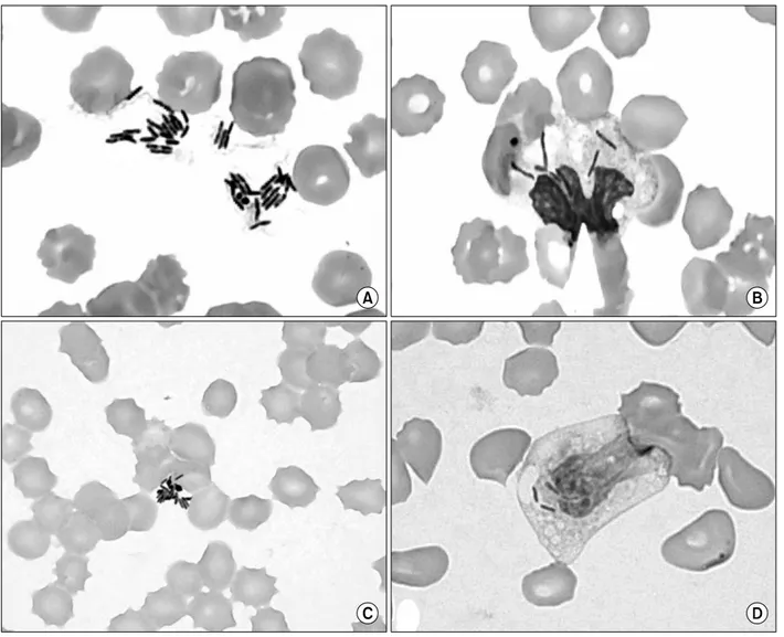
Fig. 2. Microorganisms shown on peripheral blood smears. (A) Extracellular clustering bacilli and anisocytosis and poikilocytosis of RBCs on peripheral blood smear (Wright stain, ×1,000). (B) Bacilli, vacuoles and toxic granules in cytoplasm of neutrophils (Wright stain, ×1,000). (C) Gram negative bacilli in cytoplasmic and extracellular area on peripheral blood smear (Gram stain, ×1,000). (D) Vacuolation and Gram negative bacilli in neutrophils (Gram stain, ×1,000).
of the PBS revealed severe thrombocytopenia, leukopenia with left shifted, and normochromic normocytic anemia with anisocy- tosis and poikilocytosis such as schistocytes and burr cells. There are some neutrophils with toxic granulations and vacuoles, and some intracellular and extracellular bacilli were observed (Fig. 2).
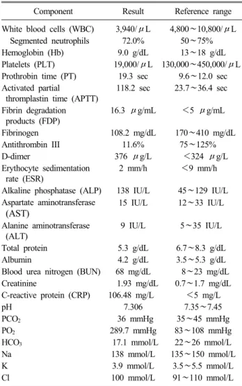
The bacilli were Gram negative and we made preliminary diag- nosis of septicemia caused by Gram negative bacilli. Two sets of blood cultures and pleural fluid cultures were obtained. At that time, the patient’s body temperature was 35.9oC, blood pressure was 70/29 mmHg, pulse rate was 126/min, and respiratory rates were 26/min. Laboratory findings revealed pancytopenia with se- vere thrombocytopenia, disseminated intravascular coagulation (DIC), and respiratory acidosis (Table 1). A chest radiograph showed pulmonary edema. The next day, multiorgan failures were developed and in spite of all intensive medical treatments includ- ing antimicrobial agent (ceftriaxone, vancomycin), the patient was expired finally. After overnight incubation, blood and pleural fluid
Table 1. Data of laboratory findings on presentation date
Component Result Reference range
White blood cells (WBC) 3,940/μL 4,800∼10,800/μL Segmented neutrophils 72.0% 50∼75%
Hemoglobin (Hb) 9.0 g/dL 13∼18 g/dL
Platelets (PLT) 19,000/μL 130,000∼450,000/μL Prothrobin time (PT) 19.3 sec 9.6∼12.0 sec Activated partial
thromplastin time (APTT)
118.2 sec 23.7∼36.4 sec
Fibrin degradation products (FDP)
16.3 μg/mL <5 μg/mL
Fibrinogen 108.2 mg/dL 170∼410 mg/dL
Antithrombin III 11.6% 75∼125%
D-dimer 376 μg/L <324 μg/L
Erythocyte sedimentation rate (ESR)
2 mm/h <9 mm/h
Alkaline phosphatase (ALP) 138 IU/L 45∼129 IU/L Aspartate aminotransferase
(AST)
15 IU/L 12∼33 IU/L
Alanine aminotransferase (ALT)
9 IU/L 5∼35 IU/L
Total protein 5.3 g/dL 6.7∼8.3 g/dL
Albumin 4.2 g/dL 3.5∼5.3 g/dL
Blood urea nitrogen (BUN) 68 mg/dL 8∼23 mg/dL
Creatinine 1.93 mg/dL 0.7∼1.7 mg/dL
C-reactive protein (CRP) 106.48 mg/L <5 mg/L
pH 7.306 7.35∼7.45
PCO2 36 mmHg 35∼45 mmHg
PO2 289.7 mmHg 83∼108 mmHg
HCO3 17.1 mmol/L 22∼26 mmol/L
Na 138 mmol/L 135∼150 mmol/L
K 3.9 mmol/L 3.5∼5.5 mmol/L
Cl 100 mmol/L 91∼110 mmol/L
cultures were positive (BactAlert 3D, bioMerieux, Inc., Durham, NC, USA) and identified P. aeruginosa and S. marcescens (Vitek2, bioMerieux, Inc., Hazelwood, MO, USA).
DISCUSSION
A rapid and definitive diagnosis is essential in the management of septicemia. Blood culture may establish the diagnosis, but it is not a factor in the initial treatment decisions because of it requires incubation time to get positive results. Several studies on the utili- ty of examining peripheral blood in the setting of probable septi- cemia have been performed. The use of buffy coat smears has been attempted to detect bacteremia[5,6]. However, these methods have never been popular as diagnostic tools because of lack of sensitivity in detecting bacteremia when being compared with conventional blood culture method.
PBS is also insensitive when compared to blood cultures and the organisms appeared on smears could not be identifed to the species level of organisms. Detection of microorganisms by clin- ical specimen examination requires a microorganism with the con-
centration of 105 CFU/mL or greater[7,8]. This degree of bacter- emia is unusual. Therefore, detection of bacteremia by routine blood smear review will not be possible in most cases and it has not been widely used for detection of bacteremia. But, the sensi- tivity of PBS review is greatly increased if the laboratory ob- servers, who have abundant experiences and receive sufficient training, are specifically directed to look for the presence of mi- croorganisms[8-10]. And this method is simple, inexpensive and safe procedure to detect a variety of organisms and to evaluate the type of inflammatory responses[9]. Typical Gram reactions, morphologies and arrangements of the observed organisms may give the presumptive identification of some certain etiological bacteria[9]. Moreover, the slide can be saved as a part of the me- dical record, examined by others, and restained if necessary[10].
So if microscopic examination of PBS by experienced laboratory observers is performed, it can provide a rapid preliminary diag- nosis of overwhelming bacteremia is suspected in such cases of neonatal sepsis[10], thereby allowing clinicians to strengthen the empirical antimicrobial regimen and it may improve the outcome of the septic process.
The results of microscopic examination are examiner depend- ent[8,10]. Careful screening of PBS only frequently reveals intra- leukocytic organisms. The criteria for diagnosing septicemia based on a PBS are as follows; cellular inclusion bodies are or- ganisms, the organisms must be intracellular, and the cells con- taining organisms must be leukocytes and not nonhematopoietic cells[3]. If only extracellular microorganisms in PBS is observed, they have to be interpreted carefully because those could be con- tamination in vitro such as artifacts from slide glasses, anti- coagulants, and staining fluid. Toxic neutrophils such as toxic granules, cytoplasmic vacuoles, and Döhle inclusion bodies ap- pear to be very predictive of systemic infection[11], therefore identification of these morphologic alterations in neutrophils could be used to distinguish true bacteremia from specimen contamination. Actually, van der Meer et al[12] demonstrated 4 cases of bacteria in PBS, two of them had a fatal outcome, but the other 2 were caused by a contamination either via the central venous catheter or in vitro, both without dramatic outcomes.
Gram negative septicemia is frequently a fatal complication threatening the newborn and infants received surgical oper- ations[13]. There are multiple factors such as contributing to the apparently increased frequency of septicemia. Direct surgical in- sults to internal organs, various vascular accesses, open wounds, and use of intravascular catheters for monitoring and endotracheal tubes and ventilators for the treatment of respiratory distress has increased the infant's exposure to bacteria.
Thrombocytopenia is frequently diagnosed in pediatric surgical patients suffering from Gram negative septicemia. The reason for decreased platelet count in postoperative septicemia has been as- cribed to several causes such as bone marrow suppression, DIC and hemophagocytosis, but the mechanisms responsible for thrombocytopenia are not clearly identified, yet[14]. Among pos- tulated mechanisms, there are such things as the following. First, platelet production may be impaired as a result of direct invasion
of bacteria in the megakaryocytes, second, circulating platelets may interact with bacteria and be destroyed by bacteria, third, pla- telets may damaged by bacterial immune complexes[15].
Thrombocytopenia is caused by infection, thrombotic thrombo- cytopenic purpura, heparin induced thrombocytopenia, DIC, drug-induced thrombocytopenia, and posttransfusion thrombocyto- penia[15]. Because of persistent thrombocytopenia is associated with high morbidity and mortality, early recognition of the cause of postoperative thrombocytopenia is essential for appropriate pre- ventive measures and management of bleeding complication.
Initial evaluation should be include CBC, reticulocyte count, hap- toglobin, prothrombin time, activated thromboplastin time, throm- bin time, fibrinogen, and fibrin degradation products. There are some specific diagnostic tools including PBS and blood culture for differential diagnosis of postoperative thrombocytopenia[15].
The degree of thrombocytopenia is very important for differ- ential diagnosis and treatment. Rowe et al[13] observed that all the major surgical infants and children with positive blood cul- tures for Gram negative septicemia had a platelet count below 150,000/μL, but the patients with Gram positive septicemia had a platelet count above 150,000/μL, and they conclude that the most rapid, simple and accurate method for the early detection of gram negative septicemia in the various pediatric surgical patients appeared to be serial platelet counts among various parameters.
The following recommendations would improve the management of pediatric surgical patients who are at high risk to develop gram negative septicemia[13]. Postoperative pediatric patients at risk, especially who had major surgery such as cardiac operation as in the case of our patient, should be monitored by serial platelet counts because a fall in platelet count is very suggestive of Gram negative septicemia. Therefore, if patient has evidence of in- fection such as fever and chills, sources of infection should be sought and multiple PBS and blood cultures should be drawn to determine the pathogens. Removal of intravascular catheters, dis- continuance of total parenteral nutrition and the initiation of anti- biotic therapy should be considered. If a fall in platelet count be- low 150,000/μL and even minimal clinical signs are present pres- ent in the postoperative infants or children, antibiotics admin- istration should be considered and other supportive or special measures such as platelet transfusion should be taken to combat infection. In spite of antibiotics and other treatments, a fall or a persistently low platelet count over several days after operation suggests ineffective therapy. Sources of continued infection should be sought and the antibiotic program should be changed according to the antimicrobial susceptibility test[13].
In conclusion, although PBS is not a routine procedure for de- tecting bacteremia, this procedure could be useful before blood culture when overwhelming Gram negative bacteremia is sus-
pected in pediatric surgical patients with persistent thrombocyto- penia as in the case of our patient.
REFERENCES
1. Fife A, Hill D, Barton C, Burden P. Gram negative septicaemia diagnosed on peripheral blood smear appearances. J Clin Pathol 1994;47:82-4.
2. Mirza I, Wolk J, Toth L, Rostenberg P, Kranwinkel R, Sieber SC.
Waterhouse-Friderichsen syndrome secondary to Capnocytophaga canimorsus septicemia and demonstration of bacteremia by pe- ripheral blood smear. Arch Pathol Lab Med 2000;124:859-63.
3. Nakamura H, Saitou M, Kinjo S, Kaneshima H, Higa F, Tateyama M, et al. Overwhelming pneumococcal bacteremia revealed by a peripheral blood smear in a 74-year-old healthy woman. Intern Med 2007;46:303-6.
4. Sohn HE and Chung HR. Bacteremia diagnosed on peripheral blood smear before blood cultures become positive: a case report.
Korean J Clin Pathol 1999;19:27-30.
5. Ristuccia PA, Hoeffner RA, Digamon-Beltran M, Cunha BA.
Detection of bacteremia by buffy coat smears. Scand J Infect Dis 1987;19:215-7.
6. Rodwell RL, Leslie AL, Tudehope DI. Evaluation of direct and buffy coat films of peripheral blood for the early detection of bacteraemia. Aust Paediatr J 1989;25:83-5.
7. Shanholtzer CJ, Schaper PJ, Peterson LR. Concentrated gram stain smears prepared with a cytospin centrifuge. J Clin Microbiol 1982;16:1052-6.
8. Branda JA, Ferraro MJ, Kratz A. Sensitivity of peripheral blood smear review for the diagnosis of Candida fungemia. Arch Pathol Lab Med 2007;131:97-101.
9. Misawa S. Rapid diagnosis of infectious diseases; features and limitations of the microscopic examination of clinical specimens.
Rinsho Biseibutshu Jinsoku Shindan Kenkyukai Shi 1999;10:
121-31.
10. Graham BS. Detection of bacteremia and fungemia: microscopic examination of peripheral blood smears. Infect Control 1984;5:448- 52.
11. Kroft SH. Infectious diseases manifested in the peripheral blood.
Clin Lab Med 2002;22:253-77.
12. van der Meer W, Verwiel JM, Gidding CE, de Metz M, de Keijzer MH. Bacteria in blood smears: overwhelming sepsis or trivial contamination. Acta Haematol 2002;107:220-3.
13. Rowe MI, Buckner DM, Newmark S. The early diagnosis of gram negative septicemia in the pediatric surgical patient. Ann Surg 1975;182:280-6.
14. François B, Trimoreau F, Vignon P, Fixe P, Praloran V, Gastinne H. Thrombocytopenia in the sepsis syndrome: role of hemopha- gocytosis and macrophage colony-stimulating factor. Am J Med 1997;103:114-20.
15. Chang JC. Review: Postoperative thrombocytopenia: with etiologic, diagnostic, and therapeutic consideration. Am J Med Sci 1996;311:
96-105.
=국문초록=
수술 후 혈소판감소증을 보인 환아의 말초혈액도말표본에서 진단된 균혈증 1예
전북대학교 의학전문대학원 1진단검사의학교실, 2소아과학교실, 전북대학교 3임상의학연구소, 4의과학연구소
김정태1, 이재현1, 이혜수1,3,4, 조용곤1,3,4, 김달식1,3,4, 최삼임1,3,4, 조수철2,3,4
말초혈액도말표본에서 세균의 관찰은 균혈증의 조기 진단 및 치료에 도움을 줄 수 있는 매우 간단한 방법이나, 실제로 말초혈액도말표본에 의해서 균혈증을 진단하는 경우는 매우 드물다. 저자들은 수술 후 지속적인 혈소판감소증을 보인 환아의 말초혈액도말표본에서 그람음성균혈증을 진단하였기에 보고하고자 한다. 환아는 6개월 2주 된 남아로 청색증을 주소로 입원하여 대혈관전위, 동맥관개존, 심방사이막결손 등의 선천성심장병으로 진단받고 수술을 받았다. 수술 후 농 축혈소판의 수혈에도 불구하고 혈소판수가 지속적으로 감소되어 말초혈액도말검사를 시행한 결과, 혈소판 감소, 백혈구 좌방이동, 백혈구 내 독성과립과 독성공포와 함께 백혈구에 탐식되거나 탐식되지 않은 막대균들이 관찰되었다. 막대균 들은 그람음성막대균으로, 이 균들은 뒤이은 혈액배양에서 Pseudomonas aeruginosa와 Serratia marcescens로 최종 동정되 었다. 말초혈액도말검사로 진단된 균혈증은 국내에서는 본 증례가 두 번째이다. 본 증례와 같이 수술 후 지속적인 혈소 판감소증을 보인 환아에서 말초혈액 도말표본의 관찰은 균혈증을 신속히 진단하는 데 도움이 될 것으로 생각한다. [대한 임상미생물학회지 2010;13:182-186]
교신저자 : 이혜수, 561-182, 전북 전주시 덕진구 금암동 634-18 전북대학교병원 진단검사의학과
Tel: 063-250-1218, Fax: 063-250-1200 E-mail: leehs@jbnu.ac.kr